

INTRODUCTION
Microglia constitute the innate immune cell population of the central nervous system (CNS). Since the first studies by del Rio-Hortega, it has generally been accepted that microglia in the adult CNS derive from cells of mesodermal origin with ameboid morphology (ameboid microglia) that colonize all regions of CNS during development (del Rio-Hortega, Reference del Rio-Hortega and Penfield1932; Cuadros and Navascués, Reference Cuadros and Navascués1998; Chan et al., Reference Chan, Kohsaka and Rezaie2007; Prinz and Mildner, Reference Prinz and Mildner2011; Prinz et al., Reference Prinz, Priller, Sisodia and Ransohoff2011). Ameboid microglia have the ability to migrate long distances, proliferate and perform phagocytosis. Once they have reached their final destination in the nervous parenchyma, they differentiate into ramified mature cells (ramified microglia) that are evenly dispersed throughout the CNS. Each ramified microglial cell continuously explores the environment by extending and retracting its cell processes, which show contact inhibition with the processes of other microglial cells, leading to a characteristic spatial arrangement in which microglial cells occupy non-overlapping territories (Davalos et al., Reference Davalos, Grutzendler, Yang, Kim, Zuo and Jung2005; Nimmerjahn et al., Reference Nimmerjahn, Kirchhoff and Helmchen2005). In the pathological CNS, ramified microglia acquire an activated phenotype (activated microglia) characterized by a macrophage-like ameboid morphology, upregulation of migratory, proliferative and phagocytic capacities, and the production of neurotoxic and/or neurotrophic molecules (Hanisch and Kettenmann, Reference Hanisch and Kettenmann2007).
Since their discovery, microglial cells have been associated with neuronal cell death for the following reasons:
1. Developing microglial cells spread throughout the nervous parenchyma, coinciding with a period of naturally occurring neuron cell death in different CNS regions (Perry et al., Reference Perry, Hume and Gordon1985; Ashwell, Reference Ashwell1990, Reference Ashwell1991; Ferrer et al., Reference Ferrer, Bernet, Soriano, del Rio and Fonseca1990; Perry and Gordon, Reference Perry and Gordon1991; Marín-Teva et al., Reference Marín-Teva, Cuadros, Calvente, Almendros and Navascués1999).
2. Developing microglial cells are frequently observed to phagocytose dying neurons and show phagosomes containing neuronal debris (Ferrer et al., Reference Ferrer, Bernet, Soriano, del Rio and Fonseca1990; Marín-Teva et al., Reference Marín-Teva, Cuadros, Calvente, Almendros and Navascués1999; Peri and Nüsslein-Volhard, Reference Peri and Nüsslein-Volhard2008).
3. In response to CNS injuries, mature microglial cells activate, concentrate in areas where neuronal death occurs and remove dying cells or their fragments by phagocytosis (Neumann et al., Reference Neumann, Kotter and Franklin2009).
Besides their debris clearance function, microglial cells play a much more active role in cell death phenomena and have been shown to promote neuronal death in various CNS regions during normal development (Mallat et al., Reference Mallat, Marín-Teva and Cheret2005; Bessis et al., Reference Bessis, Bechade, Bernard and Roumier2007) and to be either neurotoxic or neuroprotective in neurodegenerative diseases (Block et al., Reference Block, Zecca and Hong2007; Hanisch and Kettenmann, Reference Hanisch and Kettenmann2007). Greater understanding of the participation of microglial cells in neuronal cell death opens up the intriguing possibility of manipulating microglial cells to eliminate or reduce the impact of neurodegeneration.
Because of differences in cellular capacities between developing and adult microglia, some aspects of the relationship between microglia and neuronal cell death will be discussed separately in relation to the developing and mature CNS. Previously, we will summarize current knowledge on microglial ontogeny in order to define the microglia lineage-related terms used throughout this review.
ONTOGENY OF MICROGLIA
Some aspects of microglia development are relatively well known (Cuadros and Navascués, Reference Cuadros and Navascués1998; Streit, Reference Streit2001), although controversial issues on the origin of their progenitors remain to be clarified (Chan et al., Reference Chan, Kohsaka and Rezaie2007; Prinz and Mildner, Reference Prinz and Mildner2011). Thus, it is generally accepted that ramified microglia are differentiated cells present in the adult CNS that arise from undifferentiated round or elongated cells endowed with short stubby processes, which are present in the developing CNS and are known as ameboid microglia. Intermediate forms between ameboid microglia and fully ramified microglia, known as poorly ramified microglia, can be recognized in the developing CNS and are considered to be transitional stages in the differentiation of microglia (Cuadros and Navascués, Reference Cuadros and Navascués1998; Dalmau et al., Reference Dalmau, Vela, Gonzalez and Castellano1998; Rezaie and Male, Reference Rezaie and Male1999). Evidence is now emerging in favor of the origin of microglia from yolk sac-derived primitive myeloid progenitors that seed the CNS during early developmental stages (Alliot et al., Reference Alliot, Godin and Pessac1999; Herbomel et al., Reference Herbomel, Thisse and Thisse2001; Streit, Reference Streit2001; Chan et al., Reference Chan, Kohsaka and Rezaie2007; Ginhoux et al., Reference Ginhoux, Greter, Leboeuf, Nandi, See and Gokhan2010; Mizutani et al., Reference Mizutani, Pino, Saederup, Charo, Ransohoff and Cardona2012; Prinz et al., Reference Prinz, Priller, Sisodia and Ransohoff2011). Once within the developing brain, these progenitors show a macrophage-like appearance and have received different names, such as primitive macrophages (Andjelkovic et al., Reference Andjelkovic, Nikolic, Pachter and Zecevic1998; Ginhoux et al., Reference Ginhoux, Greter, Leboeuf, Nandi, See and Gokhan2010), early macrophages (Cuadros et al., Reference Cuadros, Martin, Coltey, Almendros and Navascués1993; Herbomel et al., Reference Herbomel, Thisse and Thisse2001), fetal macrophages (Streit, Reference Streit2001; Ransohoff and Perry, Reference Ransohoff and Perry2009), primitive/fetal macrophages (Sorokin et al., Reference Sorokin, Hoyt, Blunt and McNelly1992) or even early microglia (Schlegelmilch et al., Reference Schlegelmilch, Henke and Peri2011). They will be designated primitive macrophages throughout this review. Based on the results published by Alliot et al. (Reference Alliot, Godin and Pessac1999) and Ginhoux et al. (Reference Ginhoux, Greter, Leboeuf, Nandi, See and Gokhan2010), it can be hypothesized that primitive macrophages in earlier CNS development give rise to ameboid microglia at more advanced developmental stages, subsequently differentiating into ramified microglia. However, the extent to which the population of primitive macrophages contributes to the later ameboid microglia pool remains elusive, because an unquestionable connection between the two cell populations has not been conclusively established. With these reservations, we refer to primitive macrophages and ameboid microglia as two separate developmental stages of the microglial lineage in this review.
It can be useful to include both primitive macrophages and ameboid microglia in the term primitive macrophages/ameboid microglia because, except in the avascular retina of birds, ameboid microglial cells that colonize the developing CNS are mixed with preexisting primitive macrophages from which they are generally indistinguishable (Cuadros et al., Reference Cuadros, García-Martín, Martin and Ríos1991; Navascués et al., Reference Navascués, Moujahid, Almendros, Marín-Teva and Cuadros1995; Marín-Teva et al., Reference Marín-Teva, Cuadros, Calvente, Almendros and Navascués1999; Santos et al., Reference Santos, Calvente, Tassi, Carrasco, Martín-Oliva and Marín-Teva2008). In fact, primitive macrophages may have three possible fates in later development. First, they may disappear during the course of development, being a transient population that exists only as long as is necessary to accomplish their role during development. Second, their progeny may give rise to ameboid microglial cells and then to ramified microglia. Third, they may be a distinct cell population from that of ameboid microglia that also progressively differentiates into ramified microglia, contributing to the idea that microglial cells are a heterogeneous population with different origins (Provis et al., Reference Provis, Diaz and Penfold1996; Rezaie and Male, Reference Rezaie and Male1999; Olah et al., Reference Olah, Biber, Vinet and Boddeke2011). In this context, it was recently reported that primitive macrophages substantially contribute to the adult microglia population (Ginhoux et al., Reference Ginhoux, Greter, Leboeuf, Nandi, See and Gokhan2010), although whether they are precursors of ameboid microglia is an issue that requires further experimental research.
PRIMITIVE MACROPHAGES/AMEBOID MICROGLIA AND NEURONAL CELL DEATH DURING DEVELOPMENT: PHAGOCYTIC CLEARANCE OF CELL DEBRIS VERSUS MICROGLIA-PROMOTED CELL DEATH
Developmental cell death of neurons
Programmed cell death (PCD) is a key process in the proper development of the nervous system (Oppenheim, Reference Oppenheim1991; Kuan et al., Reference Kuan, Roth, Flavell and Rakic2000; Yuan and Yankner, Reference Yuan and Yankner2000). During vertebrate development, up to 50% of many types of neurons undergo cell death (Oppenheim, Reference Oppenheim1991; Raff et al., Reference Raff, Barres, Burne, Coles, Ishizaki and Jacobson1993), which is critical to establish a definitive pattern of neuronal connections. This neuronal death is commonly known as neurotrophic cell death, because it affects projecting neurons and other types of differentiated neurons and is regulated through competition for the limited amount of neurotrophic factors (NGF, BDNF, etc.) released by the target cells innervated by these neurons (Barde, Reference Barde1989; Oppenheim, Reference Oppenheim1989; Raff et al., Reference Raff, Barres, Burne, Coles, Ishizaki and Jacobson1993; Snider, Reference Snider1994). PCD is also observed earlier in development, before neurotrophic cell death, in populations of undifferentiated cells such as proliferating neuroepithelial cells and newly postmitotic neuroblasts, and has specific functions (e.g. morphogenic sculpting of the early nervous system) and involves different regulatory mechanisms (de la Rosa and de Pablo, Reference de la Rosa and de Pablo2000; Kuan et al., Reference Kuan, Roth, Flavell and Rakic2000; Yeo and Gautier Reference Yeo and Gautier2004; Valenciano et al., Reference Valenciano, Boya and de la Rosa2009). In any case, the PCD of neural cells during development leads to the formation of membrane bound apoptotic bodies that are rapidly phagocytosed and digested by different types of ‘professional’ and ‘non-professional’ phagocytes.
Phagocytosis of neuronal cell debris
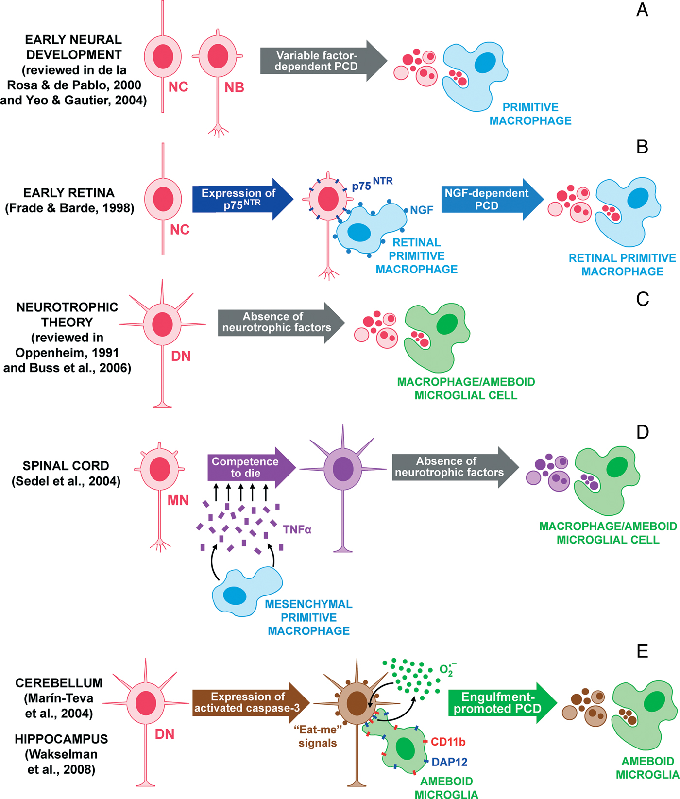
During early neural development, cell death debris is phagocytosed by yolk sac-derived primitive macrophages (see Fig. 1A,B) that colonize and ‘patrol’ the neuroepithelium (Cuadros et al., Reference Cuadros, Coltey, Carmen Nieto and Martin1992, Reference Cuadros, Martin, Coltey, Almendros and Navascués1993; Sorokin et al., Reference Sorokin, Hoyt, Blunt and McNelly1992; Herbomel et al., Reference Herbomel, Thisse and Thisse1999, Reference Herbomel, Thisse and Thisse2001; Lichanska and Hume, Reference Lichanska and Hume2000), as well as by neighboring neuroepithelial cells (García-Porrero and Ojeda, Reference García-Porrero and Ojeda1979; Kálmán, Reference Kálmán1989).
Relationship between microglial cells and neuronal cell death during normal development, as demonstrated in diverse studies on different parts of the developing nervous system. (A) At early phases of development, primitive macrophages throughout the CNS phagocytose the cell debris produced during the PCD of undifferentiated cells such as proliferating neuroepithelial cells (NCs) and postmitotic neuroblasts (NBs). (B) In the early retina, some NCs expressing the p75 neurotrophin receptor (p75NTR) die from the effect of the NGF produced by primitive macrophages (Frade and Barde, Reference Frade and Barde1998). (C) In more advanced development, when PCD is predominantly due to the lack of neurotrophic factors, the clearance of cell debris from differentiated neurons (DNs) is accomplished by primitive macrophages or ameboid microglial cells. (D) Differentiating motoneurons (MNs) in the spinal cord acquire competence to die through signaling by the TNF-α released from primitive macrophages in adjacent somites (Sedel et al., Reference Sedel, Bechade, Vyas and Triller2004). Two days after TNF-α signaling, committed MNs die and cell debris is phagocytosed by primitive macrophages/ameboid microglial cells invading the spinal cord. (E) Microglial-promoted PCD in some populations of DNs in the cerebellum (Marín-Teva et al., Reference Marín-Teva, Dusart, Colin, Gervais, van Rooijen and Mallat2004) and hippocampus (Wakselman et al., Reference Wakselman, Bechade, Roumier, Bernard, Triller and Bessis2008). Neurons initiating PCD express activated caspase-3 and show putative ‘find-me’ and ‘eat-me’ signals that attract microglial cells. Recognition of eat-me signals by microglial cells, which express both the β2-integrin CD11b and the immunoreceptor DAP12, triggers the neuron engulfment. Then, the final stages of PCD in the engulfed neurons are promoted by microglial-derived apoptotic effectors such as superoxide ions.
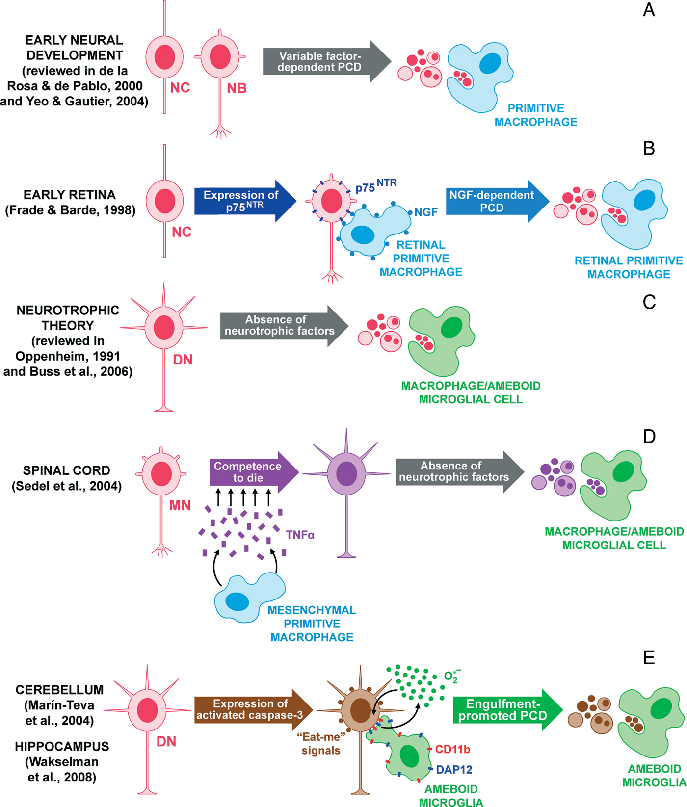
During neurotrophic cell death in more advanced development, the apoptotic neuronal cells are phagocytosed by primitive macrophages/ameboid microglial cells (Hume et al., Reference Hume, Perry and Gordon1983; Perry et al., Reference Perry, Hume and Gordon1985; Ashwell et al., Reference Ashwell, Hollander, Streit and Stone1989; Ashwell, Reference Ashwell1990, Reference Ashwell1991; Ferrer et al., Reference Ferrer, Bernet, Soriano, del Rio and Fonseca1990; Perry and Gordon, Reference Perry and Gordon1991; Thanos, Reference Thanos1991; Pearson et al., Reference Pearson, Payne and Cunningham1993; Egensperger et al., Reference Egensperger, Maslim, Bisti, Hollander and Stone1996; Moujahid et al., Reference Moujahid, Navascués, Marín-Teva and Cuadros1996; Marín-Teva et al., Reference Marín-Teva, Cuadros, Calvente, Almendros and Navascués1999; Upender and Naegele, Reference Upender and Naegele1999; Rakic and Zecevic, Reference Rakic and Zecevic2000; Peri and Nüsslein-Volhard, Reference Peri and Nüsslein-Volhard2008) (Fig. 1C–E) and by neuroectoderm-derived non-professional phagocytes such as neuroblasts, retinal Müller cells, cerebellar Bergmann glia and spinal cord astrocytes (O'Connor and Wyttenbach, Reference O'Connor and Wyttenbach1974; Egensperger et al., Reference Egensperger, Maslim, Bisti, Hollander and Stone1996; Marín-Teva et al., Reference Marín-Teva, Cuadros, Calvente, Almendros and Navascués1999; Parnaik et al., Reference Parnaik, Raff and Scholes2000).
Naturally occurring neuronal cell death has long been proposed as the most likely stimulus for the entry and spread of primitive macrophages/ameboid microglia into nervous parenchyma in different species, based on their temporal and spatial coincidence during development and the frequent observations of microglial cells in the process of phagocytosing dying neurons or with phagosomes containing neuronal debris (Perry et al., Reference Perry, Hume and Gordon1985; Ashwell, Reference Ashwell1990, Reference Ashwell1991; Perry and Gordon, Reference Perry and Gordon1991; Moujahid et al., Reference Moujahid, Navascués, Marín-Teva and Cuadros1996; Peri and Nüsslein-Volhard, Reference Peri and Nüsslein-Volhard2008). However, it has not yet been demonstrated that primitive macrophages/ameboid microglia are attracted to PCD sites in the developing CNS. In contrast, some studies found that migrating ameboid microglial cells followed well-defined routes that were not altered when they passed close to regions of abundant cell death, only phagocytosing apoptotic bodies that they met in their path (Marín-Teva et al., Reference Marín-Teva, Cuadros, Calvente, Almendros and Navascués1999; Rezaie and Male, Reference Rezaie and Male1999; Calderó et al., Reference Calderó, Brunet, Ciutat, Hereu and Esquerda2009). Hence, phagocytosis by primitive macrophages/ameboid microglia of apoptotic bodies from differentiated neurons during development appears to be a consequence of a chance encounter rather than a regulated process. This idea is supported by the active removal of cell death debris by other non-professional phagocytes during development, as reported above.
Developmental roles of microglial cells in neuronal death
Besides their role as scavengers, primitive macrophages/ameboid microglia may perform other physiological tasks during development, including cell death regulation in some neuronal populations, axonal guidance and synaptogenesis (Mallat and Chamak, Reference Mallat and Chamak1994; Mallat et al., Reference Mallat, Marín-Teva and Cheret2005; Bessis et al., Reference Bessis, Bechade, Bernard and Roumier2007). Primitive macrophages/ameboid microglia are able to produce and release, in vivo and in vitro, a wide array of factors ranging from cytotoxic mediators (cytokines, proteases, free radicals or glutamate agonists) to trophic factors, which can exert deleterious as well as beneficial effects on the surrounding neurons (Piani et al., Reference Piani, Frei, Do, Cuenod and Fontana1991; Thery et al., Reference Thery, Chamak and Mallat1991; Chao et al., Reference Chao, Hu, Molitor, Shaskan and Peterson1992; Mallat and Chamak, Reference Mallat and Chamak1994; Flavin et al., Reference Flavin, Zhao and Ho2000; Combs et al., Reference Combs, Karlo, Kao and Landreth2001). Frade and Barde (Reference Frade and Barde1998) showed that nerve growth factor (NGF) produced during early development by microglial cells (as designated by the authors), which were present in the vitreous body and scattered in the retinal neuroepithelium between embryonic day 3 (E3) and E6, induces cell death of undifferentiated neurons expressing neurotrophin receptor p75 at E3 (Fig. 1B). However, these so-called microglial cells would be primitive macrophages that disappear from the embryonic retina before the entry of ameboid microglia (Cuadros et al., Reference Cuadros, García-Martín, Martin and Ríos1991; Navascués et al., Reference Navascués, Moujahid, Almendros, Marín-Teva and Cuadros1995; Marín-Teva et al., Reference Marín-Teva, Cuadros, Calvente, Almendros and Navascués1999; Cuadros and Navascués, Reference Cuadros and Navascués2001). Another role for primitive macrophages in the cell death of developing neurons was revealed in an interesting study of rat spinal cord (Sedel et al., Reference Sedel, Bechade, Vyas and Triller2004), which found that embryonic motoneurons undergo a developmental switch between E12 and E13, i.e. 2 days before the onset of neurotrophic PCD, when they acquire the competence to die. This process is driven by tumor necrosis factor α (TNF-α) released from primitive macrophages that invade the surrounding somitic mesenchyme at these developmental stages (Fig. 1D). However, it was not demonstrated that these mesenchymal primitive macrophages were microglial progenitors and would therefore differentiate into mature ramified microglia. A recent study (Rigato et al., Reference Rigato, Buckinx, Le-Corronc, Rigo and Legendre2011) showed that mesenchymal primitive macrophages progressively colonize the embryonic mouse spinal cord from E10.5 to E15.5, when they adopt a poorly ramified morphology, suggesting that the primitive macrophages might truly be microglial progenitors; however, the authors did not confirm this hypothesis with studies at more advanced ages. Primitive macrophages/ameboid microglia accumulated within the lateral motor columns at the onset of the developmental cell death of motoneurons (E13.5–E14.5) and made contacts with dying motoneurons expressing activated-caspase-3. These observations suggested that microglial cells play an active role in motoneuronal PCD (Rigato et al., Reference Rigato, Buckinx, Le-Corronc, Rigo and Legendre2011), as previously reported in the early mouse postnatal cerebellum (Marín-Teva et al., Reference Marín-Teva, Dusart, Colin, Gervais, van Rooijen and Mallat2004; Fig. 1E), in which ameboid microglial cells promote the death of Purkinje cells at postnatal day 3 as shown in a novel model of microglial depletion in cerebellar organotypic cultures by selective targeting with liposomes. At this developmental age, two-thirds of Purkinje cells with activated caspase-3 are engulfed by microglial cells, which promote apoptosis in Purkinje cells by producing superoxide ions in a manner comparable to the respiratory burst described for the elimination of pathogens by phagocytes (Chanock et al., Reference Chanock, el Benna, Smith and Babior1994; Marín-Teva et al., Reference Marín-Teva, Dusart, Colin, Gervais, van Rooijen and Mallat2004). The mechanism of microglial-promoted PCD of developing Purkinje cells in the mouse cerebellum is consistent with the engulfment-promoted PCD described in the nematode Caenorhabditis elegans, in which ‘death-committed’ cells that express CED-3 (homolog to vertebrate caspase-3) can be rescued by inactivation of engulfment genes (Hoeppner et al., Reference Hoeppner, Hengartner and Schnabel2001; Reddien et al., Reference Reddien, Cameron and Horvitz2001) and might constitute a conserved mechanism from nematodes to vertebrates (Marín-Teva et al., Reference Marín-Teva, Dusart, Colin, Gervais, van Rooijen and Mallat2004; Mallat et al., Reference Mallat, Marín-Teva and Cheret2005). However, it is not known how death-committed developing Purkinje cells are recognized as prey.
How do microglial cells recognize their prey for microglial-promoted neuronal death?
New insight into the mechanisms of cellular recognition underlying microglial-promoted neuronal death was provided by a study in the hippocampus of newborn mice (Wakselman et al., Reference Wakselman, Bechade, Roumier, Bernard, Triller and Bessis2008), which showed the developmental death of neurons to be triggered by microglia expressing the β2-integrin CD11b and DAP12. DAP12 is a transmembrane-anchored signaling adaptor containing an immunoreceptor tyrosine-based activation motif that is associated with the triggering receptor expressed on myeloid cells 2 (TREM2) and cooperates in promoting the production of superoxide ions on contact with neurons expressing activated caspase-3, as previously shown in the developing cerebellum (Fig. 1E). Nevertheless, there are no data on molecules recognized by microglial cells on the surface of death-committed developing neurons that might act as ‘eat-me’ signals for the activation of engulfment-promoted PCD. The presentation of ‘eat-me’ signals on the dying cell is an important part of its recognition and engulfment by professional phagocytes (Savill and Fadok, Reference Savill and Fadok2000; Lauber et al., Reference Lauber, Blumenthal, Waibel and Wesselborg2004; Ravichandran, Reference Ravichandran2010). In mammals, the best-characterized ‘eat-me’ signal is phosphatidylserine (PS), which is observed on the plasma membrane of dying cells (Fadok et al., Reference Fadok, Bratton, Rose, Pearson, Ezekewitz and Henson2000). PS is known to bind adhesive ‘bridging’ molecules present in extracellular fluid such as C3bi, which can be recognized for phagocytosis by complement-receptor-3 (CR3/CD11b) expressed by macrophages (Mevorach et al., Reference Mevorach, Mascarenhas, Gershov and Elkon1998; Savill and Fadok, Reference Savill and Fadok2000). In this context, a recent report demonstrated that CD11b-expressing microglial cells uptake single-walled carbon nanotubes coated with PS (Konduru et al., Reference Konduru, Tyurina, Feng, Basova, Belikova and Bayir2009). Therefore, one reason why CD11b expression is required by microglial cells for developmental death in the hippocampus (Wakselman et al., Reference Wakselman, Bechade, Roumier, Bernard, Triller and Bessis2008) may be that death-committed neurons express PS, which could then be recognized by CD11b integrins with the possible participation of C3bi. However, other mechanisms cannot be ruled out. Thus, in primary rat cultures of neurons and glia under inflammatory conditions, microglial cells induce engulfment-promoted cell death in neurons displaying the ‘eat-me’ signal PS, which is recognized by another ‘bridging’ molecule, the milk fat globule epidermal growth factor-8 (MFG-E8) and its microglial vitronectin receptor (Neher et al., Reference Neher, Neniskyte, Zhao, Bal-Price, Tolkovsky and Brown2011). Interestingly, the externalization of PS on neurons in this experimental system was reversible, and the inhibition of phagocytosis prevented the neuronal death. Another possible ‘eat-me’ signal is the recently discovered endogenous cellular ligand for TREM2 (Hsieh et al., Reference Hsieh, Koike, Spusta, Niemi, Yenari and Nakamura2009), which is upregulated on apoptotic neurons, mediates signal transduction by TREM2/DAP12 on microglia and promotes the phagocytosis of dying neurons (Takahashi et al., Reference Takahashi, Rochford and Neumann2005; Hsieh et al., Reference Hsieh, Koike, Spusta, Niemi, Yenari and Nakamura2009; Neumann et al., Reference Neumann, Kotter and Franklin2009). Hence, both PS and the ligand for TREM2 may be considered putative ‘eat-me’ signals for microglial-promoted PCD and warrant further investigation.
An additional issue to be clarified is how microglial cells are directed toward specific neuronal populations during development to carry out engulfment-promoted PCD. Recent studies suggested an important role for soluble mediators (‘find-me’ signals) such as the phospholipid lysophosphatidylcholine (LPC), the chemokine and adhesion molecule CX3CL1 (also known as fractalkine), and the nucleotides ATP and UTP, which are released by apoptotic cells and attract phagocytes (Lauber et al., Reference Lauber, Blumenthal, Waibel and Wesselborg2004; Ravichandran, Reference Ravichandran2010). LPC has an attractive chemotactic effect on human monocytic cells and primary macrophages (Lauber et al., Reference Lauber, Bohn, Krober, Xiao, Blumenthal and Lindemann2003). Moreover, LPC is released from apoptotic cells due to the caspase-3-mediated activation of calcium-independent phospholipase A2 in dying cells and is recognized by the phagocyte receptor G2A (Lauber et al., Reference Lauber, Bohn, Krober, Xiao, Blumenthal and Lindemann2003; Peter et al., Reference Peter, Waibel, Radu, Yang, Witte and Schulze-Osthoff2008). Interestingly, activated caspase-3 is expressed in engulfed neurons during microglia-promoted PCD (Marín-Teva et al., Reference Marín-Teva, Dusart, Colin, Gervais, van Rooijen and Mallat2004; Wakselman et al., Reference Wakselman, Bechade, Roumier, Bernard, Triller and Bessis2008), suggesting that LPC released by these death-committed neurons might attract microglial cells in order to complete their PCD by means of phagocytosis. On the other hand, it has been shown that fractalkine is released before the loss of plasma membrane integrity from apoptotic lymphocytes via a caspase-regulated mechanism and has an attractive effect on macrophages that express the fractalkine receptor CX3CR1 (Truman et al., Reference Truman, Ford, Pasikowska, Pound, Wilkinson and Dumitriu2008). It is well known that microglial cells are the only CNS cells that express CX3CR1 (Jung et al., Reference Jung, Aliberti, Graemmel, Sunshine, Kreutzberg and Sher2000). In addition, it is interesting to note that fractalkine secreted from neurons that have been damaged by glutamate promotes microglial phagocytosis through the release of MFG-E8 (Noda et al., Reference Noda, Doi, Liang, Kawanokuchi, Sonobe and Takeuchi2011), which is the ‘bridging’ molecule implicated in the engulfment-promoted cell death of neurons displaying the ‘eat-me’ signal PS described above (Neher et al., Reference Neher, Neniskyte, Zhao, Bal-Price, Tolkovsky and Brown2011). Taken together, these studies suggest that fractalkine may be another possible ‘find-me’ signal released by early apoptotic neurons with a role in microglial-promoted PCD. Finally, it was recently reported that the caspase-dependent release of ATP and UTP during the early stages of apoptosis in thymocytes acts as a ‘find-me’ signal to promote phagocytic clearance by human monocytes (Elliott et al., Reference Elliott, Chekeni, Trampont, Lazarowski, Kadl and Walk2009). In relation to microglial cells, it has been extensively shown that extracellular ATP/ADP has chemotactic effects on these cells through P2Y12 purinergic receptors (Davalos et al., Reference Davalos, Grutzendler, Yang, Kim, Zuo and Jung2005; Nimmerjahn et al., Reference Nimmerjahn, Kirchhoff and Helmchen2005; Haynes et al., Reference Haynes, Hollopeter, Yang, Kurpius, Dailey and Gan2006; Ohsawa and Kohsaka, Reference Ohsawa and Kohsaka2011). The ectonucleotidase triphosphate-diphosphohydrolase (NTPDase), an enzyme (also designated CD39) that hydrolyzes ATP to AMP (see next section), also appears to play an important role in ATP-induced microglia chemotaxis; thus, ATP fails to stimulate P2Y receptor-mediated migration in CD39-deficient microglia but this migration can be restored by addition of soluble CD39 (Farber et al., Reference Farber, Markworth, Pannasch, Nolte, Prinz and Kronenberg2008). In addition, another study demonstrated that activation of P2Y6 receptors in microglial cells by UTP/UDP released from damaged neurons activates the clearance of dying cells by phagocytosis (Koizumi et al., Reference Koizumi, Shigemoto-Mogami, Nasu-Tada, Shinozaki, Ohsawa and Tsuda2007). Hence, ATP/ADP could act as a ‘find-me’ signal, whereas UTP/UDP would function as an ‘eat-me’ signal. Microglia might therefore be attracted by ATP/ADP released in very small amounts by early apoptotic neurons and subsequently recognize UTP/UDP, leading to the phagocytosis of dying cells. Nevertheless, the involvement of all of these ‘find-me’ molecules in microglial-promoted PCD needs further investigation, and it cannot be ruled out that several of them may participate synergistically.
MICROGLIA AND NEURONAL CELL DEATH IN THE ADULT CNS: INTERACTION AFTER MICROGLIAL ACTIVATION
Microglial surveillance in the adult: recognition of injury signals
In the adult, microglia are considered to ‘sense’ pathological events associated with CNS injury or disease, e.g. the death or ‘poor health’ of particular neurons (Kreutzberg, Reference Kreutzberg1996). To perform this task, microglial cells need (i) to continuously check the nervous parenchyma and (ii) to be equipped with the appropriate detection kit.
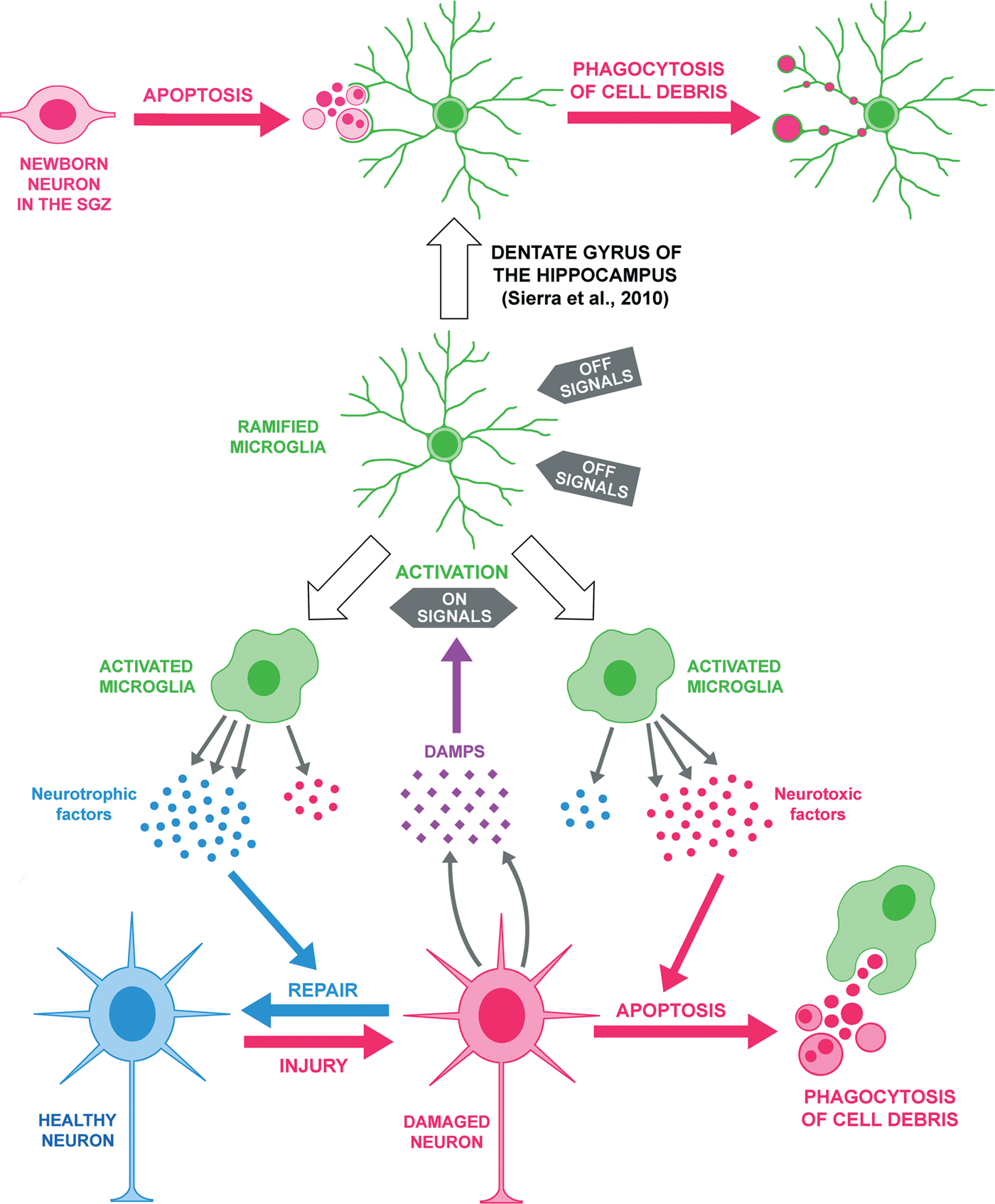
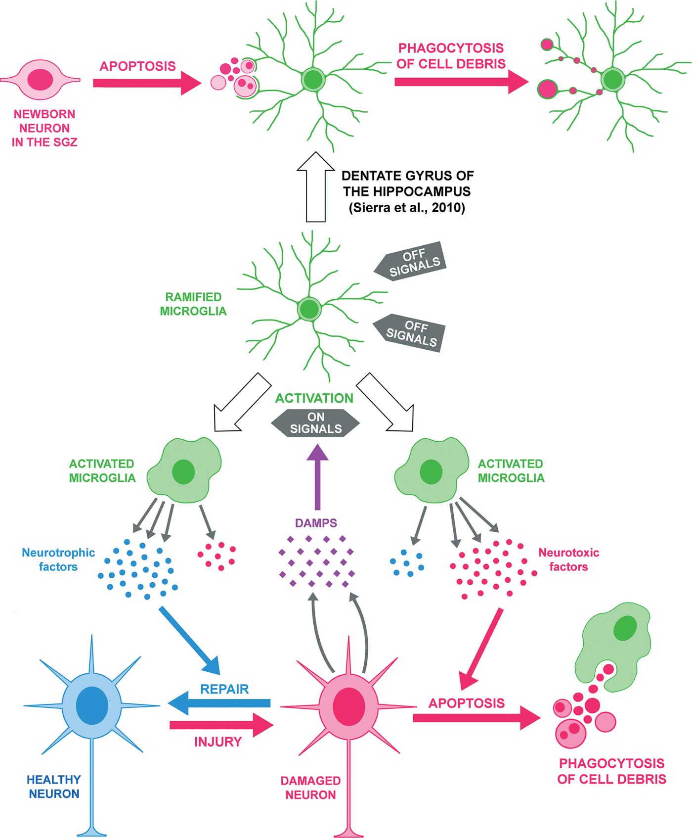
For (i), microglial processes are in continuous motion (Davalos et al., Reference Davalos, Grutzendler, Yang, Kim, Zuo and Jung2005; Nimmerjahn et al., Reference Nimmerjahn, Kirchhoff and Helmchen2005; Liang et al., Reference Liang, Lee, Wang, Ma, Fontainhas and Fariss2009), and microglia are able to move rapidly to target affected regions (Hanisch and Kettenmann, Reference Hanisch and Kettenmann2007). For (ii), microglial cells possess a panoply of receptors to recognize injury signals, which are classified in two main types: PAMPs (pathogen associated molecular patterns) and DAMPs (damage associated molecular patterns). PAMPs warn of the presence of exogenous material, such as components of bacterial cell walls (e.g. LPS) or repeats of bacterial or viral nucleic acids (Kumar et al., Reference Kumar, Kawai and Akira2011). For their part, DAMPs warn of internal damage to cells of the organism (Fig. 2), including molecules released by damaged cells and molecules modified as a consequence of tissue damage, such as oxidized lipoproteins or fragments of extracellular matrix molecules (Kono and Rock, Reference Kono and Rock2008; Piccinini and Midwood, Reference Piccinini and Midwood2010; Saijo and Glass, Reference Saijo and Glass2011). Many endogenous molecules, which serve diverse functions in healthy tissue, are not DAMPs in the normal CNS but acquire DAMP characteristics upon injury, when they are released from their intracellular stores and gain free access to specific receptors on the microglial cell surface. This process takes place not only in necrotic cells but also in the secondary necrosis of apoptotic cells that are not rapidly cleared by phagocytes (Kono and Rock, Reference Kono and Rock2008). Nevertheless, not all molecules released through the loss of cell membrane integrity are DAMPs, and various criteria have been established to identify a given molecule as a true DAMP (Bianchi, Reference Bianchi2007; Kono and Rock, Reference Kono and Rock2008). Several molecules have been reported to meet these criteria, including the nuclear protein HMGB1, heat-shock proteins, uric acid and ATP (Bianchi, Reference Bianchi2007; Kono and Rock, Reference Kono and Rock2008; Kettenmann et al., Reference Kettenmann, Hanisch, Noda and Verkhratsky2011; Saijo and Glass, Reference Saijo and Glass2011).
Roles of microglia in neuronal cell death in the adult CNS. In the healthy CNS, microglial cells can remove cell debris without changing the original ramified phenotype. In this case, no overt morphological activation is observed, as described in the dentate gyrus of the adult hippocampus, in which newborn neurons in the subgranular zone (SGZ) undergo death by apoptosis and are rapidly cleared through phagocytosis by unchallenged microglia (upper part of the figure). In large CNS injuries (lower part), the amount of ‘danger’ signals (DAMPs) released by damaged neurons promotes microglial activation, likely by altering the balance between ‘on’ and ‘off’ signals. It is worth noting that microglia can adopt different activated immunophenotypes. Thus, activated microglia may predominantly release either neurotrophic factors that help in the repair of damaged neurons (left) or neurotoxic factors that induce damaged neurons to continue the cell death process (right). Then, cell debris is phagocytosed by the activated microglial cells that induced the neuronal death.
Microglial cells possess an array of PAMP and DAMP receptors that allow them to detect most, if not all, pathological situations. For example, the microglial membrane contains purinergic receptors that bind ATP and its derivatives (Kettenmann et al., Reference Kettenmann, Hanisch, Noda and Verkhratsky2011), and they also bear toll-like receptors (TLRs), a family of receptors in the cell membrane or intracellular membranes that recognize both specific PAMPs and DAMPs; an account of TLR receptors and its ligands is given in Lehnardt (Reference Lehnardt2010) and Piccinini and Midwood (Reference Piccinini and Midwood2010). The presence of these and other receptors in microglial cells orchestrates their response to an exogenous or endogenous challenge.
It is evident that the concepts of DAMP molecules and ‘find-me’ signals (discussed in the previous section) are closely related, because both notions emerged from analyses of the need for the selective clearance of damaged cells by phagocytes in developing and adult organisms. However, the concept of the find-me signal is based on the premise that a chemotactic gradient would serve to attract phagocytes (Ravichandran, Reference Ravichandran2010), whereas DAMPs have been proposed to activate local cells of the immune system (Kono and Rock, Reference Kono and Rock2008). In view of this close relationship, it is not surprising that identical molecules and receptors, such as ATP and purinergic receptors, have been identified as players in both signaling systems.
In addition to their activation capability via ATP signaling, microglial cells can also participate in regulating the availability of ligand in the extracellular microenvironment through the action of ectonucleotidases on their surface, such as NTPDase and ecto-5′-nucleotidase (5′-NT). NTPDase dephosphorylates ATP via ADP to AMP, whereas 5′-NT catalyses the hydrolysis of AMP to adenosine. The importance of the presence of either ATP or adenosine in the extracellular milieu derives from the specificity of each ligand for the different purinoceptors present on every type of glia, giving rise to different effects on neurons. Thus, P1 (A1–A3) purinoceptors are activated by adenosine, ionotropic P2X purinoceptors are activated by ATP, and metabotropic P2Y purinoceptors are differentially activated by ATP and other nucleotides (Kettenmann et al., Reference Kettenmann, Hanisch, Noda and Verkhratsky2011). Importantly, it has been reported that an excess of ATP in the extracellular space of the CNS is neurotoxic (Ryu et al., Reference Ryu, Kim, Choi, Oh, Lee and Kim2002; Gandelman et al., Reference Gandelman, Peluffo, Beckman, Cassina and Barbeito2010), whereas the activation of adenosine A1 receptors can protect neurons against damage produced by CNS injury (Braun et al., Reference Braun, Zhu, Krieglstein, Culmsee and Zimmermann1998; Stone, Reference Stone2002; Cunha, Reference Cunha2005). In the normal CNS, NTDPase is present in both astrocytes (Wink et al., Reference Wink, Braganhol, Tamajusuku, Lenz, Zerbini and Libermann2006) and ramified microglia (Dalmau et al., Reference Dalmau, Vela, Gonzalez and Castellano1998; Braun et al., Reference Braun, Sevigny, Robson, Enjyoji, Guckelberger and Hammer2000), whereas 5′-NT is expressed in astrocytes (Zimmermann and Braun, Reference Zimmermann and Braun1996) but not in ramified microglia (Braun et al., Reference Braun, Lenz, Gillardon, Zimmermann and Zimmermann1997; Dalmau et al., Reference Dalmau, Vela, Gonzalez and Castellano1998). An increase in NTDPase has been described in activated microglia after CNS injury (Braun et al., Reference Braun, Zhu, Krieglstein, Culmsee and Zimmermann1998; Nedeljkovic et al., Reference Nedeljkovic, Bjelobaba, Subasic, Lavrnja, Pekovic and Stojkov2006). 5′-NT also increases in reactive astrocytes (Bjelobaba et al., Reference Bjelobaba, Parabucki, Lavrnja, Stojkov, Dacic and Pekovic2011) and is newly expressed in activated microglia (Braun et al., Reference Braun, Lenz, Gillardon, Zimmermann and Zimmermann1997; Dalmau et al., Reference Dalmau, Vela, Gonzalez and Castellano1998), thereby contributing to the active degradation of ATP into adenosine, which might rescue a proportion of neurons from death. The above reports indicate a complex scenario of interactions after CNS injury to ensure that released ATP, whose excess is potentially neurotoxic, is degraded to a potentially neuroprotective molecule such as adenosine. Further research is warranted to elucidate the precise cellular and molecular interactions, which involve different cell types (mainly microglia and astrocytes) and various purinergic receptors and ectonucleotidases.
Triggering microglial activation: ‘on’ and ‘off’ signals
When alerted to a CNS injury, microglial cells progressively transform from a ‘resting state’ to a fully activated state characterized by morphological transformation, increased migratory ability to accumulate in injured regions, enhanced proliferation, active phagocytosis and the release of cytokines and other factors (Raivich et al., Reference Raivich, Bohatschek, Kloss, Werner, Jones and Kreutzberg1999). Recent studies refuted the idea that normal ‘unchallenged’ microglia are merely resting cells with no special function in the CNS (Hanisch and Kettenmann, Reference Hanisch and Kettenmann2007), finding that ‘resting’ microglia actively inspect the nervous parenchyma (Davalos et al., Reference Davalos, Grutzendler, Yang, Kim, Zuo and Jung2005; Nimmerjahn et al., Reference Nimmerjahn, Kirchhoff and Helmchen2005; Liang et al., Reference Liang, Lee, Wang, Ma, Fontainhas and Fariss2009; Wake et al., Reference Wake, Moorhouse, Jinno, Kohsaka and Nabekura2009). Although microglial cells are expected to respond to small injuries (e.g. degeneration of single neuron or tiny ruptures in blood vessels) without overt phenotypic transformation, the microglial response to more serious injuries (e.g. death of a large number of neurons or extensive blood hemorrhage in the CNS) induces evident changes in their immunophenotype and behavior, e.g. the acquisition of migratory and phagocytic abilities (Hanisch and Kettenmann, Reference Hanisch and Kettenmann2007). A recent investigation in the neurogenic niche of the subgranular zone of the dentate gyrus of the adult mouse hippocampus (Sierra et al., Reference Sierra, Encinas, Deudero, Chancey, Enikolopov and Overstreet-Wadiche2010) demonstrated that ramified microglia are not challenged by the death of neuroprogenitors in the adult CNS. The authors showed that the apoptotic death of numerous newborn neuroprogenitors in transit to neuroblasts did not activate ramified microglia, which were able to efficiently engulf apoptotic neuroprogenitors through terminal or en passant branches (Fig. 2). This finding indicates that overt activation is not a prerequisite for microglial phagocytosis.
The terms ‘resting microglia’ and ‘activated microglia’ refer to different activity stages of the same cell. Some effort has been devoted to determining the factors that induce the microglial cells to adopt one or other state. Currently, it is largely believed that there are constitutive signals in the normal brain that represent a ‘brake’ on microglia activation, the so-called ‘off’ signals (van Rossum and Hanisch, Reference van Rossum and Hanisch2004; Biber et al., Reference Biber, Neumann, Inoue and Boddeke2007), such as fractalkine paired with its receptor CX3CR1 and CD200 paired with CD200R1 (Fig. 2). In a pathological situation, the ‘on’ signals (e.g. increased extracellular ATP) prevail over ‘off’ signals, resulting in the activation of microglial cells (Biber et al., Reference Biber, Neumann, Inoue and Boddeke2007). However, the picture appears to be more complex, because some authors have described an increased expression of fractalkine in pathological situations (Shan et al., Reference Shan, Hong-Min, Yi, Jun-Peng, Yue and Yan-Hong2011), and the microglial state may be determined by the balance between ligand and receptor (Taylor et al., Reference Taylor, Calder, Albon, Erichsen, Boulton and Morgan2011).
Activation is not an ‘all or nothing’ switch (Raivich et al., Reference Raivich, Bohatschek, Kloss, Werner, Jones and Kreutzberg1999), and microglial cells can show different levels of activation. Therefore, not all activated microglia show the same immunophenotype, and morphological observations and detection of microglial cell markers may be inadequate to distinguish between different modes of activation (Perry et al., Reference Perry, Nicoll and Holmes2010). The particular characteristics of the activated microglial phenotype in each case can vary as a function of intrinsic factors, e.g. age, microglial density or previous microglial history, and extrinsic factors, e.g. the severity and type of injury or cell context (Lai and Todd, Reference Lai and Todd2008; Sawada et al., Reference Sawada, Sawada and Nagatsu2008; Olah et al., Reference Olah, Biber, Vinet and Boddeke2011). This heterogeneity of activated microglial phenotypes, similar to that of monocyte-derived macrophages (Gordon and Taylor, Reference Gordon and Taylor2005), would explain the different effects of microglial activation in different CNS damage settings.
Phagocytosis and release of a molecular panoply by activated microglia
In the best characterized event after their activation, microglial cells phagocytose and eliminate the debris resulting from cell death, acting as scavenger cells to clean the nervous parenchyma. The phagocytic role of microglial cells is well documented in numerous systems and is considered decisive to maintain homeostasis in the nervous parenchyma and trigger CNS repair after injury (Neumann et al., Reference Neumann, Kotter and Franklin2009; Walter and Neumann, Reference Walter and Neumann2009). In support of this view, it has been reported that the decline in microglial phagocytic capacity (e.g. in the aged brain) hampers regenerative response of the nervous tissue and results in more frequent pathological situations (Neumann et al., Reference Neumann, Kotter and Franklin2009; von Bernhardi et al., Reference von Bernhardi, Tichauer and Eugenin2010).
As previously noted, two types of receptors are involved in microglial phagocytosis, depending on the material to be phagocytosed. Receptors for PAMPs recognize microbes and other exogenous material, hence supporting the removal of pathogens, whereas receptors for DAMPs (e.g. receptors recognizing the ‘eat-me’ signal PS) participate in the phagocytosis of dead cells (Konduru et al., Reference Konduru, Tyurina, Feng, Basova, Belikova and Bayir2009; Neher et al., Reference Neher, Neniskyte, Zhao, Bal-Price, Tolkovsky and Brown2011). PAMP receptor-mediated pathogen phagocytosis stimulates a pro-inflammatory response, while dead cell clearance can be performed through either pro- or anti-inflammatory responses. It should be noted that microglia phagocytose not only neurons dying as a result of pathological or traumatic events but also neurons dying by apoptosis in the normal CNS (Wirenfeldt et al., Reference Wirenfeldt, Babcock and Vinters2011). In the first case, the phagocytosis is accompanied by inflammatory events, whereas the phagocytosis of apoptotic neurons in the healthy CNS occurs without inflammation.
Interestingly, it has been reported that the neuron degeneration after inflammatory activation of microglia may be attributable to direct phagocytosis by microglial cells that previously induced neurons to express PS ‘eat-me’ signals (Neher et al., Reference Neher, Neniskyte, Zhao, Bal-Price, Tolkovsky and Brown2011; Neniskyte et al., Reference Neniskyte, Neher and Brown2011). The authors found that neurons were able to reverse PS signaling and return to a healthy state when phagocytosis was blocked. Given that this activated microglia-induced neuronal death shares common features with the microglial engulfment-promoted PCD that occurs during development, as reported above, it can be hypothesized that similar mechanisms might participate in both developmental and inflammatory neurodegeneration.
In addition to phagocytosis, metabolic routes releasing an array of molecules that can induce or promote neuron degeneration are upregulated in activated microglia (Brown and Neher, Reference Brown and Neher2010). These molecules include reactive oxygen species (Block et al., Reference Block, Zecca and Hong2007) and nitric oxide (Brown, Reference Brown2010) as well as pro- and anti-inflammatory cytokines and chemokines (Hanisch, Reference Hanisch2002). The release of the pro-inflammatory cytokines TNF-α and IL1β induces classical activation and recruitment of immune cells that frequently result in increased neuronal damage. Nevertheless, it should be emphasized that pro-inflammatory cytokines have a versatile spectrum of effects on neurons ranging from neuroprotection to neurodegeneration (Hanisch, Reference Hanisch2002; Hanisch and Kettenmann, Reference Hanisch and Kettenmann2007; Loane and Byrnes, Reference Loane and Byrnes2010). For instance, TNF-α signaling may have a role in promoting neuronal death by activation of pro-apoptotic proteins, but it can also participate in neuronal survival by activation of the nuclear factor NF-κB (Liu et al., Reference Liu, Hsu, Goeddel and Karin1996). In fact, the release of pro-inflammatory cytokines is initially intended to prevent further damage to the nervous tissue, but they can become toxic to neurons and glial cells under certain conditions (Smith et al., Reference Smith, Das, Ray and Banik2012). The conditions that trigger neuroprotective or neurodegenerative TNF-α signaling remain to be clarified. Activated microglia have also been reported to produce anti-inflammatory cytokines such as IL-4 and IL-10 (Park et al., Reference Park, Lee, Joe, Kim and Jin2005; Henry et al., Reference Henry, Huang, Wynne and Godbout2009), which appear to reduce neuron degeneration in numerous situations (Lyons et al., Reference Lyons, Downer, Crotty, Nolan, Mills and Lynch2007; Xiong et al., Reference Xiong, Barreto, Xu, Ouyang, Xie and Giffard2011). Chemokines are also released by activated microglia and other cell types to attract immune cells intrinsic or extrinsic to the CNS. For instance, CCL2 (also called MCP-1) produced by astrocytes and activated microglia recruits immune cells to the damaged brain (D'Mello et al., Reference D'Mello, Le and Swain2009; Schilling et al., Reference Schilling, Strecker, Schabitz, Ringelstein and Kiefer2009; Conductier et al., Reference Conductier, Blondeau, Guyon, Nahon and Rovere2010), whereas CCL2 released by retinal Müller cells promotes the concentration of microglia in regions affected by retinal detachment (Nakazawa et al., Reference Nakazawa, Hisatomi, Nakazawa, Noda, Maruyama and She2007) and stimulates the recruitment of monocytes to retinas with light-induced photoreceptor degeneration (Rutar et al., Reference Rutar, Natoli, Valter and Provis2011).
It is conceivable that phagocytosis and the release of molecules are related processes, with one influencing the other. In fact, microbial phagocytosis by microglial cells is accompanied by the release of pro-inflammatory factors, which are not released when microglial cells engulf apoptotic cells or oligodendrocyte membrane vesicles called exosomes (Neumann et al., Reference Neumann, Kotter and Franklin2009; Fitzner et al., Reference Fitzner, Schnaars, van Rossum, Krishnamoorthy, Dibaj and Bakhti2011).
Neurotoxic versus neuroprotective roles of activated microglia
Although microglial cells have mainly been considered ‘cleaners’ that remove cell debris produced by degeneration, numerous observations strongly suggest that they fulfill a more active role in determining cell life/death in the damaged CNS. Thus, microglial activation has different outcomes in the nervous parenchyma, increasing neuron degeneration (neurotoxic) in some cases but reducing neuron death (neuroprotective) in others (Mallat and Chamak, Reference Mallat and Chamak1994; Hanisch and Kettenmann, Reference Hanisch and Kettenmann2007). The neurotoxic or neuroprotective role of activated microglia will depend on the balance between different types of neurotrophic and neurotoxic molecules released by them (Fig. 2) as well as the microenvironmental context.
Various observations support the ‘negative’ role of microglial activation: (i) microglia depletion or blockage of the microglial response diminishes or totally abolishes cell death in vivo (Mount et al., Reference Mount, Lira, Grimes, Smith, Faucher and Slack2007) and in vitro (Gao et al., Reference Gao, Zhou, Zhang, Wilson, Kam and Hong2011); (ii) the damage produced by some pathological situations, e.g. lipoteichoic acid or LPS treatments or retinal detachment, requires the presence of microglial cells (Qin et al., Reference Qin, Liu, Wang, Wei, Block and Wilson2004; Nakazawa et al., Reference Nakazawa, Hisatomi, Nakazawa, Noda, Maruyama and She2007; Liu and Bing, Reference Liu and Bing2011; Neher et al., Reference Neher, Neniskyte, Zhao, Bal-Price, Tolkovsky and Brown2011); and (iii) microglia-conditioned medium is sufficient to induce neuronal degeneration in some systems (Flavin et al., Reference Flavin, Zhao and Ho2000; Hur et al., Reference Hur, Lee, Kim, Kim and Cho2010). Therefore, it has been proposed that initially injured neurons originate a cascade of degeneration through the activity of microglia and other cell types, such as astrocytes (Ilieva et al., Reference Ilieva, Polymenidou and Cleveland2009; Cameron and Landreth, Reference Cameron and Landreth2010).
The ‘negative’ role of microglial activation has been widely reported to be a decisive factor in different neurodegenerative diseases (Block et al., Reference Block, Zecca and Hong2007; Hanisch and Kettenmann, Reference Hanisch and Kettenmann2007; Lull and Block, Reference Lull and Block2010; Prinz and Mildner, Reference Prinz and Mildner2011; Saijo and Glass, Reference Saijo and Glass2011, among others). These diseases are characterized, among other features, by progressive and chronic neuronal death and elevated levels of pro-inflammatory factors released from activated microglia. Microglial activation-derived inflammation is now thought to be an amplifier of neurodegeneration, which is initiated by a single stimulus that varies among different diseases (α-synuclein in Parkinson's disease or β-amyloid in Alzheimer's disease). Once early neuronal death is triggered by the initial neurotoxic stimulus, DAMP signals released in the extracellular milieu by dead neurons (and the neurotoxic stimulus itself) contribute to stimulate reactive microgliosis; this is a process of chronic microglial activation in response to neuronal damage that is perpetuated by further microglial activation associated with acute inflammation and neurotoxicity (Block and Hong, Reference Block and Hong2007; Lull and Block, Reference Lull and Block2010; Gao et al., Reference Gao, Zhou, Zhang, Wilson, Kam and Hong2011). In order to halt this vicious cycle, there must be mechanisms to regulate the resolution of the inflammation-mediated activation of microglial cells and their transition to non-activated phenotypes (Colton, Reference Colton2009; Saijo and Glass, Reference Saijo and Glass2011). Defects in these mechanisms appear to contribute to chronic neurodegenerative diseases.
The ‘positive’ role of microglial activation has also been widely reported. Thus, the removal of microglial cells exacerbated the damage in a number of CNS injuries (Imai et al., Reference Imai, Suzuki, Oda, Ninomiya, Ono and Sano2007; Simard and Rivest, Reference Simard and Rivest2007; Lalancette-Herbert et al., Reference Lalancette-Hebert, Gowing, Simard, Weng and Kriz2007; Streit et al., Reference Streit, Miller, Lopes and Njie2008). Moreover, the addition of microglia-conditioned media reduced the toxicity of various treatments (Morgan et al., Reference Morgan, Taylor and Pocock2004; Polazzi et al., Reference Polazzi, Altamira, Eleuteri, Barbaro, Casadio and Contestabile2009). Microglia were also found to produce neurotrophic factors (Elkabes et al., Reference Elkabes, DiCicco-Bloom and Black1996; Batchelor et al., Reference Batchelor, Liberatore, Wong, Porritt, Frerichs and Donnan1999; Nakajima et al., Reference Nakajima, Honda, Tohyama, Imai, Kohsaka and Kurihara2001; Bessis et al., Reference Bessis, Bechade, Bernard and Roumier2007; Olah et al., Reference Olah, Amor, Brouwer, Vinet, Eggen and Biber2012) that may improve the growth of neurons and neurites (Batchelor et al., Reference Batchelor, Liberatore, Wong, Porritt, Frerichs and Donnan1999) and remyelination (Olah et al., Reference Olah, Amor, Brouwer, Vinet, Eggen and Biber2012). In this regard, it has been reported that microglial cells also have a role in neurogenesis and in neuroblast migration and differentiation (Aarum et al., Reference Aarum, Sandberg, Haeberlein and Persson2003; Walton et al., Reference Walton, Sutter, Laywell, Levkoff, Kearns and Marshall2006; Sierra et al., Reference Sierra, Encinas, Deudero, Chancey, Enikolopov and Overstreet-Wadiche2010).
In several systems, microglial activation switches from being fundamentally neurotoxic to being neuroprotective during the microglial response (Colton, Reference Colton2009; Loane and Byrnes, Reference Loane and Byrnes2010). It is likely that the early microglial response after injury predominantly removes affected cells, although some healthy cells can also be eliminated, whereas a late response is mainly directed to repair and reconstitute the tissue. It has been proposed that these differences may be due to the resulting balance between two different activation types: classical (M1), which induces activated microglia to produce cytotoxic mediators and pro-inflammatory cytokines and is considered neurotoxic; and alternative (M2), which promotes downregulation of the above factors and upregulation of MHCII expression and is considered neuroprotective. M1-activated cells would predominate during early microglial activation, whereas M2-activated cells (microglia and macrophages) would be more abundant during later stages (Loane and Byrnes, Reference Loane and Byrnes2010; David and Kroner, Reference David and Kroner2011). However, the defense stance of microglial activation is primarily protective, and inflammatory factors released by activated microglia are repair-oriented, although they first charge the injured region a neurotoxic price. Therefore, the dynamics and mechanisms of microglial activation would be much more complex than represented by a simple M1 (neurotoxic)/M2 (neuroprotective) sorting, and their elucidation requires further research.
Discrepancies in the literature on the neurotoxic or neuroprotective role of activated microglia may have different explanations. First, some of the differences observed may be due to the comparison of data from different experimental models; the mode of microglial activation is not the same in all cases and varies between different strains of the same species (Wei and Lin, Reference Wei and Lin2009). Second, as reported above, the predominant microglial response is distinct at different times after injury; therefore, the neurotoxic versus neuroprotective controversy may be in part related to differences in the time since the injury. Third, the sensitivity to treatments that induce microglial reaction varies among different CNS regions and different neurons (Tansey and Goldberg, Reference Tansey and Goldberg2010). Fourth, there are several types of microglial cells; thus, different subtypes of microglial cells have been characterized that release different substances (Kawahara et al., Reference Kawahara, Yoshida, Koga, Yokoo, Kuniyasu and Gotoh2009). Fifth, microglial reactions are influenced by age; thus, the increase in neurodegenerative diseases at higher age has been linked to the more neurotoxic behavior of aging microglia (von Bernhardi et al., Reference von Bernhardi, Tichauer and Eugenin2010). Therefore, although much is already known, further research is necessary to clarify the precise factors and microenvironmental circumstances that induce the ‘positive’ and ‘negative’ roles of activated microglia.
IS THE ROLE OF MICROGLIA IN NEURONAL DEATH DURING DEVELOPMENT COMPARABLE WITH THAT IN THE ADULT CNS?
Ameboid microglia during development and activated microglia at adulthood share some morphological, cytochemical and physiological characteristics. It might be thought that ameboid microglia perform phagocytosis of apoptotic neurons during development in a similar way to that carried out by activated microglia in the adult CNS. However, it has been reported that ameboid microglia of the developing CNS are functionally different from activated microglia of the adult CNS, because ameboid microglia do not release the pro-inflammatory cytokines produced by activated microglia, such as IL1β (Hurley et al., Reference Hurley, Walter, Semple-Rowland and Streit1999). Therefore, it appears reasonable to question whether the role of ameboid microglia in neuronal death during development is truly equivalent to that of microglia in the adult CNS.
It is noteworthy that ameboid microglia during development are able to activate in response to neuron damage in the same way as ramified microglia in the adult CNS. This notion is supported by studies showing immunophenotypical changes of ameboid microglia, compatible with their activation, in response to cell death after injury in the developing CNS. Thus, activation of ameboid microglia was demonstrated in numerous parts of the developing CNS after different models of injury: in the retina after stab wound injury (Sánchez-López et al., Reference Sánchez-López, Cuadros, Calvente, Tassi, Marín-Teva and Navascués2005) or hypoxia (Sivakumar et al., Reference Sivakumar, Foulds, Luu, Ling and Kaur2011); in the neocortex and the hippocampus after N-methyl-d-aspartate-mediated excitotoxicity (Acarin et al., Reference Acarin, Gonzalez, Castellano and Castro1996); in the olfactory bulb after deprivation of the olfactory stimulus (Fiske and Brunjes, Reference Fiske and Brunjes2000); in the cerebral cortex, corpus callosum and striatum after hypoxia–ischemia by carotid artery ligation (Fukui et al., Reference Fukui, Kinugasa, Fukuda, Fukuda, Tskitishvili and Hayashi2006); in the spinal cord after axotomy by sciatic nerve crush (Morioka and Streit, Reference Morioka and Streit1991); in the facial nucleus after facial nerve transection (Graeber et al., Reference Graeber, Lopez-Redondo, Ikoma, Ishikawa, Imai and Nakajima1998); and in the diencephalon, cerebral cortex and hippocampus after intraperitoneal injection of a teratogen (Hao et al., Reference Hao, Dheen and Ling2001). Injury-induced activation of ameboid microglia gives rise to the release of pro-inflammatory cytokines (Fukui et al., Reference Fukui, Kinugasa, Fukuda, Fukuda, Tskitishvili and Hayashi2006; Hristova et al., Reference Hristova, Cuthill, Zbarsky, Acosta-Saltos, Wallace and Blight2010; Sivakumar et al., Reference Sivakumar, Foulds, Luu, Ling and Kaur2011), chemokines such as MCP-1 (Sivakumar et al., Reference Sivakumar, Foulds, Luu, Ling and Kaur2011) and cytotoxic factors such as nitric oxide (Hao et al., Reference Hao, Dheen and Ling2001). Microglial activation and massive inflammatory reaction were also demonstrated in association with abnormal neurodegeneration in the developing forebrain of cyclin-dependent kinase 5 conditional knockout mice (Takahashi et al., Reference Takahashi, Ohshima, Hirasawa, Pareek, Bugge and Morozov2010). It can be concluded from the above studies that injury-induced activation of ameboid microglia during development is similar to that of ramified microglia in the adult CNS, including immunophenotypical changes and the release of pro-inflammatory cytokines and other cytotoxic factors.
Interestingly, two recent studies (Hristova et al., Reference Hristova, Cuthill, Zbarsky, Acosta-Saltos, Wallace and Blight2010; Rigato et al., Reference Rigato, Buckinx, Le-Corronc, Rigo and Legendre2011) revealed that some ameboid microglia become transiently activated during the normal development of the non-injured CNS, although the process of microglial activation showed differences with that in the developing or adult CNS after injury. Rigato et al. (Reference Rigato, Buckinx, Le-Corronc, Rigo and Legendre2011) reported the accumulation of ameboid microglial cells in the lateral motor columns of embryonic mouse spinal cord at the onset of the motoneuron cell death. These cells were transiently activated between E13.5 and E14.5, as shown by the expression of Mac-2 (a carbohydrate-binding protein on the surface of activated microglia and macrophages), coinciding with the developmental cell death of motoneurons. A similar transient microglial activation, closely related to a phagocytic role, was reported by Hristova et al. (Reference Hristova, Cuthill, Zbarsky, Acosta-Saltos, Wallace and Blight2010) during normal postnatal mouse development in the periventricular white matter (corpus callosum, fornix and external capsule), where rounded microglia expressing different activation markers accumulated at birth. These activation markers decreased during subsequent postnatal maturation of microglia, which were no longer activated after the first postnatal week. However, activated ameboid microglia in the developing periventricular white matter appeared to differ from activated microglia in the adult CNS, because the former did not release the pro-inflammatory cytokine TNF-α but rather produced neuroprotective factors such as insulin-like growth factor-1 and macrophage colony stimulating factor, which would favor developmental processes. Further research is warranted to elucidate the reasons for functional differences between the transient anti-inflammatory activation during normal development and the damage-induced pro-inflammatory activation of ameboid microglia in the injured developing CNS.
In addition to ameboid microglia activation in the developing white matter, the study by Hristova et al. (Reference Hristova, Cuthill, Zbarsky, Acosta-Saltos, Wallace and Blight2010) also demonstrated that no microglial activation was detected in several regions of gray matter (cerebral cortex, hippocampus, striatum or thalamus) during postnatal development, despite the neuronal death that occurs at this time. This regional heterogeneity in the activation of ameboid microglia may be attributable to differences in the types of cellular debris among different regions. For instance, rounded activated ameboid microglia appear to phagocytose axonal debris in the developing periventricular white matter, whereas non-activated ameboid microglia would phagocytose apoptotic neuronal bodies in gray matter regions. Nevertheless, the reasons why ameboid microglia are activated in some CNS regions but not others during normal development have not yet been fully clarified.
As stated above, ameboid microglia in the developing gray matter are able to phagocytose neurons that are actively induced to die after microglial engulfment. This was demonstrated to occur in the developing cerebellar cortex (Marín-Teva et al., Reference Marín-Teva, Dusart, Colin, Gervais, van Rooijen and Mallat2004) and hippocampus (Wakselman et al., Reference Wakselman, Bechade, Roumier, Bernard, Triller and Bessis2008). Based on the findings of Hristova et al. (Reference Hristova, Cuthill, Zbarsky, Acosta-Saltos, Wallace and Blight2010) in the developing hippocampus, these ameboid microglial cells would be in a non-activated state but are nevertheless able to kill engulfed neurons through the action of superoxide ions. This mechanism, which links the execution of neuron death to the clearance of dead cells, would probably hamper secondary necrosis of apoptotic neurons and subsequent activation of ameboid microglia. In addition, in vitro experiments showed that the interaction of externalized PS in apoptotic neurons with specific PS receptors in microglial cells inhibits metabolic pathways for the production of pro-inflammatory substances in these cells (De Simone et al., Reference De Simone, Ajmone-Cat and Minghetti2004). This would contribute to a silent clearance of dying neurons by non-activated ameboid microglia.
On the other hand, it has been hypothesized that homeostasis of the normal CNS can require the clearance of single neurons that must eventually be removed from their circuitry, and microglia would be constantly engaged in this process (Hanisch and Kettenmann, Reference Hanisch and Kettenmann2007). How does this clearance of neurons dying normally in the adult CNS take place? The study by Sierra et al. (Reference Sierra, Encinas, Deudero, Chancey, Enikolopov and Overstreet-Wadiche2010) dealt with the clearance by microglia of naturally dying neurons in the dentate gyrus of the normal adult hippocampus, as previously discussed. They reported that ramified microglia phagocytose newborn neuroprogenitors without being activated. Neurogenesis in the adult hippocampus is a natural process in which many newborn neuroprogenitors must be eliminated, because they cannot be incorporated into the normal circuitry. In this case, ramified microglia are not challenged but would rather sense neuroprogenitor death as a process that is essential to maintain CNS homeostasis and does not need a pro-inflammatory response. Thus, apoptotic neuroprogenitors do not appear to release signals for microglial activation, probably as a consequence of the rapid clearance of apoptotic cells by ramified microglia, thereby avoiding secondary necrosis that would release DAMP signals to the extracellular milieu (Kono and Rock, Reference Kono and Rock2008). It is tempting to hypothesize that ramified microglia themselves may induce the death of neuroblasts after making contact with them, in a similar way to engulfment-induced neuron apoptosis during development. The observation of microglial cells in close contact with dying granule cells in the rat dentate gyrus (Ribak et al., Reference Ribak, Shapiro, Perez and Spigelman2009) is compatible with this hypothesis. It can be concluded from the study of Sierra and colleagues that ‘natural’ neuronal death in the adult CNS does not trigger microglial activation and that cell debris is rapidly cleared through silent and safe phagocytosis by non-activated ramified microglia, similarly to the action of non-activated ameboid microglia during development. Likewise, the so-called ‘natural’ death of single mature neurons would produce apoptotic fragments that might be rapidly phagocytosed by ramified microglia, thereby contributing to the avoidance of microglial activation and of a pro-inflammatory response in the normal brain. Although it is not easy to study the ‘natural’ death of single neurons, because of its sporadic occurrence, further research is required to clarify this issue.
CONCLUDING REMARKS
Microglial cells respond to the death of neurons (or their progenitors) by phagocytosing cell debris in a physiological context of either pro-inflammatory activation, anti-inflammatory activation or non-activation, depending on the characteristics of the neuron death. Pro-inflammatory activation is a typical response of ramified or ameboid microglia to the death of damaged neurons after injury or disease in the adult or developing CNS. As reviewed here, numerous studies are available on mechanisms involved in the damage-induced pro-inflammatory activation of ramified microglia in the adult CNS, but less is known about the pro-inflammatory activation of ameboid microglia in the developing CNS. Anti-inflammatory activation of ameboid microglia takes place transiently during development in white matter regions, where cell bodies are absent but axons of neurons dying by natural PCD are present. Further studies are required to obtain a more complete characterization of the transient anti-inflammatory activation of ameboid microglia during normal development and to elucidate differences with the better-known damage-induced pro-inflammatory microglial activation in the injured developing or adult CNS. Microglia in a non-activated state phagocytose cell bodies of neurons or neuroprogenitors dying in gray matter regions by natural PCD, which is frequent during CNS development but can also occur in the adult CNS. In both cases, microglia phagocytose cell debris without altering their morphological phenotype, which is ameboid during development and ramified at adulthood. A few studies on the phagocytosis of naturally dying neurons by non-activated ameboid microglia during CNS development have led to the concept of engulfment-promoted neuron death, which would explain the absence of microglial activation. Nevertheless, further research is necessary to determine whether this process takes place throughout the developing CNS. Future studies must also examine the possibility of the microglial engulfment-promoted natural death of neurons in the adult CNS, related and unrelated to adult neurogenesis.
In short, future research should focus on some as yet unresolved issues, including (i) characterization of the transient anti-inflammatory activation of ameboid microglia in the CNS white matter during normal development; (ii) the possible existence of microglial engulfment-promoted neuron death throughout different parts of the developing CNS gray matter, besides the already studied cerebellar cortex and hippocampus; and (iii) the possible existence in the adult CNS of similar microglial engulfment-promoted death of either neuroprogenitors born during adult neurogenesis or single mature neurons that must eventually be removed from the circuitry.
ACKNOWLEDGEMENTS
This work was supported by a grant from the Spanish Ministry of Science and Innovation (BFU2010-19981). The authors thank Richard Davies for improving the English.
Statement of interests
None.