INTRODUCTION
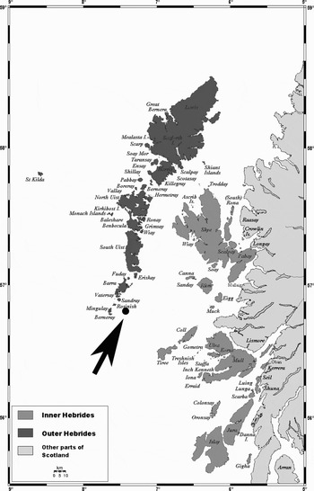
Coldwater reefs off the east coast of Mingulay Island, southern Hebrides, north-west Scotland (Figure 1), were investigated in July 2006 during a joint multidisciplinary effort of the Royal Netherlands Institute of Sea Research (NIOZ) and the Scottish Association of Marine Sciences (SAMS). Bottom samples were obtained by video-grabbing, boxcoring, and dredging, while side-scan and multibeam transects were made over the reefs (Maier, Reference Maier2006). One of the projects concerned the species composition of sponges occurring in and near the reefs, as a continuation of a series of investigations made in coldwater reefs elsewhere in the north-east Atlantic (see e.g. Van Soest & Lavaleye, Reference van Soest and Lavaleye2005; Van Soest et al., Reference van Soest, Cleary, de Kluijver, Lavaleye, Maier and van Duyl2007), forming a part of the Dutch funded BIOSYS 2005–2007 programme.
Map of the Hebrides off the Scottish west coast showing the approximate location of the Mingulay reefs and the sampling stations (map obtained through Wikipedia from the internet source: www.aquarius.ifm-geomar.de, and was slightly modified).

Excavating or ‘boring’ sponges play a distinct role in coldwater reef systems as major bioeroders (Beuck & Freiwald, Reference Beuck, Freiwald, Freiwald and Roberts2005; Beuck et al., Reference Beuck, Vertino, Stepina, Karolczak and Pfannkuche2007). Not unexpectedly they were found to be frequent dwellers in the Mingulay reefs, typically occupying recently dead coral branches, but they were not found in the living parts of the corals. However, recent sophisticated techniques applied by Beuck et al. (Reference Beuck, Vertino, Stepina, Karolczak and Pfannkuche2007) demonstrated the cryptic presence of sponge borings infesting living parts of Lophelia in reefs on the Porcupine Seabight. Five species were encountered: Alectona millari Carter, Reference Carter1879, Pione vastifica (Hancock, Reference Hancock1849), both quite common, Cliona lobata Hancock, Reference Hancock1849, which occurred only rarely, and two species not previously known from Scottish waters, a Cliona species that appeared to be undescribed, and a southern deep-water species Spiroxya levispira (Topsent, Reference Topsent1898). The former species was at first assigned to alpha forms of Cliona aff. celata Grant, Reference Grant1826, because it seemed as if no microscleres were present, its colour is a distinct yellow or orange, and the papilla-size appears similar to that of C. celata. However, upon closer examination, not very numerous, thick spirasters were detected in some individuals. Subsequent studies demonstrated that the microscleres were confined to the papillar skeleton of all the alleged ‘celata’ specimens. Below, the new species of Cliona and the newly recorded Spiroxya levispira are described and extensively illustrated.
MATERIALS AND METHODS
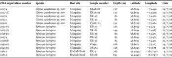
Bottom samples containing dead and living reef corals, mostly Lophelia pertusa but occasionally also Madrepora oculata, were obtained by three sampling methods (Maier, Reference Maier2006): video-grabbing (cf. Mortensen et al., Reference Mortensen, Roberts and Sundt2000 for an extensive description of this method), boxcoring using a 50 cm diameter cylinder, and trawling (10 minutes bottom time). Table 1 lists all the samples from which bioeroding sponges were obtained. Sponges were usually obtained from subsamples of coral branches assigned for sponge research. Occasionally the entire sample was made available for sponge studies. Sponges detected on board were pre-identified using crude slide preparations (cf. Van Soest & Lavaleye, Reference van Soest and Lavaleye2005) and preserved in 96% ethanol. Unsorted trawl subsamples and boxcore subsamples were directly preserved in larger containers and analysed onshore in Amsterdam. Dead coral branches were examined using a low power microscope and opened up using hammer and chisel. Sponge identification was done from teased tissue preparations. Spicules were dissociated by boiling in concentrated nitric acid and the residue was washed five times in distilled water and mounted on slides and SEM stubs for examination at high magnification.
Locality data of studied samples.

SYSTEMATIC DESCRIPTIONS
The classification used here follows the Systema Porifera (Hooper & van Soest, Reference Hooper and van Soest2002):
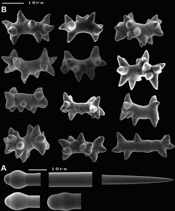
Cliona caledoniae sp. nov., scanning electron microscopy images of spicules. (A) Tylostyle and variations of tyles; (B) spirasters, showing from upper left to lower right spicules with spiral turns and conical rays spread randomly to amphiaster-like forms with straight rhabd and apically concentrated rays.

TYPE MATERIAL
Holotype: ZMA Por. 20174, Mingulay, Station BX48/06, 56.8054°N 7.4419°W, 127 m, in Lophelia pertusa, 14/7/06.
Paratypes: ZMA Por. 20171C, same data as the holotype; ZMA Por. 20270 & 20271, Mingulay, Station BX127, 82 m, in Lophelia pertusa, 56.8033°N 7.4471°W, 20/7/06; ZMA Por. 20320, Station VG28/05, 131 m, in Lophelia pertusa, 56.8219°N 7.3964°W, 12/7/06.
DESCRIPTION
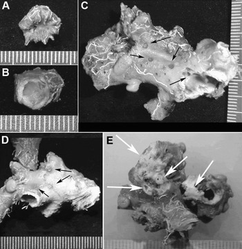
Excavating sponge, with exclusively alpha-habit (distinctly separate non-confluent papillae protruding from the corals). Papillae yellow, or orange-yellow, rather conspicuous, flush with the surface, often slightly elevated beyond the coral surface (Figure 4D), but in preserved material the papillae tend to be contracted below the substratum level (Figure 4C). The papillae turn brown in alcohol. Both optically ‘closed’ papillae, presumably exhalant, and optically perforated papillae, presumably oscular, occur. The holotype has only a single papilla (Figure 4A) hidden in the calyx of a dead coral polyp and sponge tissue has permeated and replaced most of the limestone skeleton (Figure 4B), leaving only a thin-walled coral. Papilla size variable, 1–4 mm diameter. Papillae unevenly distributed over the coral surface (Figure 4C–D). Excavations are extensive chambers, up to 5 mm or more in diameter, filled with soft tissue of a pale brown or off-white colour. The branching corals and coral polyps are hollowed out to leave only a thin limestone wall.
SKELETON
The papillae are provided with the usual dense palisade of tylostyles, points outward, with intermingled microscleres in a moderate density. The intra-coral tissue contains scattered loose tylostyles, and microscleres are absent.
SPICULES
Tylostyle megascleres and spiraster microscleres. Tylostyles (Figure 2A), robust, with usually slightly subterminal tyle, 246–360.9–426 × 8–9.8–12 µm (N = 25); tyles 11–12.3–15 µm diameter.
Spirasters (Figure 2B), thick, blunt-rayed, with short axis, occasionally almost straight and with the rays concentrated at the extremities. Length: 19–24.3–31 × 5–6.8–9 µm (N = 25), excluding rays which are 3–5.1–7 µm; number of rays 10–11.8–17.
ECOLOGY
Excavating dead Lophelia and Madrepora corals at 82–131 m depth.
ETYMOLOGY
Named after Caledonia, the Roman name for Scotland.
REMARKS
The new species is distinguished from the known north-east Atlantic Clionaidae species by its combination of commonplace tylostyles and a single category of thick-rayed amphiaster-like spirasters. Cliona lobata is the only species in the area with similar overall spiculation of tylostyles and spirasters, but it has two categories of spirasters the larger of which are longer and thinner (up to 65 µm), with up to 9 spiral curves (Topsent, Reference Topsent1900) and its papillae are usually not larger than 0.5 mm in diameter. Cliona celata has similar papilla size as the new species but these are usually much closer together, and the species lacks microscleres entirely (Rützler, Reference Rützler, Hooper and van Soest2002a). Excavations in the alpha-form of this species usually are thin, branching corridors and small galleries, not large chambers entirely filled with tissues as described above. No specimens of Cliona celata were encountered in the Mingulay reefs, although initially the present species was mistaken for it. Pione vastifica, very common in the Mingulay reefs, possesses next to tylostyles and thin microrhabds also rugose microxeas, usually in two size-classes. Macroscopically, Pione vastifica and Cliona caledoniae sp. nov. are similar and microscopic examination is necessary to identify either with certainty.
Other excavating sponges in the Mingulay reefs (Spiroxya levispira, cf. below, and Alectona millari) belong to a different family, Alectonidae, and are clearly distinct in spiculation from the Clionaidae. Further excavating species, e.g. Aka infesta (Johnson, Reference Johnson1899), A. nodosa (Johnson, Reference Johnson1899) and A. insidiosa (Johnson, Reference Johnson1899), found in North Atlantic coldwater reefs (see e.g. Van Soest & Lavaleye, Reference van Soest and Lavaleye2005 and Schönberg & Beuck, Reference Schönberg and Beuck2007), but so far not found in the Mingulay reefs, differ in lacking microscleres and having only oxeas as spicules.
Elsewhere in European waters, several additional Cliona species are reported (Topsent, Reference Topsent1888, Reference Topsent1900; Rützler, Reference Rützler1973; Rosell & Uriz, Reference Rosell and Uriz2002). Cliona janitrix Topsent, Reference Topsent1932 and C. amplicavata Rützler, Reference Rützler1974 do not have spirasters. Cliona viridis (Schmidt, Reference Schmidt1862) has two categories of spirasters, long thin ones with 2–5 bends, 10–53 µm and short straight ones with long spines, 13–30 µm. The short spirasters have the spines much sharper and subdivided compared with those of C. caledoniae sp. nov. It occurs predominantly in shallow waters in the Mediterranean–Atlantic area. Cliona schmidti (Ridley, Reference Ridley and Gunther1881) likewise has two categories of spirasters, longer/thinner (25–90 µm) and shorter/thicker (35–62 µm). The shorter/thicker spirasters are not unlike those of C. caledoniae sp. nov. in shape but are longer and thicker, neatly separated in size. The colour of C. schmidti is characteristically dark purple, which is retained in alcohol-preserved specimens. Similar spiculation is found in Cliona carteri sensu Rützler, Reference Rützler1973 (probably not Ridley, Reference Ridley and Gunther1881) and C. rhodensis Rützler & Bromley, Reference Rützler and Bromley1981 from Tunisia, with small spirasters resembling those of C. caledoniae sp. nov. but possessing an additional long type with several bends.
Thick knobby spirasters occur in Cliona thoosina Topsent, Reference Topsent1888 described from several localities in the Mediterranean, and this species shares the yellow colour and tylostyle size with the new species, but the spirasters occur in three size-categories, up to 65 µm in length. Addtional similar species are the Mediterranean Cliona cretensis Pulitzer-Finali (Reference Pulitzer-Finali1983), but this has the typical thin spirasters of the majority of Cliona species, and C. tremitensis Sarà, Reference Sarà1961, which has a complement of oxea megascleres (which may mean it is not a Cliona but a Volzia) and microscleres longer and slimmer than but not unlike those of C. caledoniae sp. nov.
Interestingly, the microscleres of C. caledoniae sp. nov. are reminiscent of the knobby amphiasters described for several Cliothosa hancocki (Topsent, Reference Topsent1888) individuals from widely disparate areas (Rützler, Reference Rützler, Hooper and van Soest2002a). Like the thick spirasters of C. caledoniae sp. nov., these knobby amphiasters of C. hancocki are concentrated in the papillae. Also the excavations of the new species are similar to those of C. hancocki (cf. also Calcinai et al., Reference Calcinai, Bavestrello and Cerrano2005). The resemblance is superficial, but needs further comment. Cliothosa hancocki was originally described from various Pacific and Indian Ocean localities (Topsent, Reference Topsent1888, Reference Topsent1905; Lindgren, Reference Lindgren1898; Annandale, Reference Annandale1915; Schönberg, Reference Schönberg2000; Calcinai et al., Reference Calcinai, Bavestrello and Cerrano2005) but subsequently from the Mediterranean (Adriatic, Tunisia) (Lendenfeld, Reference von Lendenfeld1897 (as Vioa ramosa); Topsent, Reference Topsent1928; Rützler, Reference Rützler1973). The closest locality to Mingulay from which this species was recorded is the Adriatic (Lendenfeld, Reference von Lendenfeld1897; see also Topsent, Reference Topsent1932), so occurrence in Mingulay is not unimaginable. The nodulous amphiasters of Cliothosa hancocki are characterized by blunt cone-shaped rays and a very short axis, making them similar to thick spirasters. The difference between Cliona and Cliothosa is that the latter has only amphiasters, while the former has spirasters sometimes combined with amphiaster-like microscleres. Cliothosa typically has thin-rayed amphiasters with branched rays as the major synapomorphy for the genus, while the nodulous amphiasters are apparently rare or absent. In theory, the thin-rayed amphiasters of Cliothosa could also be rare in some individuals (cf. Topsent, Reference Topsent1932), and then such Cliothosa individuals would resemble Cliona species with thick, blunt-rayed spirasters such as the present new species. Further similarities with Cliothosa hancocki are the exclusively alpha-stage, the colour and the size of papillae and galleries. However, we do not think the specimens of our new species are deficient Cliothosa specimens, because: (1) exhaustive search did not reveal the presence of thin-rayed amphiasters with branching rays; (2) the axis of most microscleres is curved; (3) the rays are distributed all over the axis and do not appear to be concentrated at both ends so technically they are not amphiasters; and (4) length of the microscleres (19–31 µm) is clearly greater than that of reported nodulous amphiaster sizes in Cliothosa hancocki (10.5–25 µm)
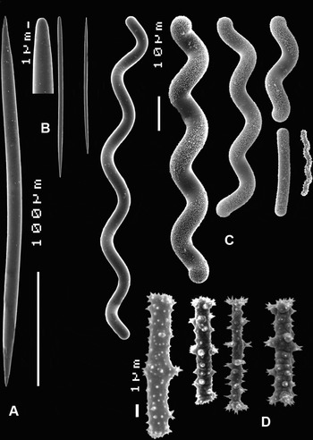
Spiroxya levispira (Topsent, Reference Topsent1898), scanning electron microscopy images of spicules. (A) Large oxea; (B) smaller anisoxeas with magnified bluntly rounded end; (C) various sizes and forms of spiral strongyles; (D) spined microrhabds.

Habitus images of Cliona caledoniae sp. nov. (A–D) and Spiroxya levispira (Topsent, Reference Topsent1898) (E). (A–B) Holotype of C. caledoniae sp. nov. infesting dead Lophelia corallite; (C–D) papillae (arrows) of paratypes of C. caledoniae sp. nov.; (C: ZMA 20271, D: 20270); (E) S. levispira excavations (arrows) (ZMA Por. 20418A).

Cliona levispira Topsent, Reference Topsent1898: 235, figure 2; Topsent, Reference Topsent1904: 105, pl. XII figure 1; Pouliquen, Reference Pouliquen1972: 751, pl. 9 figure 4; Boury-Esnault, Reference Boury-Esnault, Pansini and Uriz1994: 61, figure 37.
Scantilla spiralis Johnson, Reference Johnson1899: 462, figure 5.
Scantiletta levispira; Rosell & Uriz, Reference Rosell and Uriz1997: 361; Rosell & Uriz, Reference Rosell and Uriz2002: 60, figure 3.
Spiroxya levispira; Rützler, Reference Rützler, Hooper and van Soest2002b: 289, figure 10D–E; Longo et al., Reference Longo, Mastrototaro and Corriero2005: Table 2; Van Soest et al., Reference van Soest, Cleary, de Kluijver, Lavaleye, Maier and van Duyl2007: Table 2.
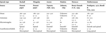
Spicule measurements and characteristics of all European Spiroxya levispira records.

MATERIAL EXAMINED
ZMA Por. 20210A, Mingulay, Station BX123, 162 m, 56.8059°N 7.429°W, 19/7/06; ZMA Por. 20255A, Mingulay, Station BX180, 144 m, 56.8034°N 7.4467°W, 22/7/06; ZMA Por. 20269A & 20274 & 20280B, Mingulay, Station BX127, 82 m, 56.8033°N 7.4471°W, 20/7/06; ZMA Por. 20350A, Mingulay, Station VG49, 168 m, 56.8071°N 7.4303°W, 14/7/06; ZMA Por. 20418A, Mingulay, Station DR182, 128 m, 56.8243°N 7.3686°W, 22/7/06; ZMA Por. 20611, Rockall Bank, Station BX72, 762 m, 55.44437°N 16.07597°W, 4/7/05; ZMA Por. 20612, Rockall Bank, Station BX168, 691 m, 55.44432°N 16.07457°W, 12/7/05.
DESCRIPTION
Excavating sponge, with exclusively alpha-habit, papillae inconspicuous, flush with the surface, 2–4 mm in diameter, giving access to relatively large ellipsoid cavities (Figure 4E), up to approximately 10 mm diameter, filled with pale-coloured, off-white or beige, soft tissue.
SKELETON
Radiate arrangement of megascleres at the surface of papillae, with tracts of oxeas lining the cavities and with confused arrangement of spicules in the soft interior mass. Smaller oxeas are concentrated in the surface, larger oxeas in the cavities.
SPICULES
Two size-classes of oxeas, spiral microstrongyles and acanthomicrorhabds. Oxeas I: fusiform, sharply pointed, 186–280.3–330 × 9–12 µm (N = 25); oxeas II, anisoxeote, with one end usually rounded or blunt, the other end sharply pointed, 126–169.0–198 × 4–5 µm (N = 25); spiral microstrongyles, large size variation, 15–138 × 5–6 µm (N = 50); acanthomicrorhabds, straight, amphiaster-like, usually centrotylote or spined most prominently in the middle, 9–11.9–15 × 1–1.2–2 µm (N = 25).
ECOLOGY
Excavating dead Lophelia and Madrepora corals at 82–762 m depth (present study); elsewhere 5–2165 m. The shallow record is from Mediterranean cave habitats (Pouliquen, Reference Pouliquen1972), which are known to contain a deepwater fauna.
REMARKS
The present record is the most northern location known for the species and the first report of it from the waters of the British Isles. The species is predominantly reported from southern European localities, Azores, Madeira, Morocco and the western Mediterranean (Topsent, Reference Topsent1898, Reference Topsent1904; Johnson, Reference Johnson1899; Boury-Esnault et al., Reference Boury-Esnault, Pansini and Uriz1994; Pouliquen, Reference Pouliquen1972; Rosell & Uriz, Reference Rosell and Uriz2002; Longo et al., Reference Longo, Mastrototaro and Corriero2005). The species was recently reported from Rockall Bank deepwater coral reefs, in the ocean west of Ireland (Van Soest et al., Reference van Soest, Cleary, de Kluijver, Lavaleye, Maier and van Duyl2007). Together with the Mingulay specimens these recent records comprise a considerable range extension. Most of the reported specimens show closely similar spicular characters (see Table 2). Compared to the description of the holotype from deep water off the Azores, the oxeas I of the Mingulay specimens are notably shorter and do not appear to overlap in length. The Madeira specimen of Johnson (Reference Johnson1899 as Scantilla spiralis) was not fully described, but the figures provided by this author make it likely that they concern the same species.
A second species of Spiroxya, S. heteroclita Topsent, Reference Topsent1896 is recorded from Lophelia reefs west and south of Ireland (Stephens, Reference Stephens1915; Beuck et al., Reference Beuck, Vertino, Stepina, Karolczak and Pfannkuche2007). This species differs clearly from S. levispira in possessing spirally spined straight strongyles, lacking acanthomicrorhabds, and having oxeas up to 900 µm (Rützler, Reference Rützler, Hooper and van Soest2002b).
The acanthomicrorhabds are named amphiasters by most past authors (e.g. Rosell & Uriz, Reference Rosell and Uriz2002), but microstructure of the Mingulay and Rockall microscleres does not justify the name amphiaster as there are no proper rays, merely spines and clusters of spines (cf. Figure 3D). Calcinai et al. (Reference Calcinai, Cerrano and Bavestrello2002) called the microscleres of their species Scantiletta (=Spiroxya) corallophila spiny microrhabds and we fully agree with that and recommend naming them acanthomicrorhabds in the definition of the genus Spiroxya. These microrhabds appear to be the only spicule type with some morphological diversification over the range of Spiroxya levispira, as the microrhabds shown by Rosell & Uriz, Reference Rosell and Uriz2002 have a different appearance with fewer much more prominent spines and less microspination. Further studies along the lines of Picton & Goodwin (Reference Picton and Goodwin2007), who looked at differences of local sponge populations in a restricted area, are needed to establish whether such subtle but apparently consistent differences in microstructures of spicules and relatively small size differences are of taxonomic importance.
ACKNOWLEDGEMENTS
Connie Maier and Fleur van Duyl (NIOZ, Texel) are thanked for inviting the first author to participate in the cruise to Mingulay Reefs as part of the BIOSYS programme financed by the Netherlands Organization for Science (NWO). Andy Davies (Scottish Association for Marine Sciences, SAMS, Oban) manipulated the video-grab which brought up coral material with a minimum of damage. Colleagues of the NIOZ secured the boxcore and dredge samples. The captain and crew of RV ‘Pelagia’ are acknowledged for their skills at operating the sampling gear and for their seamanship.