INTRODUCTION
Phytomonas sp. are flagellated trypanosomatid parasites of plants, that can be found in latex, phloem, fruits and seed albumin of many plant families with a wide geographical distribution. Phloem parasites cause crop diseases of economic significance in coconut, oil palms, cassava and coffee plantations in Latin America (Camargo, Reference Camargo1999). Conversely, the vast majority of laticiferous trypanosomatids apparently do not promote any pathological effect, and some authors even see them as symbionts (Dollet, Reference Dollet2001). Such is also the case of Phytomonas encountered in fruits and seeds. Phytomonas sp. are thought to be transmitted to the plants through the bite of phytophagous hemipterans of the Coreidae, Pentatomidae and Lygaeidae families (Dollet, Reference Dollet1984; Sbravate et al. Reference Sbravate, Campaner, Camargo, Conchon, Teixseira and Camargo1989).
The successful culturing of the tomato isolate Phytomonas serpens (Jankevicius et al. Reference Jankevicius, Jankevicius, Campaner, Conchon, Maeda, Teixeira, Freymuller and Camargo1989) allowed the identification of some characteristics of the life cycle and metabolism of this organism. Krebs cycle and mitochondrial functions, such as cytochrome-mediated respiration and ATP production, are missing in this flagellate and cell energetics are based predominantly on glycolysis (Maslov et al. Reference Maslov, Nawathean and Scheel1999; Nawathean and Maslov, Reference Nawathean and Maslov2000; Bringaud et al. Reference Bringaud, Rivière and Coustou2006).
Multiple horizontal gene transfer (HGT) events have been implicated in the acquisition of structural and functional characteristics of the trypanosomatids that allowed their adaptation to a vast range of hosts (Opperdoes and Michels, Reference Opperdoes and Michels2007). Among structural and biochemical peculiarities found only in members of the order Kinetoplastida the kinetoplast DNA, mitochondrial RNA editing, the sequestration of glycolysis inside glycosomes and unique oxidative-stress protection mechanisms can be listed. Opperdoes and Michels (Reference Opperdoes and Michels2007) have proposed that the trypanosome-specific evolution of novel processes and cell organization could only have been made possible by the acquisition of a large number of foreign genes, most probably from plants, viruses and bacteria.
In a previous study we have characterized the complete gene encoding a pyruvate decarboxylase (PDC)/indolepyruvate decarboxylase (IPDC) of P. serpens, which exhibits high amino acid sequence similarity with enzymes of phytobacteria and the trypanosomatids Leishmania spp. and Crithidia fasciculata (Ienne et al. Reference Ienne, Pappas, Benabdellah, González and Zingales2012). PDC and IPDC are homotetrameric enzymes and their amino acid sequence has extensive similarity that precludes the direct assignment of a functional role based solely on the protein sequence (Koga, Reference Koga1995).
A phylogenetic analysis showed that the clade that contains the trypanosomatid proteins behaves as a sister group of IPDC of γ-proteobacteria. We have proposed that the PDC/IPDC gene was acquired by an ancestral trypanosome from a donor phytobacteria via a HGT event (Ienne et al. Reference Ienne, Pappas, Benabdellah, González and Zingales2012). Interestingly, two recent studies based on extensive phylogenetic analysis concluded that the genomes of some trypanosomatids seem to have incorporated at least three γ-proteobacterial genes that encode enzymes required for haeme biosynthesis (Korený et al. Reference Korený, Lukes and Oborník2010; Alves et al. Reference Alves, Voegtly, Matveyev, Lara, da Silva, Serrano, Buck, Teixeira and Camargo2011).
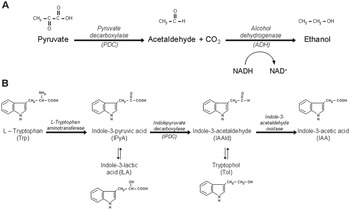
PDC and IPDC belong to the decarboxylase family of thiamine diphosphate (ThDP)-dependent enzymes (Duggleby, Reference Duggleby2006). For the comprehensive comparison of protein sequences and structures the ThDP-dependent Enzyme Engineering Database (TEED) was established (http://www.teed.uni-stuttgart.de). ThDP-dependent decarboxylases catalyse the non-oxidative decarboxylation of α-keto acids to aldehydes. Based on their substrate specificity, the α-keto acid decarboxylases can be subdivided into various groups. Pyruvate decarboxylases (PDC – EC 4.1.1.1) function in alcoholic fermentation and convert pyruvate to acetaldehyde (Fig. 1A). Indolepyruvate decarboxylases (IPDC – EC 4.1.1.74) are key enzymes in the biosynthesis of the plant hormone indole-3-acetic acid (IAA), the most abundant naturally occurring auxin, which is derived from the aromatic amino acid L-tryptophan (Truelsen, Reference Truelsen1973). The ThDP-dependent IPDC catalyses the formation of indole-3-acetaldehyde (IAAld) from indole-3-pyruvic acid (IPyA) (Koga, Reference Koga1995) (Fig. 1B). The pathway shown in Fig. 1B is considered the major route for IAA production in bacteria and plants. Besides decarboxylation of indolepyruvate, IPDCs can decarboxylate a number of α-keto acids, including pyruvate, benzoylformate and phenylpyruvate with different catalytic efficiencies (Fedorov et al. Reference Fedorov, Doronina and Trotsenko2010). The direct involvement of these enzymes in IAA biosynthesis was shown for Azospirillum brasilense, Enterobacter cloacae, Pantoea agglomerans (Schütz et al. Reference Schütz, Golbik, Tittmann, Svergun, Koch, Hübner and König2003a ; Spaepen et al. Reference Spaepen, Vanderleyden and Remans2007) and Methylobacterium extorquens (Fedorov et al. Reference Fedorov, Doronina and Trotsenko2010).
(A) Alcohol fermentation. (B) Indole-3-pyruvate pathway for IAA biosynthesis in bacteria (adapted from Koga et al. Reference Koga, Adachi and Hidaka1991). Indole-3-lactic acid (ILA) and tryptophol (Tol) are originated by reduction of IPyA and IAAld, respectively.

In a previous study we have detected PDC activity in P. serpens cell extracts and suggested that this enzyme, that participates in the alcoholic fermentation, would reoxidize a portion of the NADH produced in the very active glycolytic pathway of the flagellate. This would confer an adaptive advantage for the parasite survival in the rich carbohydrate media of the plant host (Ienne et al. Reference Ienne, Pappas, Benabdellah, González and Zingales2012). This is particularly important since, as mentioned above, in Phytomonas ATP is produced mainly via substrate level phosphorylation instead of the classical cytochrome-based respiratory chain (reviewed by Bringaud et al. Reference Bringaud, Rivière and Coustou2006).
Because ThDP-dependent decarboxylases can decarboxylate various α-keto acids, and the P. serpens enzyme behaves as a sister group of γ-proteobacteria IPDC, the present study aimed at verifying whether P. serpens produces IAA. Since IAA regulates many important processes of plant growth and development (Woodward and Bartel, Reference Woodward and Bartel2005; Teale et al. Reference Teale, Paponov and Palme2006; Spaepen et al. Reference Spaepen, Vanderleyden and Remans2007), the auxin production could confer additional adaptive characteristics in the Phytomonas-plant interaction.
Here, by using high performance liquid chromatography we provide evidence that P. serpens is able to produce IAA in vitro and in vivo and describe the putative destination of the auxin in tomato fruits inoculated with the flagellate.
MATERIALS AND METHODS
Growth of the organisms
Phytomonas serpens TCC 060 (Trypanosomatids Culture Collection of the Universidade de São Paulo) was kindly provided by Dr Marta M. G. Teixeira (Instituto de Ciências Biomédicas, USP). The flagellates were grown in Grace's insect medium (Sigma-Aldrich, USA) supplemented with 10% foetal calf serum (Grace-FCS) at 28 °C. Cell density was estimated in a Neubauer chamber. When specified, the media were supplemented with L-tryptophan (Trp) or glucose (Glc). Conditioned media were obtained from 4-day cultures, when the flagellates reached the late exponential growth phase. The cultures were centrifuged at 5900 g at 4 °C for 7 min and the supernatant media were filtered (0·22 μm Millipore® membranes) for the elimination of parasites and cell debris. The media were stored at −20 °C.
Detection and quantification of IAA and related indole compounds
For the analysis of IAA, Trp, tryptophol (Tol), indole-3-acetaldehyde (IAAld) and indole-3-pyruvic acid (IPyA), 1 mL conditioned media was deproteinized with 4 mL methanol. Indole-propionic acid was added as an internal standard (IS) for recovery estimation. The sample was centrifuged at 13 000 g, at 25 °C for 10 min; the supernatant concentrated to 1 mL under N2 flow and filtered (0·45 μm Millipore® membranes). The compounds were chromatographed in a HPLC (Hewlett-Packard, model 1100, USA) equipped with a reverse phase C18 Luna column (5 μm, 300×4 mm) (Supelco, Sigma-Aldrich, USA). The injection volume was 40 μL. The elution programme started with methanol : 1% acetic acid (20 : 80, v/v), followed by a linear gradient from 0 to 100% methanol. The analytes were monitored by UV absorbance and fluorescence in a HP-1046A detector at 270 nm excitation wavelength (λ) and 350 nm emission (325 nm to IPyA), and confirmed by retention time with authentic standards (Sigma-Aldrich). Calibration curves were constructed for each analyte by plotting the peak areas of the compound/IS vs known amount (ng) of each standard. Values were adjusted to 107 cells.
Detection and quantification of ethanol in conditioned media
For ethanol quantification, 50 μL conditioned media were deproteinized with 25 μL acetonitrile. n-Propanol (16 μg in acetonitrile) was added as IS. The sample was centrifuged at 13 000 g at 25 °C for 10 min. The supernatant was analysed in a GC-FID system (Hewlett-Packard, model 6890). A Supelcowax column (30 m, I.D. 0·25 mm, 0·50 μm internal film thickness) was used with helium as carrier gas (1 mL min−1 flow rate). The injections (3 μL) were made with an automatic injector (ALS-7683, Hewllett-Packard) in Splitless mode. The column was maintained at 35 °C for 1 min, followed by a ramp temperature of 5 °C min−1 up to 150 °C and 20 °C min−1 up to 250 °C. The injector and detector temperatures were 250 and 200 °C, respectively. The ethanol content of the samples was calculated by the area ratio compound/IS. A calibration curve was generated using the analyte/IS peak area vs known amounts of the ethanol standard. Values were adjusted to 107 cells.
Plant material and growth conditions
Germination and cultivation of tomato seedlings (Solanum lycopersicum L. cv Micro-Tom) were performed in vitro according to Pino-Nunes et al. (Reference Pino-Nunes, de O. Figueira, Tulmann Neto, Zsögön, Piotto, Silva, Bernardi and Peres2009) with some modifications. Tomato seeds (kindly provided by Dr Lázaro E. P. Peres, Escola Superior de Agricultura Luiz de Queiroz, Universidade de São Paulo) were sterilized with 95% ethanol for 30 s, transferred to 50% sodium hypochlorite for 15 min and washed with sterile distilled water. For the hypocotyl elongation assay, hypocotyls were obtained from light-grown (de-etiolated) seedlings. The seeds were inoculated in glass bottles containing 100 mL MS medium (Murashige and Skoog, Reference Murashige and Skoog1962) with half the concentration of macronutrients and 0·2% Phytagel (Sigma-Aldrich) and kept for 7 days under a 12-h photoperiod, light intensity of 200 μmol m−2 s−1 and constant temperature of 25±2 °C. For tomato infections, the plants were grown from seeds in a greenhouse and kept under controlled conditions from anthesis to the day of harvest (light intensity 300 μmol m−2 s−1; 60–80% relative humidity; constant temperature of 25±2 °C; 16/8 h photoperiod).
Hypocotyl elongation assay
The bioassay was adapted from Kelly and Bradford (Reference Kelly and Bradford1986). Seven days after germination, the tomato seedlings had their hypocotyls removed and sliced into ∼12 mm segments. The segments were washed in distilled water and their exact length was measured with a caliper rule. Twenty hypocotyl segments were placed in Petri dishes (10 cm) and incubated with 6 mL: (i) filtered P. serpens-conditioned media; (ii) Grace-FCS media and (iii) Grace-FCS media plus 9 μ m IAA (Sigma-Aldrich). The closed Petri dishes were kept at room temperature (25 °C) for 24 h on the bench, after which the length of the hypocotyls was measured using an electronic digital caliper. Two biological replicates with 20 hypocotyl segments each (technical replicates) were performed on two different occasions (biological replicates). The results were subjected to statistical analysis (see below).
Inoculations of Micro-Tom tomatoes
Red ripe fruits (about 40 days after anthesis) kept on the plant were injected via needle inoculation according to Jankevicius et al. (Reference Jankevicius, Jankevicius, Campaner, Conchon, Maeda, Teixeira, Freymuller and Camargo1989) with 20 μL PBS (control) and 20 μL PBS containing: (i) P. serpens (107 cells); (ii) Trypanosoma cruzi epimastigote forms (107 cells) (kindly provided by Marcelo Nunes Silva, Instituto de Química, Universidade de São Paulo); (iii) 1·34 μg IAA (Sigma-Aldrich). For each experimental point six tomatoes were injected. The inoculation sites were sealed with warm melted paraffin to prevent secondary infections. After inoculation, the plants were kept in a growth chamber under controlled conditions as described above. Three tomato fruits were collected on days 7 and 14 post-inoculation (pi).
Detection and quantification of free and conjugated IAA in inoculated Micro-Tom tomatoes
Individual tomato fruits were weighed, homogenized in a blender for 30 s and subsequently passed through a sieve (∼0·07 mm pore) to retain the seeds and epicarp. The pulp was weighed (1·8 g fresh weight average), frozen and kept at −80 °C until use. The tomato pulp of each fruit (200 mg fresh weight) was homogenized in 1 mL isopropanol : acetic acid (95 : 5, v/v). One microgram of [2H5]-IAA (D-IAA) (OlChemim, Czech Republic) IS was added to quantify losses during the purification process. The sample was kept under agitation at 4 °C for 2 h and centrifuged at 14 000 g for 10 min at 4 °C. The supernatant was divided into three equal aliquots for analysis, respectively, of free IAA, IAA-ester and IAA-amide conjugates. Extraction of free IAA was performed according to Ludwig-Müller et al. (Reference Ludwig-Müller, Georgiev and Bley2008). The methylated sample was analysed on a Hewlett Packard 6890 gas chromatograph/HP5973 mass selective detector, as described (Purgatto et al. Reference Purgatto, Nascimento, Lajolo and Cordenunsi2002). For the IAA-ester-conjugate quantification, samples were stirred in 1 N NaOH for 1 h at room temperature (Chen et al. Reference Chen, Miller, Patterson and Cohen1988). Under these mild conditions, ester-, but not amide-conjugates of IAA are hydrolysed. At the end of the incubation period, the pH of the hydrolysate was adjusted to 2·5. Methylation and GC-MS analysis were performed as described above. The quantification of IAA-amide conjugates was performed as previously reported (Chen et al. Reference Chen, Miller, Patterson and Cohen1988, adapted by Purgatto et al. Reference Purgatto, Nascimento, Lajolo and Cordenunsi2002). Sample methylation and GC-MS analysis were as described above for free IAA quantification.
Sequence alignment and modelling
The amino acid sequence of P. serpens IPDC/PDC (GenBank AEX33309); IPDC of E. cloacae (GenBank P23234) and P. agglomerans (GenBank AAB06571); and PDC of Saccharomyces cerevisiae (GenBank CAA54522) and Zymomonas mobilis (GenBank CAA42157) were aligned with CLUSTALW version 1.83 (Thompson et al. Reference Thompson, Higgins and Gibson1994) and visualized with BioEdit (Biological Sequence Alignment Editor for Windows) (Hall, Reference Hall1999). The three-dimensional structure of the P. serpens protein was constructed using the automated mode of homology modelling servers (PS)2-V2 (Chen et al. Reference Chen, Hwang and Yang2009) and Swiss-Model (Peitsch, Reference Peitsch1995; Arnold et al. Reference Arnold, Bordoli, Kopp and Schwede2006; Kiefer et al. Reference Kiefer, Arnold, Künzli, Bordoli and Schwede2009). The overall quality of the models was assessed by Ramachandran plots using the RAMPAGE server (Lovell et al. Reference Lovell, Davis, Arendall, de Bakker, Word, Prisant, Richardson and Richardson2003). PyMOL (http://www.pymol.org) was used to visualize and align the established three-dimensional structures of E. cloacae IPDC (PDB ID 1ovmA, Schütz et al. Reference Schütz, Sandalova, Ricagno, Hübner, König and Schneider2003b ) and Z. mobilis PDC (PDB ID 2wva, Pei et al. Reference Pei, Erixon, Luisi and Leeper2010).
Statistical analysis
The data regarding the length of the hypocotyls were analysed with SigmaPlot to determine the significance using paired observations (paired t-test). The test was applied with a confidence level of 95% (P = 0·05) to accept the null hypothesis (H0: there is no significant difference in the lengths of hypocotyls before and after the test). Data regarding the quantification of free and conjugated IAA were subjected to statistical analysis (SigmaPlot) to determine the significance of the results (Mann–Whitney test).
RESULTS
Detection and quantification of IAA and related indole compounds in P. serpens-conditioned media
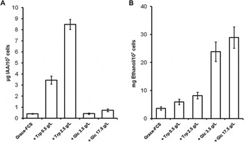
To verify whether P. serpens produces IAA, the concentration of the auxin and related indole compounds (IPyA, IAAld and Tol, see Fig. 1B) was determined in P. serpens-conditioned media by HPLC (Fig. 2A; Table 1). Commercial standards were readily separated and detected as distinct peaks using a reverse phase C18 Luna column (data not shown). No corresponding peaks were identified in the original culture medium. To test the importance of Trp as precursor for IAA production, the flagellates were grown in media supplemented with two Trp concentrations (Fig. 2A; Table 1). The production of indole compounds was very low in Grace-FCS medium, which contains 0·1 g L−1 Trp. Addition of 0·5 and 2·5 g L−1 Trp promoted 8·4- and 20·6-fold increase, respectively, of IAA concentration, as well as Tol accumulation (2·65- and 9·79-fold increase, respectively) (Table 1). We could not detect IAAld in the media and the concentration of IPyA was low and invariant with Trp addition, likely due to the high instabilities of the two compounds and/or rapid conversion, as previously reported (Moore and Shaner, Reference Moore and Shaner1968; Gibson et al. Reference Gibson, Schneider and Wightman1972; Koga et al. Reference Koga, Adachi and Hidaka1992).
Quantification of IAA (A) and ethanol (B) in P. serpens-conditioned media. Trp (L-tryptophan) and Glc (glucose) concentrations in Grace-FCS normal medium are, respectively, 0·1 and 0·7 g L−1.

Accumulation of IAA, IPyA, Tol and ethanol in P. serpens-conditioned media a

a Data represent the mean±s.d. of biological triplicates of technical duplicates and refer to 4-day P. serpens cultures containing a total amount of 107 parasites.
Addition of Glc did not enhance auxin production (Fig. 2A) and promoted a slight enhancement (∼2·5-fold) of Tol accumulation (Table 1). Overall, the data indicate that P. serpens synthesizes and excretes IAA in the media and that the production of the auxin and Tol increases with Trp concentration.
Ethanol accumulation in P. serpens-conditioned media
The accumulation of ethanol by Phytomonas cultured in glucose-rich media has been documented (Sanchez-Moreno et al. Reference Sanchez-Moreno, Lasztity, Coppens and Opperdoes1992; Chaumont et al. Reference Chaumont, Schanck, Blum and Opperdoes1994). We quantified ethanol in the above-mentioned conditioned media by GC-FID (Fig. 2B; Table 1). As expected, a 5-fold increase of Glc concentration in regard to the control medium (0·7 g L−1 Glc) stimulated about 6-fold ethanol accumulation. Further increase to 17·5 g L−1 Glc produced a non-significant increase. Trp supplementation promoted only a 2·0-fold increase of ethanol production.
Bioassay of IAA functionality
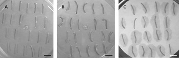
Some bioassays are available for assessing IAA functionality (see references in Woodward and Bartel, Reference Woodward and Bartel2005). Since P. serpens is a tomato isolate, the tomato hypocotyl elongation assay developed by Kelly and Bradford (Reference Kelly and Bradford1986) was chosen to investigate the auxin activity in P. serpens-conditioned media. As controls, 1-week-old Micro-Tom hypocotyls were also incubated with Grace-FCS medium and Grace-FCS plus 9 μ m exogenous IAA. This IAA concentration was calculated based on the equivalent amount of auxin accumulated in P. serpens-conditioned media (1·64±0·16 μg mL−1; 4×10⁷ cells mL−1; Table 1). The data shown in Table 2 refer to the average length of 20 hypocotyl segments (two biological replicates with 20 segments each) before and after 24 h incubation. Phytomonas serpens-conditioned media promoted ∼13% increase of the hypocotyls’ length after the incubation period, whereas only 0·9% increase was observed with Grace-FCS media. Exogenous IAA promoted a ∼10% hypocotyl growth stimulation. Student's t-test (paired analysis) was performed with a confidence interval of 95%, and 19 degrees of freedom (tc = 2·86094), and indicated that the percentage of hypocotyl elongation promoted by P. serpens-conditioned media and exogenous IAA was statistically significant (t = 8·88 and 6·37, respectively). We also verified that after incubation with either media the hypocotyls showed a curvature (Fig. 3). Interestingly, stem bending tests have been used also to establish whether a given compound exerts auxin-like effects (Woodward and Bartel, Reference Woodward and Bartel2005). Taken together the data suggest that IAA produced by P. serpens is functional. The greater stimulation caused by P. serpens media may be due to the presence of a IAA concentration higher than 9 μ m. Nevertheless we cannot rule out that additional molecules produced by the flagellate may have some contribution to the hypocotyls’ growth.
Elongation of hypocotyl segments of S. lycopersicum L. cv Micro-Tom incubated for 24 h with: (A) Grace-FCS medium; (B) P. serpens-conditioned medium; (C) Grace-FCS+9 μ m IAA. The bars correspond to 1 cm.

Effect of P. serpens-conditioned media on Micro-Tom hypocotyl elongation a

a The data represent the average of 20 hypocotyl segments (biological replicates). The s.e. is indicated in parentheses.
b Percentage of length increase in relation to T 0.
Quantification of free and conjugated IAA in infected tomatoes
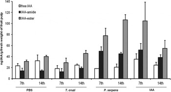
Next we evaluated whether IAA accumulated in Micro-Tom tomatoes inoculated with 107 P. serpens cells. As controls red ripe fruits were alternatively injected with PBS, 107 T. cruzi cells or 1·34 μg exogenous IAA. After inoculation the fruits were kept on the plants, maintained in a growth chamber under controlled conditions and collected on the days 7 and 14 pi. No visible morphological alterations of the plants or fruits were observed. Optical microscope inspection of the pulp of the fruits confirmed the presence of P. serpens and T. cruzi motile parasites (data not shown). Free IAA was quantified in the pulp of the fruits by GC-MS analysis (Fig. 4). Since in plants it is well established that IAA occurs either as a hormonally active free form or in bound forms in which the carboxyl group is conjugated to sugars or myo-inositol via ester linkages and to amino acids or peptides via amide linkages (reviewed by Ludwig-Müller, Reference Ludwig-Müller2011), we also quantified IAA conjugates (ester and amide) in the pulp of the injected fruits (Fig. 4). Table 1 in the supplementary material shows the concentration values of free and conjugated auxin in the injected fruits and the statistical analysis of the data.
Content of free and conjugated IAA (amide and ester-linked) in the pulp of Micro-Tom tomato fruits inoculated with PBS; 107 T. cruzi cells; 107 P. serpens cells; or 1·34 μg IAA on days 7 and 14 pi. Data are mean±s.d. (n = 3).

Considering days 7 and 14 pi, fruits inoculated with PBS or T. cruzi cells showed similar kinetics of the accumulation of free IAA, IAA-amide and IAA-ester conjugates. On the other hand, on day 7 day pi, P. serpens promoted a 3·5-fold and 2·5-fold increase in the abundance of IAA-amide and IAA-ester, respectively, in relation to PBS injection. Inoculation of 1·34 μg IAA also increased the amount of IAA-amide (3·6-fold) and IAA-ester (3·3-fold) in the same period. Interestingly, the kinetics of IAA-ester conjugates accumulation between days 7 and 14 pi indicated a 36% increase in the level of these compounds in P. serpens-infected tomatoes, whereas ∼50% reduction was detected during the same time frame in fruits inoculated with exogenous IAA. Of note, the levels of IAA-ester in the fruits were higher than those of IAA-amide irrespective of the nature of the inoculated sample.
Structural characteristics of the active site of P. serpens PDC/IPDC
The observation that P. serpens secretes both ethanol and IAA in the culture media suggested that the PDC/IPDC enzyme could be bifunctional, participating in the alcohol fermentation process and in the IAA biosynthetic pathway. In previous work, we measured PDC activity in P. serpens extracts (Ienne et al. Reference Ienne, Pappas, Benabdellah, González and Zingales2012). In the supplementary material (Fig. 1S) we show that IPyA functions as a competitive inhibitor of PDC activity, increasing the KM for pyruvate with no alteration of Vmax. This circumstantial evidence supports the proposition that P. serpens enzyme could display both PDC and IPDC activities, harbouring pyruvate and indole-pyruvate in the active site.
Based on crystal structure, kinetic data, sequence alignments and mutagenesis studies the elements involved in ThDP-dependent decarboxylases substrate specificity have been identified (Dobritzsch et al. Reference Dobritzsch, Konig, Schneider and Lu1998; Schütz et al. Reference Schütz, Golbik, Tittmann, Svergun, Koch, Hübner and König2003a , Reference Schütz, Sandalova, Ricagno, Hübner, König and Schneider b ; Duggleby, Reference Duggleby2006; Pei et al. Reference Pei, Erixon, Luisi and Leeper2010; Andrews and McLeish, Reference Andrews and McLeish2012).
The active site residues of PDC and IPDC enzymes are highly conserved, suggesting that the interactions with the cofactor ThDP and the catalytic mechanisms are very similar (Schütz et al. Reference Schütz, Golbik, Tittmann, Svergun, Koch, Hübner and König2003a , Reference Schütz, Sandalova, Ricagno, Hübner, König and Schneider b ). Comparison of the crystal structures of Z. mobilis PDC and E. cloacae IPDC carried out by Schütz and coworkers (Schütz et al. Reference Schütz, Golbik, Tittmann, Svergun, Koch, Hübner and König2003a , Reference Schütz, Sandalova, Ricagno, Hübner, König and Schneider b ) lead to the conclusion that the substrate binding site of IPDC contains a large hydrophobic pocket which can accommodate the bulky indole moiety of the substrate, whereas in most PDC enzymes this pocket is smaller due to the presence of large, hydrophobic residues (Schütz et al. Reference Schütz, Sandalova, Ricagno, Hübner, König and Schneider2003b ).
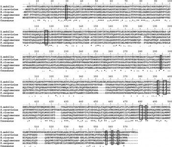
To investigate the nature of the amino acid residues putatively related to the substrate specificity in P. serpens enzyme, we have aligned the amino acid sequence of Z. mobilis and S. cerevisiae PDCs; E. cloacae and P. agglomerans IPDCs; and P. serpens PDC/IPDC (Fig. 5). The alignment confirmed previous conclusions regarding the presence of Gln392 in the active site of the IPDC enzymes replaced by Thr in the PDC proteins (Schütz et al. Reference Schütz, Sandalova, Ricagno, Hübner, König and Schneider2003b ). Phytomonas serpens enzyme showed Gln at 392 position (Fig. 5). The alignment also confirmed conservation of the localization of the four ionizable residues in PDC and IPDC proteins (Asp30; His116; His117; Glu481), which seem to play a more prominent role in catalysis than in substrate binding (Andrews and McLeish, Reference Andrews and McLeish2012).
Alignment of PDC and IPDC protein sequences by Clustal W (1·83 version) and BioEdit sequence editor. Core ionizable residues and those thought to influence substrate specificity are surrounded by rectangles (see Fig. 6 and Table 2 in Supplementary material). In Clustal consensus: asterisks denote identical residues; two dots, conserved substitutions; and one dot, semi-conserved substitutions.

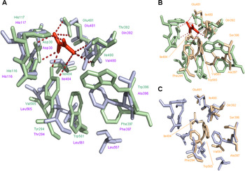
The established 3D structures of pyruvate-complexed Z. mobilis PDC (PDB ID 2wva; Pei et al. Reference Pei, Erixon, Luisi and Leeper2010) and E. cloacae IPDC (PDB ID 1ovmA; Schütz et al. Reference Schütz, Sandalova, Ricagno, Hübner, König and Schneider2003b ) were used to infer the size of the active site of P. serpens enzyme and the location of potentially important residues (Fig. 6). The 3D model of P. serpens enzyme was constructed by (PS)2-V2 and Swiss-Model servers. In both the template structure automatically selected by homology with P. serpens protein was E. cloacae IPDC (PDB ID 1ovmA). (PS)2-V2 used an alignment with 43·95% and E-value 1·4e-25, while the Swiss-Model used an alignment with 43·33% and E-value 0·00e-1. Ramachandran plots of the two models showed that the most consistent structure was the (PS)2-V2 model (supplementary material – Fig. 2S). In this model, 94·9% of the residues were in the favoured region; 4% in the allowed region; and 1·1% in the outlier. The (PS)2-V2 model was aligned with the established 3D structures of E. cloacae and Z. mobilis and confirmed the positions of the active site residues in P. serpens enzyme (Fig. 6).
Alignment of the three-dimensional structures of Z. mobilis PDC (PDB 2wva; shown in pale green), E. cloacae IPDC (PDB 1ovmA; in light blue) and P. serpens PDC/IPDC (in light orange, predicted by the homology modelling (PS)2-V2). The figures were visualized and aligned with PyMOL. Alignments: (A) Z. mobilis and E. cloacae; (B) Z. mobilis and P. serpens; (C) E. cloacae and P. serpens. Only the region containing the core ionizable residues (Asp, His, His, Glu) and those putatively related to substrate specificity are shown (see Table 2 in supplementary material). Pyruvate substrate is shown in red, and its contact with amino acid residues was based on a previous report (Andrews and McLeish, Reference Andrews and McLeish2012). In Z. mobilis the core ionizable residues Asp30, His116 and His117 (positions indicated in the alignment shown in Fig. 5) correspond to the V monomer; the residues related to substrate specificity along with the ionizable residue Glu481, correspond to the Z monomer. In E. cloacae the residues correspond to A3 and B3 monomers, respectively. The core ionizable residues are not shown in the P. serpens structure given their conserved position.

This model suggests that the P. serpens active site has an intermediate size between E. cloacae and Z. mobilis. In fact, inspection of the number of residues with cyclic side chains in the substrate binding site of the enzymes (supplementary material Table 2) indicated six residues in Z. mobilis PDC, three in E. cloacae IPDC and four in P. serpens protein. The substitution of Thr392 of Z. mobilis by Gln both in E. cloacae and P. serpens proteins seems to leave free the side chain of Gly for the interaction and accommodation of the indole ring of IPyA. In turn, the position of the side chain of Trp565 of P. serpens enzyme most likely reduces the size of the active site, giving ambiguous characteristics to the substrate binding site.
DISCUSSION
We report here for the first time the production of IAA by P. serpens, a flagellated trypanosomatid highly abundant in tomato fruits in many countries. The functionality of the auxin produced by P. serpens was confirmed in the hypocotyl elongation test. Under the conditions assayed both P. serpens-conditioned media and exogenous IAA (9 μ m) promoted 10–13% increase of the tomato hypocotyl length. Coincidentally, 13% increase of hypocotyl elongation was reported for the tomato Lycopersicon esculentum cultivar VFN8 following incubation with 10 μ m IAA (Kelly and Bradford, Reference Kelly and Bradford1986). In that report, stimulation of elongation was detectable at 0·1 μ m and saturated typically at 1 to 10 μ m IAA.
We have shown that free IAA concentration had no significant variation in tomato fruits inoculated with P. serpens, T. cruzi or exogenous IAA. On the other hand, in P. serpens and IAA inoculated tomatoes the abundance of IAA-amide and IAA-ester compounds increased on day 7 pi. Interestingly the kinetics of IAA-ester accumulation in the fruits inoculated with P. serpens or IAA was clearly different. In the flagellate infection IAA-ester raised on day 14 pi suggesting the continuous production of the auxin and conversion to the conjugated form, whereas in fruits inoculated with exogenous IAA the concentration of IAA-ester decreased, suggesting degradation or utilization of the conjugate. These observations support previous conclusions indicating a tight regulation of both steady-state levels of IAA and metabolic processes leading to auxin conjugation in tomato fruits (Catalá et al. Reference Catalá, Ostin, Chamarro, Sandberg and Crozier1992). In fact, labelled IAA added to tomato pericarp discs (L. esculentum Mill.) was converted to IAA-Glc, IAA-Asp and other non-characterized intermediates. The Asp-conjugate was not hydrolysed to yield free IAA, in contrast to what was observed for IAA-Glc (Catalá et al. Reference Catalá, Ostin, Chamarro, Sandberg and Crozier1992).
Most auxin conjugations are reversible via the action of specific hydrolases with the subsequent release of free IAA (Seidel et al. Reference Seidel, Walz, Park, Cohen and Ludwig-Müller2006; Rosquete et al. Reference Rosquete, Barbez and Kleine-Vehn2012). Whereas little is known about the hydrolysis of auxin-sugar conjugates, the process is better understood for amino acid conjugates, in which IAA amidohydrolases have been implicated in the deconjugation of auxin amides. Yet, conjugation with Asp and Glu seems to be irreversible, eventually implying alternative functions for these conjugates (reviewed by Rosquete et al. Reference Rosquete, Barbez and Kleine-Vehn2012). In the future it will be of interest to characterize the routes taken by the IAA conjugates after the P. serpens tomato infection.
In a previous study (Ienne et al. Reference Ienne, Pappas, Benabdellah, González and Zingales2012) we observed that the P. serpens enzyme exhibits high amino acid sequence similarity with proteins of the trypanosomatids Leishmania spp. and C. fasciculata. Contrary to what was verified for P. serpens we did not detect PDC activity in Leishmania major extracts (Ienne et al. Reference Ienne, Pappas, Benabdellah, González and Zingales2012). Here, we have investigated the accumulation of IAA and intermediates in L. major conditioned 199 media. Negative results were obtained even after addition of different Trp concentrations to the cultures (data not shown). We have reported a 6-fold increase in the abundance of the PDC/IPDC gene copy number and corresponding transcripts in P. serpens as compared with L. major, which suggests that the acquisition of this gene may represent an adaptive advantage for Phytomonas sp. (Ienne et al. Reference Ienne, Pappas, Benabdellah, González and Zingales2012). Since, to our knowledge, no reports describe ethanol production in Leishmania, the function of the PDC/IPDC gene in the genus Leishmania deserves further investigation. On the other hand, ethanol production by Crithidia has been previously described (Cazzulo et al. Reference Cazzulo, Franke de Cazzulo, Engel and Cannata1985; Chaumont et al. Reference Chaumont, Schanck, Blum and Opperdoes1994), denoting that the PDC/IPDC gene product could participate in the alcohol fermentation pathway. We have not investigated the presence of IAA in Crithidia-conditioned media. This is an interesting topic that may be addressed in the future.
The production of IAA by various bacterial species has been widely documented. The interaction between IAA-producing bacteria and plants leads to diverse outcomes on the plant side, varying from pathogenesis to beneficial effects (Spaepen et al. Reference Spaepen, Vanderleyden and Remans2007). The consequence for the plant is usually a function of the amount of IAA and the sensitivity of the plant tissue to changes in IAA concentration. In pathogenic and non-pathogenic bacteria the biosynthesis of IAA via IPyA and IAAld is verified (Spaepen et al. Reference Spaepen, Vanderleyden and Remans2007). Tumour- and gall-inducing bacteria were the first plant-associated organisms in which IAA biosynthesis pathways were investigated and the role of the released auxin implicated in pathogenesis (Morris, Reference Morris and Davies1995). The beneficial effect of rhizobacteria on root proliferation and enhancement of plant growth has been attributed at least in part to bacterial IAA biosynthesis (Dobbelaere et al. Reference Dobbelaere, Croonenborghs, Thys, Vande Broek and Vanderleyden1999).
Since P. serpens has been found in healthy tomatoes (Camargo, Reference Camargo1999), our data suggest that the auxin produced in the fruit is converted into IAA conjugates, keeping unaltered the intracellular concentration of free IAA. As a consequence, the production of IAA by P. serpens would exert neither pathogenic nor beneficial effects to the fruit. Because P. serpens metabolism is well-adapted to consume glucose (Maslov et al. Reference Maslov, Nawathean and Scheel1999; Nawathean and Maslov, Reference Nawathean and Maslov2000), we are tempted to propose that P. serpens-tomato interaction could represent a case of commensalism. Because we have shown conservation of the PDC/IPDC gene sequence in 14 Phytomonas isolates (Ienne et al. Reference Ienne, Pappas, Benabdellah, González and Zingales2012), including the latex-associated Phytomonas françai causative of the ‘empty roots’ disease in manioc (Dollet, Reference Dollet1984), it is of interest to verify whether P. françai produces IAA and the fate of the auxin in the host plant.
Here we showed that P. serpens secreted ethanol in the culture media and that addition of Glc enhanced ethanol production. The accumulation of ethanol in the media was 104-fold higher than that of IAA. This observation supports our previous suggestion that the high PDC activity displayed by P. serpens may have contributed to the adaptation of the flagellate to the plant glucose rich media (Ienne et al. Reference Ienne, Pappas, Benabdellah, González and Zingales2012).
We have raised the possibility that P. serpens PDC/IPDC could display both PDC and IPDC activities. Three-dimensional models and analysis of the amino acid residues putatively involved in substrate recognition suggested that the active site of P. serpens enzyme has an intermediate size between PDC and IPDC proteins. Small movements in a few important residues could allow the entrance of substrates of different dimensions (Andrews and McLeish, Reference Andrews and McLeish2012).
This study showed that the levels of the auxin and Tol increased in P. serpens culture media in response to amounts of Trp, whereas the level of IPyA remained constant and very low. We have not detected IAAld in P. serpens-conditioned media suggesting its rapid conversion into IAA and Tol (see Fig. 1B) or instability. Tol is a common by-product of IAA biosynthesis via the IPyA/IAAld pathway (Shin et al. Reference Shin, Shinguu, Sano and Umezawa1991; Brandl and Lindow, Reference Brandl and Lindow1996), and could serve as a reservoir for IAA and to maintain the levels of IAAld in equilibrium in plants (Brown and Purves, Reference Brown and Purves1980). Overall we are tempted to suggest that the biosynthesis of IAA in P. serpens may occur via the Trp-IPyA-IAAld pathway, as verified in a broad range of bacteria (Spaepen et al. Reference Spaepen, Vanderleyden and Remans2007).
Data on the P. serpens genome are not available. A search in the trypanosomatid sequenced genomes (TriTrypDB) revealed Tyr and Asp aminotransferases which could convert aromatic amino acids to their corresponding ketoacids. Corroborating this suggestion, the formation IPyA in vitro and in vivo has been reported for the catabolism of Trp in African trypanosomes (el Sawalhy et al. Reference el Sawalhy, Seed, el Attar and Hall1995). Accumulation of IPyA can be justified by the fact that no orthologous PDC/IPDC genes were found in Trypanosoma brucei or T. cruzi genomes (Ienne et al. Reference Ienne, Pappas, Benabdellah, González and Zingales2012). The acquisition of the IPDC gene from donor phytobacteria could confer to P. serpens the ability to decarboxylate IPyA and proceed the biosynthesis of IAA.
SUPPLEMENTARY MATERIAL
To view supplementary material for this article, please visit http://dx.doi.org/10.1017/S0031182014000547.
ACKNOWLEDGEMENTS
We are greatly indebted to Dr Marta M. G. Teixeira for providing P. serpens culture; to Dr Lázaro E. P. Peres for providing Micro-Tom tomato seeds and to Dr Iolanda M. Cuccovia for the availability of the spectrophotometer Varian Cary WinUV. We thank Dr Georgios Pappas Jr. for helpful suggestions and Marcelo Nunes Silva for technical assistance. This work was supported by grants of Fundação de Amparo à Pesquisa do Estado de São Paulo (FAPESP) (grant number 2010/50957-1) and Conselho Nacional de Desenvolvimento Científico e Tecnológico (CNPq) (grant number 304793/2009-4) (BZ and EP). SI was recipient of a fellowship from FAPESP (grant number 2008/50209-5).