




No CrossRef data available.
Article contents
Expression of apoptosis-related factors in muscles infected with Trichinella spiralis
Published online by Cambridge University Press: 03 March 2004
Abstract
We found that the expression of mitochondrial apoptosis related genes (Bcl-2 associated protein X, BAX; apoptotic protease activating factor 1, Apaf-1; Caspase 9 and serine/threonine protein kinase, PKB) is elevated in Trichinella spiralis-infected muscles during encapsulation. Micro-dissection of the capsule and subsequent reverse transcription polymerase chain reaction (RT-PCR) confirmed that the expressions of these genes are restricted to the nurse cell. Immunocytochemistry revealed that pro-apoptosis factor (BAX, Apaf-1 and Caspase 9) are predominantly expressed in the basophilic cytoplasm (infected muscle cell origin) and anti-apoptosis factor (PKB) in the eosinophilic cytoplasm (satellite cell origin) of the nurse cell. Electron microscopy revealed that the pre-existing mitochondria in the muscle cells became swollen and disappeared immediately after newborn larva invasion, but new mitochondria of smaller size appeared in the cytoplasm. Nuclear fragmentation and condensation were observed in basophilic cytoplasm which is known to die. Together, the results suggest that the infected muscle cells transform but die through the process of apoptosis which is triggered by factors from the newly formed mitochondria. The anti-apoptosis factor may help the eosinophilic cytoplasm with its survival to ensure nurse cell function.
Information
- Type
- Research Article
- Information
- Copyright
- 2004 Cambridge University Press
INTRODUCTION
Trichinella spiralis in the muscle stage is found in a capsule which is composed of non-cellular and cellular components. The former is cyst wall composed of collagen fibres and micelle substances. The outer layer is formed by fibroblasts and the inner layer is formed by the nurse cell (Matsuo et al. 2000). The latter is within the cyst wall and is comprised of basophilic cytoplasm, eosinophilic cytoplasm and satellite cells. Sometimes host inflammatory cells may migrate. The histological analysis of the capsule formation revealed that the basophilic cytoplasm is formed by the transformation of a muscle cell after the newborn-larva invasion. The eosinophilic cytoplasm is formed by the misdifferentiation of a myoblast (satellite cell), which is thought to differentiate to the muscle cell in response to muscle damage (Wu et al. 2001).
The immature capsule, shortly after infection, has basophilic cytoplasm, while the eosinophilic cytoplasm prevailed in the mature capsule. This change in the ratio of the two kinds of cytoplasm occurs reciprocally while the capsule formation takes place. The two kinds of cytoplasm seem to differ from each other. The basophilic cytoplasm has elevated acid phosphatase activity and seems to be degraded. On the other hand, eosinophilic cytoplasm has elevated alkaline phosphatase activity and seems to be metabolically active (Boonmars et al. 2003). The eosinophilic cytoplasm is supplied from the satellite cells whose myogenic factors are activated (Wu et al. 2001).
To determine the fate of the two kinds of cytoplasm we investigated the expression of pro-apoptotic gene (Bcl-2 associated protein X, BAX; apoptotic protease activating factor 1, Apaf-1 and Caspase 9) and anti-apoptotic gene (serine/threonine protein kinase, PKB) proteins by means of quantitative PCR and immunohistochemistry in the muscle infection of T. spiralis in various stages of infection.
MATERIALS AND METHODS
Parasites and infection
Trichinella spiralis (ISS413) has been maintained in mice in our laboratory. Nude mice were orally infected with T. spiralis (800 infective larvae per mouse). The mice were sacrificed at certain time-points (8, 13, 18, 23, 28, 33, 38 and 53 days post-infection (p.i.)).
Light and electron microscopic observations
For light microscopic observation, striated muscle tissue was fixed in 10% formalin, embedded in paraffin, thin-sectioned and stained with haematoxylin and eosin (H&E staining). The muscle tissue was processed for electron microscopic observation according to the method described in a previous paper (Takahashi et al. 1988). Briefly, it was fixed with 2·5% glutaraldehyde plus 4% paraformaldehyde, and double-stained with uranyl acetate and Raynolds solution.
Primer design for quantitative reverse transcription polymerase chain reaction (RT-PCR)
The primer pairs for anti-apoptosis factor (PKB) and pro-apoptosis factor (BAX, Apaf-1 and Caspase 9) were designed based on the published sequence as summarized in Table 1.

The primer pairs for endogenous controls (mouse glyceraldehyde-3-phosphate dehydrogenase, MG3PDH and 18S rRNA) were designed based on the published sequence as summarized in Table 2.

RNA isolation from infected muscles and RT-PCR to detect apoptosis related-genes
Anaesthetized mice were killed by means of cervical dislocation and quickly dissected. Whole muscle tissues (100 mg) obtained from the hind limbs at 8, 13, 18, 23, 28, 33, 38 and 53 days p.i. were used for analysis. Uninfected muscles were used as a control. Total RNA was isolated using TRIZOL (Invitrogen, Carlsbad, CA, USA) according to the manufacturer's instructions. The isolated RNA was treated with DNase (5 units RQ1 RNase-Free DNase, Promega, Co., Madison, WI, USA) and 119 units of Ribonuclease Inhibitor (Takara Shuzo, Co., Ltd, Kyoto, Japan) in the buffer (400 mM Tris–HCl, 100 mM NaCl, 60 mM MgCl2 and 20 mM dithiothreitol, pH 7·5). The treated RNA was extracted with phenol/chloroform, precipitated with ethanol, and dissolved in RNase-free water (100 μl). Total RNA was reverse transcribed into cDNA using Oligo(dT)12–18 primers (Amersham Pharmacia Biotech, Inc. Piscataway, NJ, USA) and a Ready-to-Go First-Strands beads kit (Amersham Pharmacia Biotech). Thirty-two μl of the RNA sample (5 μl of RNA original) and 1 μl of 0·5 μg/μl Oligo(dT)12–18 were added to a Ready-to-Go tube. The tube was incubated at 37 °C for 60 min and then 95 °C for 10 min.
The PCR reaction mixture comprised 3 μl of reverse transcription products (1[ratio ]10 diluted), 3 μl of 10× PCR buffer, 3 μl of deoxynucleoside triphosphate (2·5 mM each), 6 μl of primer pairs (5 μM), 0·12 μl of Taq polymerase (5 U/μl, Takara Shuzo) and 15 μl of distilled water to give the final volume of 30 μl. PCR conditions were as follows: 22 cycles for MG3PDH, 38 cycles for BAX, 40 cycles for Apaf-1 and Caspase 9, and 28 cycles for PKB at 94 °C for 30 sec, 56 °C for 30 sec, 72 °C for 2 min followed by a final extension at 72 °C for 7 min. Aliquots of PCR products were analysed by gel electrophoresis with 1·5% agarose gel stained with 1 μg/ml ethidium bromide and photographed under UV light. The photographs were analysed for density using Scion image (Scion Co., Frederick, MD, USA). All reactions for the standard control and the experiment sample were performed in triplicate. The amount of BAX, Apaf-1, Caspase 9 and PKB genes were calculated as follows:
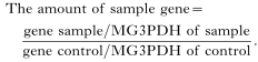
The ratio of density of the apoptosis-related genes and the standard gene were plotted in a graph (Fig. 3).
Laser capture microscopy to isolate RNA from the cyst and RT-PCR to detect apoptosis related-genes
Cryosections (8 μm in thickness) of quickly frozen muscles from 23 days p.i. were prepared and placed onto glass slides (pre-heated at 200 °C for 2 h to destroy RNase) and dried immediately with room air using a conventional dryer. The capsules were micro-dissected using the laser micro-beam of the Robot-MicroBeam system (P.A.L.M. GmbH, Bernried, Germany) equipped with a 40× dry long-distance objective lens. The control cells were collected from the unaffected area of the same sample used for the experiment. The unaffected area of the muscle was identifiable by light microscope observation. The capsules were cut together with their supporting optimal cutting temperature compound with a laser beam. One μl of mineral oil was dropped into a 0·5 ml tube cap. The test samples contained about 150–200 capsules (1×106 μm2 of tissue section), and the control was prepared from the unaffected muscle with the same square μm. Total RNA was isolated using RNeasy protect mini kit (Qiagen GmbH, Hilden, Germany). The caps of 0·5 ml tubes were reversed, and the lysis buffer was added. After the incubation in a water bath for 15 min at 37 °C with a vigorous vortex for 30 min, the manufacturer's protocol was then followed. Thirty-two μl of the RNA sample was reverse transcribed into cDNA using 1 μl of 0·5 μg/μl oligo(dT)12–18 primers (Amersham Pharmacia Biotech) and a Ready-to-Go First-Strands beads kit (Amersham Pharmacia Biotech). The tube was incubated at 37 °C for 60 min and then 95 °C for 10 min.
The PCR reaction mixture comprised 3 μl of the reverse transcription products, 1·5 μl of 10× PCR buffer, 1·5 μl of deoxynucleoside triphosphate (2·5 mM each), 3 μl of primer pairs (5 μM), 0·06 μl of Taq polymerase (5 U/μl) and 6 μl of distilled water to give a final volume of 15 μl. PCR conditions were as follows: 35 cycles at 94 °C for 30 sec, 52 °C for 30 sec, 72 °C for 2 min and followed by a final extension at 72 °C for 7 min. The aliquots of PCR products were analysed by gel electrophoresis with 1·5% agarose gel stained with 1 μg/ml ethidium bromide and photographed under UV light. The photographs were analysed for density using a Scion image. All reactions for standard control and the experiment sample were performed in triplicate. The amount of BAX, Apaf-1, Caspase 9 and PKB genes were calculated as follows:
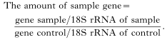
The ratio of density of the apoptosis-related genes and the standard gene were plotted in a graph (Fig. 4).
Immunohistochemical staining for apoptosis- related factors
Cryosections of 18, 23 and 48 days p.i. were fixed with 4% paraformaldehyde (PFA) in 0·1 M phosphate-buffered saline (PBS) for 5 min, washed with PBS for 15 min 3 times and then blocked with 5% skimmed milk (DIFCO Laboratories, Detroit, MI, USA) in PBS for 30 min. The primary antibody included the rabbit polyclonal antibodies to BAX (BAX-19, Santa Cruz Biotechnology Inc., Santa Cruz, CA, USA), the rabbit polyclonal antibodies to p-Akt1 (p-Akt1 (Ser473)-R, Santa Cruz Biotechnology), rabbit polyclonal antibodies to Caspase 9 (Cell Signaling Technology, Inc., Beverly, MA, USA) and the rabbit polyclonal antibodies to Apaf-1 ((H-324) Santa Cruz Biotechnology). The sections were incubated with the primary antibody (1[ratio ]100 diluted) for 1·5 h at 37 °C, washed 3 times with PBS and further processed using the Histostain-SP (ZYMED Laboratories Inc. San Francisco, CA, USA) according to the manufacturer's instructions. Slides were photographed and then stained with H&E for the histological orientation and photographed under a light microscope. The negative control study was performed using PBS instead of the primary antibody.
RESULTS
Histology of capsule formation
These data have been published previously (Matsuo et al. 2000). Therefore, only a brief summary is given here with emphasis on mitochondria and nucleus. After newborn larvae (NBL) of T. spiralis had infected the striated skeletal muscle, the infected muscle cells transformed to the nurse cell that is basophilic (Fig. 1A) by H&E staining. The earliest ultrastructural change observed was disintegration of the sarcomere structure and swelling of mitochondria (Fig. 2A). The basophilic cytoplasm was replaced by eosinophilic cytoplasm (Fig. 1B) during the capsule formation.

Fig. 1. Haematoxylin and eosin staining of Trichinella spiralis-infected muscles on 18 (A) and 38 (B) days post-infection, which showed basophilic cytoplasmic changes (A). The infected muscle cell is separated from undamaged muscle cells by a capsule wall (B). The linear alignment of nuclei of satellite cells was observed in the periphery of infected cells along their long axis (*). L, larva.

Fig. 2. Electron micrograph showing histopathology of Trichinella spiralis-infected muscles. Immediately after invasion by the newborn larvae mitochondria were swollen and disintegrated, some of them were fused with lysosomes (A). Mitochondria disappeared and were replaced by new mitochondria (arrowed, B) that were smaller than those have seen in normal muscle and located closely to the nucleus. The nucleus had an irregular shape and scattered heterochromatin (C). M, mitochondria; N, nucleus.
The nuclei in the basophilic cytoplasm exhibited an irregular shape (Fig. 2C). During the next step of capsule formation in the early stage, old mitochondria disappeared and were replaced by new mitochondria (characterized by hyper-density of their matrix and good integrity) that were much smaller in size than those seen in normal muscle cells (Fig. 2B). About 1 month after infection, the capsule had a capsule wall composed of an inner layer formed by the nurse cell and the outer layer that is formed by fibroblasts.
Kinetics of expression of apoptosis-related genes
Fig. 3 shows the level of expression of apoptosis-related genes, which were determined by semi-quantitative RT-PCR at various stages in infected or non-infected muscles (RNA was isolated from block specimens). The specific band of MG3PDH (endogenous control) was observed in all stages with the band being the same density. Specific bands of BAX, Apaf-1, Caspase 9 and PKB were also observed in all stages of infection, but the expression level at each stage was different. The expression level of BAX and Apaf-1 was increased from 13 days p.i. and reached a peak at 18 days p.i. After 28 days p.i. the BAX expression level gradually decreased, and then at 53 days p.i., the level decreased nearly to the control level. Expression levels of the Caspase 9 gene (pro-apoptosis) and PKB gene (anti-apoptosis) followed the same kinetics as BAX, although the initial increase was somewhat delayed.

Fig. 3. RT-PCR results on the expression level of apoptotsis-related genes (BAX, Apaf-1, Caspase 9, and PKB) in the infected muscle tissue. Total RNA was isolated from infected muscle tissue at various stages (0, 8, 13, 18, 23, 28, 33, 38 and 53 days post-infection) and amplification products were analysed by agarose gel electrophoresis and shown in the right panel. Using Scion Image software, the relative density of each sample band against the control band (MG3PDH) was analysed and shown in the left panel.
Detection of apoptosis-related genes in the capsule
To determine whether the origin of the BAX, Apaf-1, Caspase 9 and PKB genes is from nurse cells and/or muscle cells, RT-PCR was performed to detect the mRNA expression level in nurse cells and muscle cells separately.
Fig. 4 shows the semi-quantitative RT-PCR of the BAX, Apaf-1, Caspase 9, PKB and the control gene as 18S rRNA at 23 days p.i. in nurse cells and/or muscle cells. A specific band of 18S rRNA (endogenous control) was observed in both samples. The expression level of 18S rRNA in both samples was almost the same. Specific bands of PKB and BAX were also observed in both samples, but the higher expression level of both BAX and PKB was observed in the nurse cell sample and a lower expression level was observed in the muscle cell sample. On the other hand, a specific band of Apaf-1 and caspase 9 was observed only in the nurse cell sample.

Fig. 4. RT-PCR results on the expression level of apoptotsis-related genes (BAX, Apaf-1, Caspase 9, and PKB) in the capsules or unaffected muscle cells which were obtained from a Trichinella spiralis-infected mouse at 23 days post-infection. Total RNA was isolated from laser microdissected capsules or unaffected muscle cells (control), amplification of the sample genes (BAX, Apaf-1, Caspase 9 and PKB) and the standard gene (18S rRNA) was performed and PCR products were analysed by agarose gel electrophoresis (right panel). Using Scion Image software, the relative density of each sample band against the control band was analysed and shown in the left panel.
In situ localization of apoptosis-related factors
Immunohistochemical staining results are shown in Fig. 5 (BAX), Fig. 6 (Apaf-1), Fig. 7 (Caspase 9) and Fig. 8 (PKB). On the upper part of each figure, immunostaining slides from 18, 23 and 48 days p.i. are shown, and H&E stained sections were placed on the lower half. Strong positive staining for BAX, Apaf-1 and Caspase 9 was observed within the 18 day p.i. capsule, but not in the muscle tissues. The direct comparison of the immunostaining and H&E staining slides confirmed that the cytoplasm of the positive staining tissues was predominantly basophilic cytoplasm. PKB expression was strongly positive in the eosinophilic cytoplasm, which is derived from the satellite cell. Although these gene expressions were weaker at 23 days p.i., the immunostaining intensity for the gene products was the same. At 48 days p.i., the immunostaining intensity was negligible except for PKB (E in Fig. 8). The larval body was positively stained with only anti-BAX antibody. The control slides were negative for immunostaining, which confirmed the staining specificity.

Fig. 5. Immunohistochemical staining for BAX. Cryosections of muscles infected with Trichinella spiralis for 18 (A), 23 (C) and 48 (E) days post-infection were reacted with anti-BAX antibody. The red colour indicates the positive reaction (*). After anti-BAX staining the same section was subjected to H&E staining for histological orientation purposes (B, D, F). L, larva.

Fig. 6. Immunohistochemical staining for Apaf-1. Cryosection of muscle infected with Trichinella spiralis for 18 (A), 23 (C) and 48 (E) days post-infection was reacted with anti-Apaf-1 antibody. The red colour indicates the positive reaction (*). After anti-Apaf-1 staining the same section was subjected to H&E staining for histological orientation purposes (B, D, F). L, larva.

Fig. 7. Immunohistochemical staining for Caspase 9. Cryosection of muscle infected with Trichinella spiralis for 18 (A), 23 (C) and 48 (E) days post-infection was reacted with anti-Caspase 9 antibody. The red colour indicates the positive reaction (*). After anti-Caspase 9 staining the same section was subjected to H&E staining for histological orientation purposes (B, D, F). L, larva.

Fig. 8. Immunohistochemical staining for PKB. Cryosection of muscle infected with Trichinella spiralis for18 (A), 23 (C) and 48 (E) days post-infection was reacted with anti-akt1 antibody (akt-473). The red colour indicates the positive reaction (*). After anti-PKB staining the same section was subjected to H&E staining for histological orientation purposes (B, D, F). L, larva.
DISCUSSION
The nurse cell takes care of the parasites and is definitely not a normal constituent of the host. Why do terminally differentiated muscle cells transform to nurse cells? Why do satellite cells differentiate to nurse cells? The mechanisms of the cyst formation are one of the major biological interests related to Trichinella infections. According to our previous paper (Boonmars et al. 2003), acid phosphatase activity was observed in the basophilic cytoplasm (infected muscle cell origin) of the nurse cell, which suggested the presence of destructive processes. In addition, alkaline phosphatase activity was observed in eosinophilic cytoplasm (satellite cell origin), which suggested that this cytoplasm is metabolically active, engaging in metabolic transportation (Boonmars et al. 2003). Thus, the two kinds of cytoplasm have different fates and it is likely that apoptotic and anti-apoptotic mechanisms are involved in determining such fates. In the present study we successfully showed that apoptosis-related genes are involved in the capsule formation of T. spiralis.
Apoptotic genes are well known in cell suicide, which plays a role in various physiological processes, and deregulation of apoptosis has been implicated in pathological processes of diseases (Sandri & Carraro, 1999). The present RT-PCR experiment showed the temporal elevation of apoptosis-related genes (BAX, Apaf-1, Caspase 9 and PKB) in infected muscles concomitant with the capsule formation, although the elevation of Caspase 9 and PKB genes happened 5 days later than the other genes. Microdissection and a subsequent RT-PCR experiment confirmed that these genes were expressed predominantly in the capsule, not in the normal muscle cells.
The immunostaining results showed the precise location of products of apoptosis-related genes. Products of pro-apoptosis genes (BAX, Apaf-1 and caspase 9) were located in the basophilic cytoplasm. On the other hand, the anti-apoptosis gene (PKB) was located in the eosinophilic cytoplasm.
Following T. spiralis infection, the actin-myosin structures disappeared and mitochondria swelled and disappeared in the early phase of the infection. In other words, older mitochondria were replaced by the new mitochondria (Matsuo et al. 2000). The new mitochondria were smaller in size than those in normal muscle cells. They had a hyper-density matrix, which was in good agreement to features of mitochondrial pyknosis in apoptosis (Mancini et al. 1997; Desagher & Martinou, 2000). Three types of mitochondrial pro-apoptosis genes (BAX, Caspase 9 and Apaf-1) were found to increase in their expression in this study. Concomitantly, morphological signs characteristic of apoptosis (irregular shaped nuclei, scattered and dense heterochromatin) were seen in the basophilic cytoplasm (Matsuo et al. 2000).
Reportedly, BAX induces apoptosis by forming the membrane pore in mitochondria from which cytochrome c is released upon apoptotic signalling (Desagher & Martinou, 2000; Gottlieb, 2000; Tsujimoto & Shimizu, 2000; Parone, James & Martinou, 2002). An increase of BAX expression is probably responsible for other types of muscle disorders (Monici et al. 1998; Lee, Lee & Lee, 2001; Chuang et al. 2002; Ikezoe et al. 2002; Tews, 2002; Umaki et al. 2002). Apaf-1 is a co-apoptotic factor and caspase 9 is an initiator caspase, both of which likely play roles in cascades that lead to cell death (Saleh et al. 1999; Desagher & Martinou, 2000; Parone et al. 2002).
The PKB gene is probably expressed in satellite cell-origin cytoplasm, because the gene was expressed in the capsule and the product was in the eosinophilic cytoplasm. It is an anti-apoptosis factor and protects cells from death (Lawlor & Alessi, 2001; Nicholson & Anderson, 2002), by means of inactivation of pro-apoptotic proteins such as Bad (Datta et al. 1997; Del Peso et al. 1997) and Caspase 9 (Cardone et al. 1998; Wang et al. 2002), and inhibition of BAX conformational change (Yamaguchi & Wang, 2001).
It is a well-established concept that the nurse cell can survive for years (Steel, 1982). Although this is true our previous (Matsuo et al. 2000; Wu et al. 2001; Boonmars et al. 2003) and present results suggest that this concept needs some modifications. The infected muscle segregates its affected portion to minimize cell damage (Wu et al. 2001). This portion (basophilic cytoplasm) transforms to a nurse cell but soon dies due to the apoptotic process. In response to this muscle cell damage, the satellite cells proliferate and try to differentiate into muscle cells but differentiate instead to nurse cells (Matsuo et al. 2000). This eosinophilic cytoplasm is metabolically active (Boonmars et al. 2003), probably protected by PKB-driven anti-apoptosis mechanisms. This cytoplasm also may die soon or later but the active satellite cell is within the capsule (Matsuo et al. 2000), which likely ensures the continuous supply of new cells. Therefore, the nurse cell cyst seems to be the result of continuous destruction and production of cytoplasm. At present this is most plausible explanation why the nurse cell appears to have a long survival time.
This study was partly supported by a Grant-in-Aid for Scientific Research (15590366) from the Ministry of Education, Culture, Sports, Science and Technology of Japan.
References
REFERENCES
BOONMARS, T.,
WU, Z.,
NAGANO, I.,
NAKADA, T. &
TAKAHASHI, Y.
(2004).
Differences and similarities of nurse cells in cysts of Trichinella spiralis and T. pseudospiralis.
Journal of Helminthology 10, 000–000.Google Scholar
CARDONE, M. H.,
ROY, N.,
STENNICKE, H. R.,
SALVESEN, G. S.,
FRANKE, T. F.,
STANBRIDGE, E.,
FRISCH, S. &
REED, J. C.
(1998).
Regulation of cell death protease caspase-9 by phosphorylation.
Science 282, 1318–1321.CrossRefGoogle Scholar
CHUANG, Y. H.,
CHUANG, W. L.,
HUANG, S. P. &
HUANG, C. H.
(2002).
Over-expression of apoptosis-related proteins contributes to muscular damage in the obstructed ureter of the rat.
British Journal of Urology International 89,
106–112.CrossRefGoogle Scholar
DATTA, S. R.,
DUDEK, H.,
TAO, X.,
MASTERS, S.,
FU, H.,
GOTOH, Y. &
GREENBERG, M. E.
(1997).
Akt phosphorylation of BAD couples survival signals to the cell-intrinsic death machinery.
Cell 91, 231–241.CrossRefGoogle Scholar
DEL PESO, L.,
GONZALEZ-GARCIA, M.,
PAGE, C.,
HERRERA, R. &
NUNEZ, G.
(1997).
Interleukin-3-induced phosphorylation of BAD through the protein kinase Akt.
Science 278, 687–689.CrossRefGoogle Scholar
DESAGHER, S. &
MARTINOU, J. C.
(2000).
Mitochondria as the central control point of apoptosis.
Trends in Cell Biology 10,
369–377.CrossRefGoogle Scholar
GOTTLIEB, R. A.
(2000).
Role of mitochondria in apoptosis.
Critical Reviews in Eukaryotic Gene Expression 10,
231–239.Google Scholar
IKEZOE, K.,
NAKAGAWA, M.,
YAN, C.,
KIRA, J.,
GOTO, Y. &
NONAKA, I.
(2002).
Apoptosis is suspended in muscle of mitochondrial encephalomyopathies.
Acta Neuropathologica 103,
531–540.CrossRefGoogle Scholar
LAWLOR, M. A. &
ALESSI, D. R.
(2001).
PKB/Akt: a key mediator of cell proliferation, survival and insulin responses?
Journal of Cell Science 114,
2903–2910.Google Scholar
LEE, M. J.,
LEE, J. S. &
LEE, M. C.
(2001).
Apoptosis of skeletal muscle on steroid-induced myopathy in rats.
Journal of Korean Medical Science 16,
467–474.CrossRefGoogle Scholar
MANCINI, M.,
ANDERSON, B. O.,
CALDWELL, E.,
SEDGHINASAB, M.,
PATY, P. B. &
HOCKENBERY, D. M.
(1997).
Mitochondrial proliferation and paradoxical membrane depolarization during terminal differentiation and apoptosis in a human colon carcinoma cell line.
The Journal of Cell Biology 138,
449–469.CrossRefGoogle Scholar
MATSUO, A.,
WU, Z.,
NAGANO, I. &
TAKAHASHI, Y.
(2000).
Five types of nuclei present in the cyst of Trichinella spiralis.
Parasitology 121,
203–210.CrossRefGoogle Scholar
MONICI, M. C.,
TOSCANO, A.,
GIRLANDA, P.,
AGUENNOUZ, M.,
MUSUMECI, O. &
VITA, G.
(1998).
Apoptosis in metabolic myopathies.
Neuroreport 9,
2431–2435.CrossRefGoogle Scholar
NICHOLSON, K. M. &
ANDERSON, N. G.
(2002).
The protein kinase B/Akt signalling pathway in human malignancy.
Cellular Signalling 14,
381–395.CrossRefGoogle Scholar
PARONE, P. A.,
JAMES, D. &
MARTINOU, J. C.
(2002).
Mitochondria: regulating the inevitable.
Biochimie 84,
105–111.CrossRefGoogle Scholar
SALEH, A.,
SRINIVASULA, S. M.,
ACHARYA, S.,
FISHEL, R. &
ALNEMRI, E. S.
(1999).
Cytochrome c and dATP-mediated oligomerization of Apaf-1 is a prerequisite for procaspase-9 activation.
The Journal of Biological Chemistry 274,
17941–17945.CrossRefGoogle Scholar
SANDRI, M. &
CARRARO, U.
(1999).
Apoptosis of skeletal muscles during development and disease.
The International Journal of Biochemistry and Cell Biology 31,
1373–1390.CrossRefGoogle Scholar
STEEL, J. M.
(1982).
Trichinosis. In
Handbook Series in Zoonoses: Section C: Parasitic Zoonoses (ed. Schultz, M. G.), pp.
293–329.
CRC Press,
Boca Raton, Florida, USA.
TAKAHASHI, Y.,
UNO, T.,
FURUKI, J.,
YAMADA, S. &
ARAKI, T.
(1988).
The morphology of Trichinella spiralis: ultrastructural study of mid- and hindgut of the muscle larvae.
Parasitology Research 75,
19–27.CrossRefGoogle Scholar
TEWS, D. S.
(2002).
Apoptosis and muscle fibre loss in neuromuscular disorders.
Neuromuscular Disorders 12,
613–622.CrossRefGoogle Scholar
TSUJIMOTO, Y. &
SHIMIZU, S.
(2000).
VDAC regulation by the Bcl-2 family of proteins.
Cell Death and Differentiation 7,
1174–1181.CrossRefGoogle Scholar
UMAKI, Y.,
MITSUI, T.,
ENDO, I.,
AKAIKE, M. &
MATSUMOTO, T.
(2002).
Apoptosis-related changes in skeletal muscles of patients with mitochondrial diseases.
Acta Neuropathologica 103,
163–170.Google Scholar
WANG, S. J.,
OMORI, N.,
LI, F.,
JIN, G.,
ZHANG, W. R.,
HAMAKAWA, Y.,
SATO, K.,
NAGANO, I.,
SHOJI, M. &
ABE, K.
(2002).
Potentiation of Akt and suppression of caspase-9 activations by electroacupuncture after transient middle cerebral artery occlusion in rats.
Neuroscience Letters 331,
115–118.CrossRefGoogle Scholar
WU, Z.,
MASUO, A.,
NAKADA, T.,
NAGANO, I. &
TAKAHASHI, Y.
(2001).
Different response of satellite cells in the kinetics of myogenic regulatory factors and ultrastructural pathology after Trichinella spiralis and T. pseudospiralis infection.
Parasitology 123,
85–94.CrossRefGoogle Scholar
YAMAGUCHI, H. &
WANG, H. G.
(2001).
The protein kinase PKB/Akt regulates cell survival and apoptosis by inhibiting Bax conformational change.
Oncogene 20,
7779–7786.CrossRefGoogle Scholar
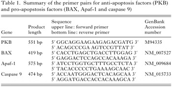
Table 1. Summary of the primer pairs for anti-apoptosis factor (PKB) and pro-apoptosis factor (BAX, Apaf-1 and Caspase 9)
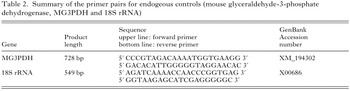
Table 2. Summary of the primer pairs for endogeous controls (mouse glyceraldehyde-3-phosphate dehydrogenase, MG3PDH and 18S rRNA)
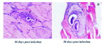
Fig. 1. Haematoxylin and eosin staining of Trichinella spiralis-infected muscles on 18 (A) and 38 (B) days post-infection, which showed basophilic cytoplasmic changes (A). The infected muscle cell is separated from undamaged muscle cells by a capsule wall (B). The linear alignment of nuclei of satellite cells was observed in the periphery of infected cells along their long axis (*). L, larva.
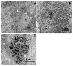
Fig. 2. Electron micrograph showing histopathology of Trichinella spiralis-infected muscles. Immediately after invasion by the newborn larvae mitochondria were swollen and disintegrated, some of them were fused with lysosomes (A). Mitochondria disappeared and were replaced by new mitochondria (arrowed, B) that were smaller than those have seen in normal muscle and located closely to the nucleus. The nucleus had an irregular shape and scattered heterochromatin (C). M, mitochondria; N, nucleus.
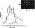
Fig. 3. RT-PCR results on the expression level of apoptotsis-related genes (BAX, Apaf-1, Caspase 9, and PKB) in the infected muscle tissue. Total RNA was isolated from infected muscle tissue at various stages (0, 8, 13, 18, 23, 28, 33, 38 and 53 days post-infection) and amplification products were analysed by agarose gel electrophoresis and shown in the right panel. Using Scion Image software, the relative density of each sample band against the control band (MG3PDH) was analysed and shown in the left panel.
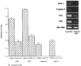
Fig. 4. RT-PCR results on the expression level of apoptotsis-related genes (BAX, Apaf-1, Caspase 9, and PKB) in the capsules or unaffected muscle cells which were obtained from a Trichinella spiralis-infected mouse at 23 days post-infection. Total RNA was isolated from laser microdissected capsules or unaffected muscle cells (control), amplification of the sample genes (BAX, Apaf-1, Caspase 9 and PKB) and the standard gene (18S rRNA) was performed and PCR products were analysed by agarose gel electrophoresis (right panel). Using Scion Image software, the relative density of each sample band against the control band was analysed and shown in the left panel.
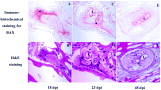
Fig. 5. Immunohistochemical staining for BAX. Cryosections of muscles infected with Trichinella spiralis for 18 (A), 23 (C) and 48 (E) days post-infection were reacted with anti-BAX antibody. The red colour indicates the positive reaction (*). After anti-BAX staining the same section was subjected to H&E staining for histological orientation purposes (B, D, F). L, larva.
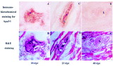
Fig. 6. Immunohistochemical staining for Apaf-1. Cryosection of muscle infected with Trichinella spiralis for 18 (A), 23 (C) and 48 (E) days post-infection was reacted with anti-Apaf-1 antibody. The red colour indicates the positive reaction (*). After anti-Apaf-1 staining the same section was subjected to H&E staining for histological orientation purposes (B, D, F). L, larva.
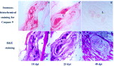
Fig. 7. Immunohistochemical staining for Caspase 9. Cryosection of muscle infected with Trichinella spiralis for 18 (A), 23 (C) and 48 (E) days post-infection was reacted with anti-Caspase 9 antibody. The red colour indicates the positive reaction (*). After anti-Caspase 9 staining the same section was subjected to H&E staining for histological orientation purposes (B, D, F). L, larva.
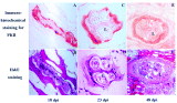
Fig. 8. Immunohistochemical staining for PKB. Cryosection of muscle infected with Trichinella spiralis for18 (A), 23 (C) and 48 (E) days post-infection was reacted with anti-akt1 antibody (akt-473). The red colour indicates the positive reaction (*). After anti-PKB staining the same section was subjected to H&E staining for histological orientation purposes (B, D, F). L, larva.
 You have
Access
- 30
- Cited by