Introduction
The genus Heterodorus Altherr, 1952 is one of the problematic nematode genera of the family Nordiidae Jairajpuri & Siddiqi, 1964. Its relationship with the genus Enchodelus Thorne, Reference Thorne1939 had been discussed for years. Altherr (1952) erected the genus Heterodorus, which was distinguished by the special structure of the female gonad, and also described its type species, H. magnificus Altherr, 1952. Siddiqi (Reference Siddiqi1969) then studied the pharyngeal region in more detail and accepted Heterodorus as a distinct genus, similar to Enchodelus. Later, most authors neglected Siddiqi's opinion and thought that Heterodorus was a junior synonym of Enchodelus. Andrássy (Reference Andrássy2009a) suggested the genus Heterodorus to be valid and included those Enchodelus-like species that have a conoid tail and few ventromedial supplements lying far anterior to the spicule. Meanwhile, he transferred 24 species with a conoid tail from Enchodelus to Heterodorus. Andrássy (Reference Andrássy2011) revised the genus Heterodorus and made a list of 25 valid species. His opinion was supported by Pedram et al. (Reference Pedram, Niknam, Guerrero, Ye and Robbins2009, Reference Pedram, Pourjam, Robbins, Ye and Peña-Santiago2011a). Lately, the first molecular study of Rhyssocolpus Andrássy, 1971, another genus of the family Nordiidae, supported a close relationship between Rhyssocolpus and Heterodorus, and suggested that both genera are not close to Enchodelus (Pedram et al., Reference Pedram, Pourjam, Robbins, Robert, Ye and Peña-Santiago2011b).
Nematodes extracted from soil samples collected from enclosure grassland, pasturable grassland and degraded grassland of several kinds of plants in Qinghai Province, China, were identified. Among these nematodes, one population from the bluegrass enclosure grassland was identified as belonging to Heterodorus. Moreover, its detailed examination, based on light microscopy, suggested that it is a new species of Heterodorus, described as Heterodorus qinghaiensis n. sp. The description of this new species could help to characterize Heterodorus and make a contribution to further studies on the relationships of Heterodorus with either Rhyssocolpus or Enchodelus.
Materials and methods
Nematodes were extracted from several 100-ml soil samples using the modified Baermann funnel method, killed by heating at 62°C for 3 min in a water bath, and fixed in 4% FG fixative (40% formaldehyde:glycerol:distilled water = 10:1:89). Fixed specimens were dehydrated by using the glycerol–ethanol method and then mounted on permanent slides (Xie, Reference Xie2005). Observations were made, and photographs of some best preserved specimens were taken, using a Zeiss Scope.A1 microscope equipped with a digital camera (AxioCam MRm; Zeiss, Jena, Germany) and specialized software (ZEN 2012, Zeiss). Measurements were made under a Nikon Eclipse 90i microscope equipped with a digitizing tablet (NIS-Elements BR 4.10; Nikon, Tokyo, Japan) and digital camera (DS-RiI, Nikon). The scanned files of drawings were obtained using an Olympus CX40 microscope (Olympus, Tokyo, Japan) with a drawing tube and managed by the software Photoshop CS3 (http://www.photoshop.com).
Results and discussion
Heterodorus qinghaiensis n. sp.
Measurements
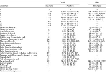
See table 1.
Morphometrics of Heterodorus qinghaiensis n. sp. All measurements are in the form: mean ± SD (range), and in μm (except for L, which is in mm). n, number of specimens observed, L, body length; a=L/maximum width; b=L/pharyngeal length; c=L/tail length; c′ = tail length/body diameter at anus, V= distance of vulva from anterior end × 100/L, G1= anterior genital branch reflection end to vulva × 100/L, G2= posterior genital branch reflection end to vulva × 100/L.

Description
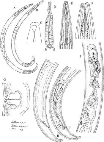
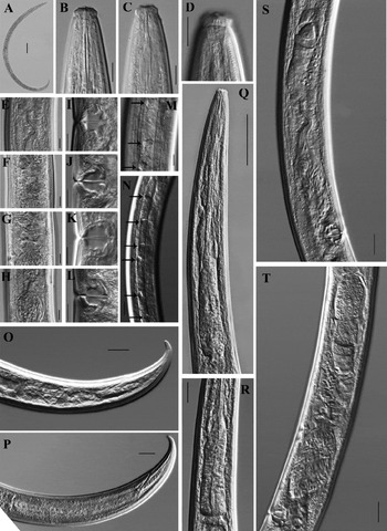
Female (figs 1 and 2): medium-sized nematode, slender. Body ventrally curved into C-shape after fixation. Cuticle two-layered, the outer one with very fine transverse striae; 1.6–2.3 μm thick in anterior region, 1.9–2.9 μm at mid-body and increasing to 2.8–4.4 μm on tail. The lateral chord is granular, with numerous large depression plaques throughout its entirety. Lip region offset by a distinct depression, about three times as wide as high, three-tenths as wide as body diameter at neck base. Lips slightly separated. Labial and cephalic papillae protrude slightly. Amphid fovea goblet-shaped, opening at the level of depression and about half to two-thirds of corresponding body diameter. Odontostyle slender and short, almost as long as lip region diameter, and with a small aperture. Odontophore rod-like, without basal flanges. Guiding ring ‘double’ but thin, situated at anterior part of odontostyle. Nerve ring situated at about 40% of the neck length. Basal expanded part of pharynx about one-third of the total neck length long. Cardia round to conoid. Pharyngeal gland nuclei locations are as follows: D = 72–77 (n= 10); AS1 = 16–23 (n= 7); AS2= 22–27 (n= 7); PS1= 35–48 (n= 10); PS2= 38–50 (n= 10) (Andrássy, 1998). Genital system amphidelphic; both branches well developed. Ovaries reflexed and relatively short, anterior 44–188 μm long and posterior 70–166 μm long, generally not reaching the sphincter level. Oocytes occupy a single row, except the terminal ones, which are found in two or more rows. Oviduct consists of a slender part, which is usually filled with sperm, and a well-developed pars dilatata; anterior 86–138 μm long and posterior 88–148 μm long. Uterus consisting of two portions; with one wider proximal portion with expanded lumen usually filled with sperm, and one narrower distal portion with narrow lumen; the anterior one is 62–134 μm long and the posterior one 85–136 μm long. A sphincter appears at the junction of oviduct and uterus, surrounding the distal portion of the uterus. Vulva transverse. Pars proximalis vaginae 9–12 × 9.0–13 μm with convex walls encircled by muscles; pars refringens vaginae with two well-developed trapezoidal sclerotized pieces that are closed to each other, each measuring about 2 × 6 μm and with a combined width of about 12.0 μm; pars distalis vaginae 1.5–2.5 μm long. One egg was observed, measuring 77 × 28 μm. Pre-rectum rather long, 2.8–7.1 times, and rectum 0.8–1.2 times, the body diameter at anus. Tail conoid, ventrally curved, with round terminus; hyaline terminal portion of tail is 11–14.5 μm long or 18–23% of total length.
Heterodorus qinghaiensis n. sp.: (A) entire female; (B) entire male; (C) amphid; (D) basal expansion of pharynx; (E) anterior region; (F) odontostyle and odontophore; (G) vulva in lateral view; (H) female posterior region; (I) male posterior region; (J) female anterior genital branch.

Heterodorus qinghaiensis n. sp. (female): (A) entire body; (B, C) odontostyles and odontophores; (D) amphid; (E–H) variability of the junction of the uterus–oviduct, showing sphincter; (I–L) vulvas in lateral view; (M, N) lateral chords, showing the relatively large, round depression plaques (arrows); (O, P) posterior body regions; (Q) anterior region; (R) basal expanded part of pharyx; (S, T) anterior genital branches. Scale bars: (A) 100 μm; (Q) 50 μm; (N–P, R–T) 20 μm; (B–M) 10 μm.

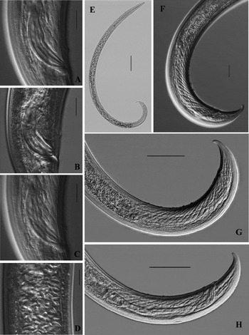
Male (figs 1 and 3): apart from the posterior region of the body, which is more ventrally curved than that of the female, its general morphology is similar to that of the female. Sperm are spindle in shape. Adanal pair of supplements located at 10–13.5 μm from the cloacal opening. Ventromedial supplements three to five, spaced, the posterior one situated at 45–60 μm from the adanal pair. Spicule dorylaimoid, relatively shorter, with lateral guiding pieces. Pre-rectum 3.8–6.2 times, and rectum 0.8–1.1 times, the body diameter at the anus. The tail is similar to that of the female.
Heterodorus qinghaiensis n. sp. (male): (A, B) spicules; (C) lateral guiding pieces; (D) sperm; (E) entire body; (F) copulatory muscles; (G, H) posterior body regions showing ventromedial supplements. Scale bars: (A–D) 10 μm; (F) 20 μm; (G, H) 50 μm; (E) 100 μm.

Habitat and locality
Soil around the roots of grasses from Graham beach, Maqen, Guoluo Prefecture, Qinghai Province, China.
Type material
Female holotype, 11 female and 9 male paratype specimens are deposited in the Laboratory of Plant Nematology/Research Center of Nematodes of Plant Quarantine, South China Agricultural University, Guangzhou, Guangdong 510642, China.
Diagnosis and relationships
The new species is characterized by the body length of 1.29–1.46 mm; a granular lateral chord with numerous large depression plaques throughout its entirety; the lip region being offset by a distinct depression; amphid goblet-shaped with an aperture about half to two-thirds of the corresponding body diameter; short odontostyle, almost as long as lip region diameter; odontophore rod-like without basal flanges; pharyngeal basal expansion about 30% of the total neck length; ovaries generally not reaching the sphincter level; vulva transverse and sclerotized; spicule 32–41 μm long; three to five ventromedial supplements; and tail conoid, ventrally curved, with a round terminus.
Based on the key to species of Heterodorus given by Andrássy (Reference Andrássy2011), H. qinghaiensis n. sp. is close to H. liangi (Ahmad, Wu & Shaheen, Reference Ahmad, Wu and Shaheen2002) Andrássy, 2009, H. brevidentatus (Thorne, Reference Thorne1939) Andrássy, 2009, H. monticola Andrássy, Reference Andrássy2011, H. morgensis (Loof, 1988) Andrássy, 2009 and H. meghalayensis (Mushtaq, Baniyamuddin & Ahmad, Reference Mushtaq, Baniyamuddin and Ahmad2007) Andrássy, 2009, in having inconspicuous or no lateral body pores, smaller odontostyle and ventrally curved tail. Heterodorus qinghaiensis n. sp. differs from these species by its lateral chord with numerous large, round depression plaques. Moreover, the new species can be distinguished from them by the following characteristics. The new species differs from H. liangi (Ahmad et al., Reference Ahmad, Wu and Shaheen2002) by a broader lip region (11–12 μm vs. 9–10 μm), dorsal pharyngeal gland nucleus located more posteriorly (72–77% vs. 64–67%), presence vs. absence of males, and longer ventrally curved tail (52–69 μm vs. 26–30 μm and almost straight). It differs from H. brevidentatus (Thorne, Reference Thorne1939; Guerrero & Peña-Santiago, Reference Guerrero and Peña-Santiago2007; Guerrero et al., Reference Guerrero, Liébanas and Peña-Santiago2007; Mushtaq et al., Reference Mushtaq, Baniyamuddin and Ahmad2007; Andrássy, Reference Andrássy2009b; Ciobanu et al., Reference Ciobanu, Popovici, Guerrero and Peña-Santiago2010) in having a larger amphid aperture (about half to two-thirds vs. one-third to half of the corresponding body diameter), a different genital system (ovaries generally not reaching the sphincter level and sperm present within genital tract vs. ovaries usually reaching and surpassing the sphincter level and sperm absent within genital tract), different pars refringens vaginae (two trapezoidal sclerotizations closed to each other vs. separated by a weakly sclerotized intermediate area), shorter pharyngeal expansion (78–90 μm vs. 91–125 μm in an American population, 93–122 μm in Spanish material and 95–129 μm in Romanian nematode specimens), and the presence vs. absence of males. From H. monticola (Andrássy, Reference Andrássy2011), it differs by having lips that are slightly separated vs. lips amalgamated, shorter odontostyle and odontophore (11–13 μm vs. 13–15 μm and 20–21 μm vs. 21–24 μm, respectively), longer basal expansion (31–34% vs. 26–30%), sclerotized vulva (vs. not sclerotized), smaller spicule (32–41 μm vs. 44–46 μm) and longer tail (52–69 μm vs. 35–48 μm). From H. morgensis (Loof, 1988), the new species differs in having a different lip region (lips slightly separated vs. lips amalgamated), a smaller spicule (32–41 μm vs. 45–56 μm). It differs from H. meghalayensis (Mushtaq et al., Reference Mushtaq, Baniyamuddin and Ahmad2007) in having a longer body (1.29–1.46 mm vs. 0.97–1.19 mm), posterior position of the vulva (V= 46.7–51.6 vs. 43–46), longer tail (c′ = 2.2–2.8 vs. 2.0–2.1), longer neck length and basal expansion (239.0–283 μm vs. 205–226 μm and 78–90 μm vs. 75–78 μm, respectively), shorter vaginal depth (15–19 μm vs. 21–24 μm) and smaller spicule (32–41 μm vs. 40–46 μm).
In conclusion, H. qinghaiensis n. sp. can be added to the key to the species of Heterodorus given by Andrássy (Reference Andrássy2011) as shown in fig. 4.
Addition of H. qinghaiensis n. sp. to the key to the species of Heterodorus given by Andrássy (Reference Andrássy2011).

Financial support
This work was supported by a Special Project of the Scientific and Technological Basis of the Ministry of Science and Technology of the People's Republic of China to H.X. (grant no. 2006FY120100).
Conflict of interest
None.