Introduction
With advances in neonatal intensive care, survival rates of infants born very preterm (VP) and very low birth-weight (VLBW) have increased (Doyle, Reference Doyle2004; Helenius, Gissler, & Lehtonen, Reference Helenius, Gissler and Lehtonen2019). However, many of these individuals continue to experience adverse long-term outcomes, with cognitive, attentional, socio-emotional and motor problems well documented (Anderson et al., Reference Anderson, De Luca, Hutchinson, Spencer-Smith, Roberts, Doyle and Victorian Infant Collaborative2011; Bhutta, Cleves, Casey, Cradock, & Anand, Reference Bhutta, Cleves, Casey, Cradock and Anand2002; Bracewell & Marlow, Reference Bracewell and Marlow2002; Talge et al., Reference Talge, Holzman, Wang, Lucia, Gardiner and Breslau2010).
Higher rates of anxiety disorders have been documented in VP and VLBW children and adolescents (Botting, Powls, Cooke, & Marlow, Reference Botting, Powls, Cooke and Marlow1997; Indredavik et al., Reference Indredavik, Vik, Heyerdahl, Kulseng, Fayers and Brubakk2004; Johnson et al., Reference Johnson, Hollis, Kochhar, Hennessy, Wolke and Marlow2010; Treyvaud et al., Reference Treyvaud, Ure, Doyle, Lee, Rogers, Kidokoro and Anderson2013), however it is less clear whether this increased risk persists beyond adolescence. Rates of VP/VLBW-born adults that meet diagnostic criteria for an anxiety disorder vary across studies, with some reporting increased risk relative to term-born peers (Van Lieshout, Boyle, Saigal, Morrison, & Schmidt, Reference Van Lieshout, Boyle, Saigal, Morrison and Schmidt2015; Walshe et al., Reference Walshe, Rifkin, Rooney, Healy, Nosarti, Wyatt and Allin2008) and others reporting no difference (Burnett et al., Reference Burnett, Davey, Wood, Wilson-Ching, Molloy, Cheong and Anderson2013; Johnson, O'Reilly, Ni, Wolke, & Marlow, Reference Johnson, O'Reilly, Ni, Wolke and Marlow2019; Westrupp, Northam, Doyle, Callanan, & Anderson, Reference Westrupp, Northam, Doyle, Callanan and Anderson2011). When applying a dimensional approach to assessment of symptoms, Johnson et al. (Reference Johnson, O'Reilly, Ni, Wolke and Marlow2019) found increased broadband internalizing and anxiety symptoms in adults born VP or VLBW compared with normal birth weight peers, while other studies have found no difference in anxiety symptoms in adulthood (Burnett et al., Reference Burnett, Davey, Wood, Wilson-Ching, Molloy, Cheong and Anderson2013; Westrupp et al., Reference Westrupp, Northam, Doyle, Callanan and Anderson2011). Prospective longitudinal cohorts provide insights into the progression of clinical anxiety in this population. In the Bavarian Longitudinal Study, rates of diagnosed anxiety were increased in the VP/VLBW group at 8 years, but not at 6 or 26 years (Jaekel, Baumann, Bartmann, & Wolke, Reference Jaekel, Baumann, Bartmann and Wolke2018). Conversely, rates of anxiety disorders were increased at 14, 19 and 26 years in those born VLBW in the Trondheim cohort (Indredavik et al., Reference Indredavik, Vik, Heyerdahl, Kulseng, Fayers and Brubakk2004; Laerum et al., Reference Laerum, Reitan, Evensen, Lydersen, Brubakk, Skranes and Indredavik2017).
Mechanisms underlying the increased vulnerability to clinical anxiety following VP birth remain poorly understood. VP birth occurs prior to completion of the third trimester, a period of rapid brain growth and maturation (Dobbing, Reference Dobbing and J1990; Volpe, Reference Volpe2009). Following this stressful birth event, infants are without placental support and rely on their own immature system to support brain development ex-utero. Neuroimaging studies have revealed high rates of white matter abnormalities and grey matter volume reductions in VP neonates compared with FT controls (Inder, Warfield, Wang, Huppi, & Volpe, Reference Inder, Warfield, Wang, Huppi and Volpe2005; Thompson et al., Reference Thompson, Lee, Egan, Warfield, Doyle, Anderson and Inder2014), and during childhood (de Kieviet, Zoetebier, van Elburg, Vermeulen, & Oosterlaan, Reference de Kieviet, Zoetebier, van Elburg, Vermeulen and Oosterlaan2012); with grey matter volume reductions occurring in a region-specific manner (Alexander et al., Reference Alexander, Kelly, Adamson, Beare, Zannino, Chen and Thompson2019; Cismaru et al., Reference Cismaru, Gui, Vasung, Lejeune, Barisnikov, Truttmann and Huppi2016; Omizzolo et al., Reference Omizzolo, Thompson, Scratch, Stargatt, Lee, Cheong and Anderson2013; Peterson et al., Reference Peterson, Vohr, Staib, Cannistraci, Dolberg, Schneider and Ment2000; Thompson et al., Reference Thompson, Warfield, Carlin, Pavlovic, Wang, Bear and Inder2007).
Smaller volume of the amygdala – a structure involved in generating a response to fear stimuli – has been reported in individuals born VP compared with FT controls in cross-sectional studies at term-equivalent age (TEA) (Cismaru et al., Reference Cismaru, Gui, Vasung, Lejeune, Barisnikov, Truttmann and Huppi2016) and at 8 years of age (Peterson et al., Reference Peterson, Vohr, Staib, Cannistraci, Dolberg, Schneider and Ment2000). Smaller hippocampal volumes have also been reported in VP individuals compared with FT controls in cross-sectional studies at 7 years and 14–15 years of age (Nosarti et al., Reference Nosarti, Al-Asady, Frangou, Stewart, Rifkin and Murray2002; Omizzolo et al., Reference Omizzolo, Thompson, Scratch, Stargatt, Lee, Cheong and Anderson2013). These regions form part of the wider, highly interconnected corticolimbic system which has been implicated in anxiety (Gold et al., Reference Gold, Steuber, White, Pacheco, Sachs, Pagliaccio and Pine2017; Mueller et al., Reference Mueller, Aouidad, Gorodetsky, Goldman, Pine and Ernst2013; Newman et al., Reference Newman, Thompson, Bartsch, Hagler, Chen, Brown and Libiger2016; Ochsner et al., Reference Ochsner, Ray, Cooper, Robertson, Chopra, Gabrieli and Gross2004; Strawn et al., Reference Strawn, Wehry, Chu, Adler, Eliassen, Cerullo and DelBello2013), and includes portions of the prefrontal and cingulate cortices. In VP compared with FT controls, we have recently reported slower volume growth from TEA to 7 years in the anterior cingulate cortex and lateral and medial orbitofrontal cortex in line with slower growth in total brain volume (Thompson et al., Reference Thompson, Matthews, Alexander, Lee, Kelly, Adamson and Anderson2020).
Thus, the current literature suggests that VP children display slower growth in corticolimbic structures early in life. However, it is unclear whether altered corticolimbic growth in children born VP is associated with anxiety. The current study extends previous findings (Thompson et al., Reference Thompson, Matthews, Alexander, Lee, Kelly, Adamson and Anderson2020) and aimed to compare corticolimbic region volume trajectories across childhood (TEA, 7 and 13 years) between VP children with and without an anxiety disorder diagnosis at 13 years. It was hypothesized that VP children diagnosed with anxiety disorders would display altered corticolimbic trajectories (especially during early childhood), compared with VP children without an anxiety disorder diagnosis at 13 years.
Methods
Participants
Participants were recruited as a part of the Victorian Infant Brain Study (VIBeS), a prospective longitudinal study consisting of infants born between July 2001 and December 2003 at the Royal Women's Hospital in Melbourne, Australia. Two hundred and twenty-four infants born VP (<30 weeks' gestation) or with VLBW (<1250 g) were recruited shortly after birth. Infants with congenital abnormalities were excluded from the study. This study was approved by the Royal Children's Hospital and Royal Women's Hospital Research Ethics committees and informed consent was obtained from children's parents/legal guardians.
Magnetic resonance imaging (MRI) acquisition
Infants were scanned at TEA (40 ± 2 weeks' gestational age) with a 1.5-Tesla General Electric Signa MRI scanner (Milwaukee, WI). Infants were fed, swaddled, fitted with earmuffs and placed in a vacuum fixation beanbag to reduce motion and scanned without sedation. T 2-weighted (1.7–3.0 mm coronal slices; repetition time 4000 ms; echo time 60/160 ms; flip angle 90°; field of view 220 × 160 mm; matrix 256 × 192, interpolated 512 × 512) whole-brain structural images were acquired. At the 7-year follow-up, structural T 1 (0.85 mm sagittal slices, repetition time 1900 ms, echo time 2.27 ms, flip angle 9°, field of view 210 × 210 mm, matrix 256 × 256) images were acquired using a 3 Tesla Trio Siemens MRI machine (Siemens, Erlangen, Germany) without sedation. At the 13-year follow-up, children were again scanned using the 3 Tesla Trio Siemens MRI machine (Siemens, Erlangen, Germany) without sedation and structural T 1 (0.9 mm3 sagittal slices, repetition time 2530 ms, echo times 1.77, 3.51, 5.32, 7.2 ms, flip angle 7°, field of view 230 × 209 mm, matrix 256 × 230, interpolated 256 × 256) images were acquired.
Image pre-processing and volumetry
T 2-weighted images acquired at TEA were bias-corrected using N4ITK (Tustison et al., Reference Tustison, Avants, Cook, Zheng, Egan, Yushkevich and Gee2010) and brain extracted using Morphologically Adaptive Neonatal Tissue Segmentation software (MANTiS) (Beare et al., Reference Beare, Chen, Adamson, Silk, Thompson, Yang and Wood2013; Beare et al., Reference Beare, Chen, Kelly, Alexopoulos, Smyser, Rogers and Thompson2016). The neonatal M-CRIB atlas (Alexander et al., Reference Alexander, Murray, Loh, Matthews, Adamson, Beare and Thompson2017), which replicates the widely used Desikan−Killiany atlas (Desikan et al., Reference Desikan, Segonne, Fischl, Quinn, Dickerson, Blacker and Killiany2006) was used to parcellate regions at TEA and ensure compatibility with regions parcellated from 7 and 13 year scans (Supplementary Fig. 1a). Each of the 10 T 2-weighted images and corresponding parcellated images comprising the neonatal M-CRIB atlas (Alexander et al., Reference Alexander, Murray, Loh, Matthews, Adamson, Beare and Thompson2017) were nonlinearly registered to each T 2-weighted image in the current sample using ANTS (Avants et al., Reference Avants, Tustison, Song, Cook, Klein and Gee2011). M-CRIB atlas labels were then applied to each brain using pSTAPLE (Akhondi-Asl & Warfield, Reference Akhondi-Asl and Warfield2013). Regional brain volumes were selected for 16 bilateral corticolimbic regions of interest [amygdala, hippocampus, parahipppocampal gyrus, entorhinal cortex, rostral and caudal anterior cingulate gyri, medial orbitofrontal cortex (mOFC), and lateral orbitofrontal cortex (lOFC); Supplementary Fig. 1b]. T 1 images acquired at 7 and 13 years of age were bias corrected using N4ITK (Tustison et al., Reference Tustison, Avants, Cook, Zheng, Egan, Yushkevich and Gee2010), brain extracted using the Brain Extraction Tool (Smith, Reference Smith2002) and parcellated using the FreeSurfer 6.0 automated parcellation and segmentation procedure (Fischl et al., Reference Fischl, Salat, Busa, Albert, Dieterich, Haselgrove and Montillo2002); manual edits to the surfaces were made according to FreeSurfer guidelines. At 7 and 13 years, volumes of limbic and prefrontal regions from the Desikan−Killiany cortical parcellation (Desikan et al., Reference Desikan, Segonne, Fischl, Quinn, Dickerson, Blacker and Killiany2006) were used, corresponding to TEA regions of interest from the M-CRIB neonatal atlas (Supplementary Fig. 1b) (Alexander et al., Reference Alexander, Murray, Loh, Matthews, Adamson, Beare and Thompson2017). Amygdala volumes from FreeSurfer's ‘aseg’ subcortical segmentation (Fischl et al., Reference Fischl, Salat, Busa, Albert, Dieterich, Haselgrove and Montillo2002) were used and hippocampal volumes were generated via the FreeSurfer 6.0 hippocampal subfields module (Iglesias et al., Reference Iglesias, Augustinack, Nguyen, Player, Player, Wright and Wald2015); these were also compatible with the neonatal regions (Alexander et al., Reference Alexander, Murray, Loh, Matthews, Adamson, Beare and Thompson2017). T 2-weighted images were used at TEA as a relatively high water content and limited myelination in the neonatal brain results in a reversal of tissue intensities on T 1- and T 2-weighted scans at this age (Holland, Haas, Norman, Brant-Zawadzki, & Newton, Reference Holland, Haas, Norman, Brant-Zawadzki and Newton1986). The quality of segmentations was assessed, and of the children who were scanned, 30 images at TEA, 7 images at 7 years and 1 image at 13 years had no usable volumetric data; this was primarily due to motion artefact. Intracranial volume (ICV) and total brain volume (TBV) were obtained by combining relevant M-CRIB regions at TEA (ICV: all regions; TBV: ICV – cerebrospinal fluid (CSF)) and with Statistical Parametric Mapping version 12 (http://www.fil.ion.ucl.ac.uk/spm/) at 7 and 13 years (ICV: grey matter + white matter + CSF; TBV: ICV – CSF).
Anxiety assessment
The 13-year follow-up included the Development and Well-Being Assessment (DAWBA; (Goodman, Ford, Richards, Gatward, & Meltzer, Reference Goodman, Ford, Richards, Gatward and Meltzer2000)), a structured clinical interview completed by parents online with regards to their child's mental health including anxiety disorders. A computer algorithm identified potential cases based on the Diagnostic and Statistical Manual of Mental Disorders (DSM-5). These cases were then reviewed by psychologists blinded to group membership and previous developmental assessments to determine if criteria for formal clinical diagnoses were met (Yates et al., Reference Yates, Treyvaud, Doyle, Ure, Cheong, Lee and Anderson2020). A summary variable was created to reflect the absence or presence of any anxiety disorder (including generalized anxiety disorder (GAD), social anxiety disorder, separation anxiety disorder, specific phobia and agoraphobia).
Social risk
A family social risk score was calculated at 13 years of age based on family structure, education of primary caregiver, primary income earner employment status and occupation, language spoken at home and maternal age at birth (Roberts et al., Reference Roberts, Howard, Spittle, Brown, Anderson and Doyle2008). Scores (0–2) were given for each domain (Roberts et al., Reference Roberts, Howard, Spittle, Brown, Anderson and Doyle2008), and summed to provide an overall risk score (0–12). Overall scores ⩾2 were categorized as higher social risk, with scores <2 indicating lower social risk.
Statistical analyses
Statistical analyses were conducted using Stata 15.0 (StataCorp, TX). Group comparisons (VP anxiety, VP no anxiety) of sample characteristics were performed using two samples t tests and Fisher's exact tests. Prior to analysis, global and regional brain volumes of participants were standardized to the mean and s.d. of the relevant brain region. Thus, standardized beta coefficients (β) are reported throughout.
Linear mixed effects (LME) models were used to assess brain growth trajectories for each corticolimbic region of interest (by hemisphere) between groups (absence v. presence of an anxiety disorder at 13 years) across childhood [TEA (0 years), 7 and 13 years]. Group, age and a group-by-age interaction were included as fixed effects in the model, fitted with a random intercept and unstructured covariance structure; which provided the lowest Akaike information criterion (AIC) and Bayesian information criterion (BIC) values during model selection (Supplementary Table 1). Age was included as a discrete variable, given the narrow distribution of ages at each follow-up relative to the intervals between follow-up assessments. The models were fitted with robust standard errors to account for clustering of multiples, given the high number of multiple births in this sample (Carlin, Gurrin, Sterne, Morley, & Dwyer, Reference Carlin, Gurrin, Sterne, Morley and Dwyer2005).
Firstly, overall group-by-age interactions were used to assess corticolimbic volume trajectories across childhood between groups (absence v. presence of anxiety disorder). For regions with significant overall group-by-age interactions, growth in early and late childhood between groups was assessed by extracting group-by-age interactions between 0–7 and 7–13 years from the LME models, respectively. In the case of significant interactions, group differences in volumes cross-sectionally (at 0, 7 or 13 years, as appropriate) were also extracted from the LME models. Secondary analyses (in regions where trajectory alterations were found) were adjusted for TBV (at the corresponding time point) and high social risk at 13 years to determine the extent to which these variables explained trajectory differences in regional brain volumes between groups. A false discovery rate (FDR) correction was applied for each set of inferences via the Benjamini and Yekutieli (Reference Benjamini and Yekutieli2001) method to all p-values to account for increased type I error resulting from multiple comparisons across different brain structures. An alpha of 0.05 was set for all analyses.
Results
Sample characteristics
From 224 VP infants recruited shortly after birth, 197 (88%) VP children returned for the follow-up at 7 years and 179 (80%) returned for the follow-up at 13 years of age (Fig. 1). Reasons for attrition included families withdrawing from the study, moving out of state/internationally and loss of contact.
Flow chart of attrition rates in current study. an = 1 participant could not be included in analysis as had completed DAWBA at 13 years however had no usable volumetric data. VP, very preterm; GA, gestational age; VLBW, very low birth weight; TEA, term equivalent age; MRI, magnetic resonance imaging; DAWBA, development and wellbeing assessment; GAD, GAD.

At 13 years of age, 13% (n = 16) of the cohort met criteria for one or more anxiety disorders, with generalized anxiety disorder (GAD) the most prevalent (n = 8, 6.4%), followed by social anxiety disorder (n = 6, 4.8%), specific phobia (n = 4, 3.2%) and separation anxiety disorder (n = 1, 0.8%). No participants met criteria for agoraphobia and three participants were diagnosed with comorbid GAD and social anxiety disorder. Children with usable MRI data for at least one time point (TEA, 7, 13 years) and DAWBA data at 13 years of age were included in the analysis (n = 124; Fig. 1). The characteristics of participants included in this study are presented in Table 1, and were similar between the anxiety and no anxiety disorder groups.
Participant characteristics

a Birthweight more than two s.d. below the mean.
b Oxygen requirement at 36 weeks.
c n = 15.
d Proven necrotizing enterocolitis and/or sepsis.
e n = 105.
f Graded according to Papile, Burstein, Burstein, and Koffler (Reference Papile, Burstein, Burstein and Koffler1978).
g Scored using Kidokoro system (Kidokoro, Neil, & Inder, Reference Kidokoro, Neil and Inder2013).
h n = 104.
i n = 87.
j DAWBA occurred at same age as 13-year MRI.
k n = 81.
l n = 106.
Note: All ages corrected for prematurity. p-values are presented for group comparisons assessed with 2 samples t tests and Fisher's exact tests.
Global brain volume trajectories across childhood and anxiety disorder in VP children
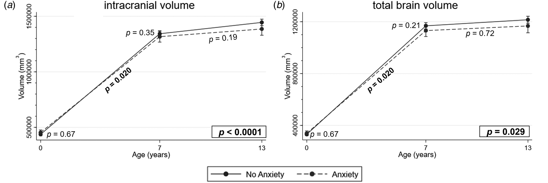
Overall trajectories, trajectories during early and late childhood and subsequent cross-sectional analysis for each region are presented in Supplementary Table 2. Overall group-by-age interactions existed for ICV (p < 0.0001; Fig. 2a) and TBV (p = 0.029; Fig. 2b), indicating altered global brain volume trajectories in VP children diagnosed with an anxiety disorder compared with those who were not. Group-by-age interactions between 0 and 7 years were found for both ICV and TBV [β = −0.461, 95% confidence interval (CI) −0.826 to 0.097, p = 0.020; β = −0.503, 95% CI −0.92 to −0.086, p = 0.020, respectively; Fig. 2a, b] indicating slower global brain growth in early childhood for VP children with an anxiety disorder diagnosis compared with VP children without a diagnosis. However, ICV (β = −0.224, 95% CI −0.485 to 0.038, p = 0.19) and TBV (β = −0.063, 95% CI −0.30 to 0.173, p = 0.72) growth appeared fairly stable from 7 to 13 years. Cross-sectional analyses at 0 and 7 years indicated no group differences in ICV or TBV at 0 (β = 0.237, 95% CI −0.153 to 0.628, p = 0.67; β = 0.165, 95% CI −0.255 to 0.585, p = 0.67, respectively; Fig. 2) or 7 years of age (β = −0.224, 95% CI −0.694 to 0.245, p = 0.35; β = −0.338, 95% CI −0.815 to 0.138, p = 0.21, respectively; Fig. 2).
Trajectory of global volumes across the first 13 years in very preterm individuals with and without anxiety disorder at 13 years. FDR corrected p-values for group-by-age interaction term in the LME model are presented in boxes in the bottom right corner for each region. Intracranial volume (ICV; a) and total brain volume (TBV; b) displayed significant overall group-by-age interactions (p < 0.05, indicated in bold) and thus were included in subsequent analysis of group-by-age interactions between 0−7 and 7−13 years (FDR corrected p-values presented below trajectory lines). Following significant group-by-age interactions between 0 and 7 years in ICV and TBV (p < 0.05 indicated in bold), group differences was explored cross-sectionally at 0 and 7 years, with no significant difference found at either time point (FDR corrected p-values presented next to 0 and 7 year data points). Error bars represent 95% confidence intervals.

Corticolimbic volume trajectories across childhood and anxiety disorder in VP children
Overall group-by-age interactions were found in the right amygdala (p = 0.0009; Fig. 3b) and left hippocampus (p = 0.029; Fig. 3c), indicating that trajectories of these regions differed between VP children with and without an anxiety disorder diagnosis. There were trends for group differences in the trajectories of the right hippocampus (p = 0.062; Fig. 3d) and left mOFC (p = 0.079; Fig. 4a), therefore, these regions were also included in secondary analysis of growth trajectories in early and late childhood. Overall group-by-age interactions in all other corticolimbic regions were not observed (Figs 3a, e–h, 4b–h), indicating no alteration in trajectory and thus were not included in subsequent analysis.
Trajectory of limbic region volumes across the first 13 years in very preterm individuals with and without anxiety disorder. FDR corrected p-values for group-by-age interaction term in the LME model are presented in boxes in the bottom right corner for each region. The right amygdala (b) and left hippocampus (c) displayed significant overall group-by-age interactions (p < 0.05, indicated in bold) and the right hippocampus (d) displayed a trend (p < 0.10, indicated by italics) and thus was included in subsequent analysis of group-by-age interactions between 0−7 and 7−13 years (FDR corrected p-values presented below trajectory lines). Following a significant group-by-age interaction between 0 and 7 years in the left and right hippocampi (c−d) (p < 0.05 indicated in bold), group differences were explored cross-sectionally at 0 and 7 years, with no significant difference found at either time point (FDR corrected p-values presented next to 0 and 7 year data points). Error bars represent 95% confidence intervals.

Trajectory of cortical region volumes across the first 13 years in very preterm individuals with and without anxiety disorder at 13 years. FDR corrected p-values for group-by-age interaction term in the LME model are presented in boxes in the bottom right corner for each region. The left medial orbitofrontal cortex (a) displayed a trend (p < 0.10, indicated by italics) and thus was included in subsequent analysis of group-by-age interactions between 0−7 and 7−13 years (FDR corrected p-values presented below trajectory lines). Following a significant group-by-age interaction between 0 and 7 years (p < 0.05 indicated in bold), group differences were explored cross-sectionally at 0 and 7 years, with no significant difference found at either time point (FDR corrected p-values presented next to 0 and 7 year data points). Error bars represent 95% confidence intervals.

Corticolimbic trajectories in early and late childhood and anxiety disorder in VP children
Slower growth in the left (β = −0.518, 95% CI −0.920 to −0.107, p = 0.020; Fig. 3c) and right (β = −0.469, 95% CI −0.832 to −0.105, p = 0.020; Fig. 3d) hippocampi and the left mOFC (β = −0.761, 95% CI −1.310 to −0.204, p = 0.020; Fig. 4a) in early childhood were observed between 0 and 7 years. There was no evidence of growth differences between groups from 0 to 7 years in the right amygdala (β = −0.20, 95% CI −0.76 to 0.367, p = 0.49; Fig. 3b). Trajectories remained similar between the groups in late childhood (7−13 years) in the left (β = −0.058, 95% CI −0.385 to 0.267, p = 0.72; Fig. 3c) and right (β = −0.055, 95% CI −0.289 to 0.179, p = 0.72; Fig. 3d) hippocampi, left mOFC (β = 0.263, 95% CI −0.038 to 0.564, p = 0.19; Fig. 4a) and right amygdala (β = −0.421, 95% CI −0.848 to 0.0005, p = 0.19; Fig. 3b).
Cross-sectional corticolimbic volumes at 0 and 7 years and anxiety disorder in VP children
Given slower growth in the left and right hippocampi and the left mOFC from 0 to 7 years in the anxiety v. no anxiety group, volumes at 0 and 7 years in these regions were assessed for group differences. Left and right hippocampi and left mOFC volumes did not differ between groups at 0 (β = 0.129, 95% CI −0.284 to 0.543, p = 0.67; β = −0.004, 95% CI −0.418 to 0.411, p = 0.99; β = −0.214, 95% CI −0.249 to 0.677, p = 0.67, respectively; Fig. 3c,d, 4a) but were smaller at 7 years in VP children with an anxiety disorder, although this did not reach statistical significance (β = −0.389, 95% CI −0.853 to 0.075, p = 0.17; β = −0.472, 95% CI −0.909 to −0.036, p = 0.085; β = −0.547, 95% CI −1.029 to −0.066, p = 0.085, respectively; Fig. 3c, d, 4a).
Corticolimbic volume trajectories across childhood and anxiety disorder in VP children after adjusting for TBV and social risk
Significant overall group-by-age interactions did not persist after adjusting for TBV and social risk (Supplementary Fig. 2).
Discussion
In this prospective longitudinal study, VP children diagnosed with an anxiety disorder at 13 years displayed altered trajectories for ICV and TBV and in various corticolimbic regions, including the left hippocampus and right amygdala, with trends in the right hippocampus and the left mOFC. More specifically, VP children with an anxiety disorder demonstrated slower growth during early childhood (0−7 years) for ICV, TBV, bilateral hippocampi and left mOFC, relative to VP children without an anxiety disorder. Cross-sectional analysis did not reveal group differences at 0 years but there was some evidence for smaller volumes in bilateral hippocampi and left mOFC at 7 years although this did not reach statistical significance, likely due to power limitations. Conversely, there was no difference between groups in volume trajectories during late childhood (7−13 years). This suggests that alterations in global and corticolimbic region volumes earlier in development may relate, at least in part, to later presentation of an anxiety disorder in VP individuals. Group differences in the trajectories of the left and right hippocampus, left mOFC and right amygdala were attenuated when controlling for TBV and high social risk, which likely reflects the alterations in TBV trajectories reported.
Global reductions in cerebral grey matter have been documented shortly after birth in VP infants (Inder et al., Reference Inder, Warfield, Wang, Huppi and Volpe2005; Padilla, Alexandrou, Blennow, Lagercrantz, & Aden, Reference Padilla, Alexandrou, Blennow, Lagercrantz and Aden2015), as well as across childhood (for review see de Kieviet et al., Reference de Kieviet, Zoetebier, van Elburg, Vermeulen and Oosterlaan2012). We have reported slower growth from TEA to 7 years in ICV, TBV and bilateral cortical grey matter and subcortical grey matter volumes in VP compared with FT controls (Monson et al., Reference Monson, Anderson, Matthews, Neil, Kapur, Cheong and Inder2016; Thompson et al., Reference Thompson, Matthews, Alexander, Lee, Kelly, Adamson and Anderson2020). The current study extends these findings, with significant group-by-age interactions indicating altered ICV and TBV trajectories in those with anxiety. Subsequent analysis identified slower growth in these global measures from TEA to 7 years in VP children who meet criteria for an anxiety disorder at 13 years compared with those who do not. Hence, altered growth in global measures of brain structure documented in the VP population in early childhood may provide a biomarker of anxiety disorder in this population.
Links between regional brain volumes and the presentation of anxiety disorder in individuals born VP are relatively unexplored. In school-age children born preterm, albeit those born between 34 and 36 gestational weeks, smaller temporal-lobe volumes have been associated with later anxiety symptoms (Rogers et al., Reference Rogers, Barch, Sylvester, Pagliaccio, Harms, Botteron and Luby2014). The current study extends this work by mapping the volume growth trajectory in key corticolimbic regions across the first 13 years, thus providing additional evidence of region-specific alterations in VP children diagnosed with an anxiety disorder. These regions are likely differentially affected by VP birth given the diversity in emergence and maturation across regions in both prenatal and postnatal life.
The amygdala has been the focus of research investigating the neurobiological underpinnings of anxiety in preterm individuals (Rogers et al., Reference Rogers, Sylvester, Mintz, Kenley, Shimony, Barch and Smyser2017), given its role in processing emotion, and reports of altered size (De Bellis et al., Reference De Bellis, Casey, Dahl, Birmaher, Williamson, Thomas and Ryan2000; Milham et al., Reference Milham, Nugent, Drevets, Dickstein, Leibenluft, Ernst and Pine2005; Schienle, Ebner, & Schafer, Reference Schienle, Ebner and Schafer2011), activation (Monk et al., Reference Monk, Telzer, Mogg, Bradley, Mai, Louro and Pine2008) and connectivity (Kim, Gee, Loucks, Davis, & Whalen, Reference Kim, Gee, Loucks, Davis and Whalen2011; Qin et al., Reference Qin, Young, Duan, Chen, Supekar and Menon2014) in the wider population of individuals experiencing anxiety. VP neonates display smaller amygdala volumes than term-born peers, which has been associated with fear processing capabilities at 1 year of age (Cismaru et al., Reference Cismaru, Gui, Vasung, Lejeune, Barisnikov, Truttmann and Huppi2016). Smaller amygdala volumes are also evident in preterm VLBW children compared with term-born children at 8 years of age (Peterson et al., Reference Peterson, Vohr, Staib, Cannistraci, Dolberg, Schneider and Ment2000). In the current study, group differences in overall volume trajectories were lateralized to the right amygdala. Alterations to the right amygdala, including volume reductions and hyperactivity have been documented in anxiety disorders such as social anxiety disorder, GAD and panic disorder (Asami et al., Reference Asami, Yamasue, Hayano, Nakamura, Uehara, Otsuka and Hirayasu2009; Hölzel et al., Reference Hölzel, Hoge, Greve, Gard, Creswell, Brown and Lazar2013; Irle et al., Reference Irle, Ruhleder, Lange, Seidler-Brandler, Salzer, Dechent and Leichsenring2010; Lipka, Miltner, & Straube, Reference Lipka, Miltner and Straube2011). Stimulation of the right amygdala has been implicated in inducing predominantly negative emotions (especially fear); contrasting the left amygdala which elicits both positive and negative emotions (Lanteaume et al., Reference Lanteaume, Khalfa, Régis, Marquis, Chauvel and Bartolomei2007). Thus, altered trajectories in the right but not left amygdala in the anxiety group may highlight a tendency towards fear conditioning. While the anxiety group appeared to diverge from the trajectory of the no anxiety group from 7 to 13 years, subsequent analysis revealed no group differences in right amygdala trajectories in early or late childhood. It is possible that this was due to low statistical power given the small anxiety group.
Findings of altered trajectory of left and right hippocampal volumes and slower growth from birth to 7 years in VP children later diagnosed with an anxiety disorder are consistent with temporal-lobe findings by Rogers et al. (Reference Rogers, Barch, Sylvester, Pagliaccio, Harms, Botteron and Luby2014) and extend on long-lasting reductions in hippocampal volumes documented in VP individuals relative to FT peers during childhood and adolescence (de Kieviet et al., Reference de Kieviet, Zoetebier, van Elburg, Vermeulen and Oosterlaan2012; Peterson et al., Reference Peterson, Vohr, Staib, Cannistraci, Dolberg, Schneider and Ment2000). The hippocampus is highly vulnerable to hypoxic and ischaemic insults (Golan et al., Reference Golan, Mane, Molczadzki, Zuckerman, Kaplan-Louson, Huleihel and Perez-Polo2009; Schmidt-Kastner & Freund, Reference Schmidt-Kastner and Freund1991), providing a possible mechanism of damage. Hippocampal alterations in individuals born VP are not apparent shortly after birth (Thompson et al., Reference Thompson, Wood, Doyle, Warfield, Lodygensky, Anderson and Inder2008) given that much of hippocampal development occurs in the first two years postnatally (Utsunomiya, Takano, Okazaki, & Mitsudome, Reference Utsunomiya, Takano, Okazaki and Mitsudome1999). However, the current study indicates that impaired hippocampal development during early childhood has functional implications. Studies investigating functional implications of hippocampal alterations following VP birth have largely focused on cognition (Thompson et al., Reference Thompson, Adamson, Roberts, Faggian, Wood, Warfield and Inder2013), given widely reported impairments in cognitive functioning (Brydges et al., Reference Brydges, Landes, Reid, Campbell, French and Anderson2018; Gimenez et al., Reference Gimenez, Junque, Narberhaus, Caldu, Salgado-Pineda, Bargallo and Botet2004; Peterson et al., Reference Peterson, Vohr, Staib, Cannistraci, Dolberg, Schneider and Ment2000; Twilhaar et al., Reference Twilhaar, Wade, De Kieviet, Van Goudoever, Van Elburg and Oosterlaan2018) and the role of this region in learning and memory processes. The current study extends these findings to include socio-emotional outcomes, namely anxiety disorder. The anterior portion of the hippocampus is involved in emotionally driven and anxiety-related behaviours (Bannerman et al., Reference Bannerman, Rawlins, McHugh, Deacon, Yee, Bast and Feldon2004). Consistent with the current findings, the hippocampus has been implicated in the pathophysiology of anxiety disorders, with reduced volumes reported in paediatric populations diagnosed with one or more anxiety disorders (Gold et al., Reference Gold, Steuber, White, Pacheco, Sachs, Pagliaccio and Pine2017; Mueller et al., Reference Mueller, Aouidad, Gorodetsky, Goldman, Pine and Ernst2013). Hippocampal morphology resembling that of an adult by 5 years of age (Kretschmann, Kammradt, Krauthausen, Sauer, & Wingert, Reference Kretschmann, Kammradt, Krauthausen, Sauer and Wingert1986). Therefore, as expected, left and right hippocampal volume trajectories remained fairly stable from 7 to 13 years in the current study. Parallel trajectories between groups in late childhood indicate that the hippocampus remained smaller in the anxiety group following slower growth from 0 to 7 years and suggest a disrupted developmental course rather than a delay in the growth of this region, in line with previous postulations for overall brain volume trajectories during this period (Thompson et al., Reference Thompson, Matthews, Alexander, Lee, Kelly, Adamson and Anderson2020).
In the current study, the anxiety disorder group displayed altered left mOFC volume trajectories compared with the no anxiety disorder group. Specifically, the anxiety disorder group displayed slower growth from TEA to 7 years of age, but not from 7 to 13 years. The final trimester of pregnancy is a critical period of OFC gyrification (Ganella et al., Reference Ganella, Burnett, Cheong, Thompson, Roberts, Wood and G.2015), thus our results suggest that early alterations to this development may have later functional implications. The mOFC has projections to the basolateral nuclei of the amygdala and plays a critical role in regulation of amygdala activation, particularly reactivity to negative emotional stimuli (Ducharme et al., Reference Ducharme, Albaugh, Hudziak, Botteron, Nguyen, Truong and Byars2014). Consistent with the current findings, self-report of generalized anxiety symptoms corresponds to less expansive surface area in early childhood and decreased cortical thickness in this prefrontal region across childhood in the general population (Newman et al., Reference Newman, Thompson, Bartsch, Hagler, Chen, Brown and Libiger2016). While these measures provide more specific indices of cortical dysmaturation, limited resolution in our neonatal scans did not enable investigation of cortical thickness or surface area longitudinally. GAD was the most prevalent anxiety disorder in the current study and similar left OFC volume decreases are noted in children diagnosed with GAD in the general population (Strawn et al., Reference Strawn, Wehry, Chu, Adler, Eliassen, Cerullo and DelBello2013).
Cross-sectional analysis at 0 and 7 years in regions where VP children with an anxiety disorder displayed altered trajectories from 0 to 7 years did not reveal group differences at either timepoint, suggesting the benefit of assessing trajectories of growth in addition to cross-sectional assessment of volumes. Alterations to growth in VP children with an anxiety disorder did not persist from 7 to 13 years. Maturation of the OFC is dynamic across childhood, with a rapid volume growth in early childhood, which ‘peaks’ at approximately 11–12 years and is followed by a decrease in adolescence (Shaw et al., Reference Shaw, Kabani, Lerch, Eckstrand, Lenroot, Gogtay and Rapoport2008). Thus, additional MRI scanning across late childhood would be beneficial to investigate this OFC trajectory in greater detail and determine how this ‘catch up’ of OFC volumes in the anxiety group occurs in the context of volume increase, peak and decrease across this period.
It must be noted, that in the current cohort, altered amygdala, hippocampal and mOFC trajectories in the anxiety group did not persist after adjustment for TBV and social risk. This suggests that growth in these regions may not contribute to a later diagnosis of anxiety disorders beyond differences in TBV and that the role of socio-environmental factors in the development of anxiety disorders should also be considered. It would be of benefit to include socio-environmental factors such as parenting, parent−child relationships and parental mental health in future models of brain growth in this population, given that such factors may moderate mental health outcomes (Clark, Woodward, Horwood, & Moor, Reference Clark, Woodward, Horwood and Moor2008; Treyvaud, Lee, Doyle, & Anderson, Reference Treyvaud, Lee, Doyle and Anderson2014; Treyvaud et al., Reference Treyvaud, Anderson, Howard, Bear, Hunt, Doyle and Anderson2009, Reference Treyvaud, Anderson, Lee, Woodward, Newnham, Inder and Anderson2010, Reference Treyvaud, Thompson, Kelly, Loh, Inder, Cheong and Anderson2020).
Grey matter alterations observed in VP individuals have been proposed to occur secondary to white matter microstructural changes (Volpe, Reference Volpe2019). Limbic and prefrontal regions are highly interconnected via white matter fibre projections, including the uncinate fasciculus, cingulum bundle and fornix. In people diagnosed with an anxiety disorder (Phan et al., Reference Phan, Orlichenko, Boyd, Angstadt, Coccaro, Liberzon and Arfanakis2009) and individuals born VP (Constable et al., Reference Constable, Ment, Vohr, Kesler, Fulbright, Lacadie and Schafer2008; Kelly et al., Reference Kelly, Thompson, Chen, Leemans, Adamson, Inder and Anderson2016; Vandewouw, Young, Shroff, Taylor, & Sled, Reference Vandewouw, Young, Shroff, Taylor and Sled2019), altered microstructural properties in the uncinate fasciculus, a key connection between the OFC, amygdala and hippocampus, have been noted. Thus, microstructural properties of limbic tracts should be investigated as a possible mechanism for altered corticolimbic volumes between individuals born VP with and without anxiety.
A major strength of the current study was the use of a large prospective longitudinal VP cohort, which enabled the characterization of brain development across the first 13 years of life in a population with a high risk of anxiety. Repeated brain imaging in the same cohort of children is preferable to multiple cross-sectional or case−control studies of different cohorts in evaluating the dynamic changes across brain development, given environmental influences. This was possible through the use of compatible MRI parcellation schemes at the neonatal (Alexander et al., Reference Alexander, Murray, Loh, Matthews, Adamson, Beare and Thompson2017) and childhood time points (Desikan et al., Reference Desikan, Segonne, Fischl, Quinn, Dickerson, Blacker and Killiany2006), which enabled comparison of brain region volumes longitudinally.
There are some limitations in the current study. While the relatively small sample size of the anxiety disorder group was representative of the population, it may have limited the potential to detect significant group differences in cross-sectional regional brain volumes. Additionally, a contemporary healthy term-born control group is not included in this study, as volume trajectories of the full-term control and the total VP group in this cohort were previously assessed (Thompson et al., Reference Thompson, Matthews, Alexander, Lee, Kelly, Adamson and Anderson2020). Due to advances in technology across the 13-year study, scanners and acquisition parameters differed across time points. While these differences could have introduced variability, assessment of total grey matter volume appears to be relatively robust across scanners (Heinen et al., Reference Heinen, Bouvy, Mendrik, Viergever, Biessels and De Bresser2016). It is unclear however, if this remains for smaller grey matter structures. Although these limitations are inherent in prospective longitudinal designs, repeated scanning of this cohort across the first 13 years of life provides a novel contribution to the current body of literature examining the neurobiological basis of anxiety disorders in this population. Grey matter development across childhood occurs in a non-linear fashion, however we did not consider it appropriate to apply a non-linear model due to the high clustering around the three time points. Thus, time was treated as a discrete variable. Follow-up during additional time points in similar studies, especially across the first 7 years of life when brain growth is most dynamic, would be beneficial.
We also acknowledge limitations in combining all DSM-5 anxiety disorders into a single outcome of anxiety disorder diagnosis. While each anxiety disorder has components of fear, the age of onset and underlying neural mechanisms may be distinct for the various anxiety disorders. Three VP children diagnosed with anxiety at 13 years also met criteria for major depressive disorder. Major depressive disorder often occurs comorbidly with anxiety disorders (Cummings, Caporino, & Kendall, Reference Cummings, Caporino and Kendall2014) with increased rates reported in the preterm cohorts (Burnett et al., Reference Burnett, Anderson, Cheong, Doyle, Davey and Wood2011). The median age of onset of depressive disorders is later than that of anxiety (Kessler et al., Reference Kessler, Berglund, Demler, Jin, Merikangas and Walters2005), thus follow-up in adulthood may enable a more detailed investigation of the trajectories of regional volumes and their role in comorbid depression, as well as separation into specific anxiety disorders.
In conclusion, the trajectory of region-specific corticolimbic volumes in VP individuals with anxiety appears to be altered in early childhood compared with VP individuals without an anxiety disorder. This builds on previous research which identified altered brain development during early childhood in VP compared with FT children and demonstrates the importance of early brain development in the socio-emotional outcome of VP children.
Supplementary material
The supplementary material for this article can be found at https://doi.org/10.1017/S0033291721002105.
Data sharing
The datasets generated during and/or analysed during the current study are available from the corresponding author on reasonable request.
Acknowledgements
We acknowledge the contributions of the Victorian Infant Brain Studies (VIBeS) and Developmental Imaging groups at the Murdoch Children's Research Institute and Michael Kean and Radiographers at the Royal Children's Hospital for the magnetic resonance images. We thank families involved in this study.
Author contribution
CPG, DKT, BA, CEK, KT, LGM, LP, DZ, RY, CA, MT, JLYC, TEI, LWD, AC and PJA have made substantial contributions to design of this study and editing the manuscript. CPG conducted data analysis and interpretation and prepared the initial manuscript draft. DZ provided statistical consultation. All authors approve the submitted version and agree to be accountable for all aspects of the work.
Financial support
This study was funded by the Australian National Health and Medical Research Council (NHMRC) (Centre for Clinical Research Excellence 546519; Centre for Research Excellence 1060733; Centre for Research Excellence 1153176; Project Grants 237117, 491209 and 1066555; Career Development Fellowships 1085754 and 1160003 [to DKT], 1141354 [to JLYC]; Early Career Fellowship 1012236 [to DKT]; Senior Research Fellowship 1081288 [to PJA]; Leadership Fellowship 1176077 [to PJA]), Financial Markets Foundation for Children Project Grant 2019-083, RMIT Vice Chancellor's Senior Research Fellowship and Australian Research Council Future Fellowship [to MT], Brigham and Women's Hospital Program for Interdisciplinary Neuroscience Fellowship (to LGM), US National Institutes of Health HD058056, United Cerebral Palsy Foundation (USA), Leila Y. Mathers Charitable Foundation (USA), the Brown Foundation (USA), Murdoch Children's Research Institute, the Royal Children's Hospital, The Royal Children's Hospital Foundation, Department of Paediatrics, The University of Melbourne, the Victorian Government's Operational Infrastructure Support Program, and an Australian Government Research Training Program Scholarship [to CG].
Conflict of interest
The authors have no conflicts of interest relevant to this article to disclose.
Financial disclosure
The authors have no financial relationships relevant to this article to disclose.
Ethical standards
The authors assert that all procedures contributing to this work comply with the ethical standards of the relevant national and institutional committees on human experimentation and with the Helsinki Declaration of 1975, as revised in 2008. Parents provided written informed consent for their child to participate.