I. INTRODUCTION
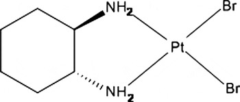
The title compound (Figure 1), systematic name cis-[dibromo(1R,2R)-1,2-diaminocyclohexane-κN,κN′]platinum(II), cis-[Pt(DACH)Br2], can be used as a precursor for preparation of platinum-based anticancer complexes. It is possible to use it for the production of not only widely used cytostatic oxaliplatin but also for the research and development of modern cytostatics of the fourth generation.
Structural formula of cis-[Pt(DACH)Br2].

We have inspected CSD database for the compound summary formula and found its analog cis-[dibromo(cis-1,2-diaminocyclohexane)]platinum(II)((Lock and Pilon, Reference Lock and Pilon1981). This compound is monoclinic [a = 15.212(4) Å, b = 11.612(5) Å, c = 13.268(5) Å, β = 121.17(2)°, unit-cell volume V = 2006(1) Å3, Z = 8, space group C2/c]. The calculated powder pattern of this compound is completely different from the measured powder pattern of the title compound.
II. SAMPLE PREPARATION
The title compound (Figure 1) was prepared from a suspension of cis-[dichloro(1R,2R)-1,2-diaminocyclohexane-κN,κN′]platinum(II) (365 mg; 0,959 mmol) in water. Then a solution of AgNO3 (326 mg, 1919 mmol) in water (30 ml) was added. Afterwards a solution of KBr (228 mg, 1916 mmol) in water (30 ml) was added at room temperature. The yellow precipitate was formed and the reaction mixture was cooled at 277 K for 12 h.
III. POWDER DIFFRACTION DATA
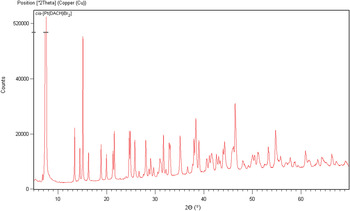
The diffraction pattern for the title compound was collected at room temperature with an X'Pert PRO θ–θ powder diffractometer with parafocusing Bragg–Brentano geometry using Cu Kα radiation (λ = 1.5406 Å, Ni filter, generator setting: 40 kV, 30 mA). An ultrafast X'Celerator detector was employed to collect XRD data over the angular range from 4 to 70° 2θ with a step size of 0.017° 2θ and a counting time of 81.28 s per step. The experimental powder diffraction pattern is depicted in Figure 2. The software package HighScore Plus V 3.0a PANalytical, Almelo, Netherlands) was used to analyze the data, to fit the background, to eliminate the Kα 2 component and the top of smoothed peak method was used to determine the peak positions and intensities of the diffraction peaks.
X-ray powder diffraction pattern of cis-Pt(C6H14N2)Br2 using Cu Kα 1 radiation (λ = 1.5406 Å).

The collected data (Table I) are consistent with a monoclinic unit-cell parameters [a = 11.862(4) Å, b = 7.222(3) Å, c = 13.353(2) Å, β = 97.813(7)°, unit-cell volume V = 1133,36 Å3, Z = 4, space group P21]. These parameters were derived using DICVOL04 (Boultif and Louër, Reference Boultif and Louër2004) with the results all being within the errors indicated. The following figures of merits were achieved F 20 = 13.5 (0.0077, 193) (Smith and Snyder, Reference Smith and Snyder1979) and M 20 = 11.6 (de Wolff, Reference de Wolff1968).
Indexed X-ray powder diffraction data for cis-Pt(C6H14N2)Br2. Only the peaks with I rel of 1 or greater are presented [a = 11.862(4) Å, b = 7.222(3) Å, c = 13.353(2) Å, β = 97.813(7)°, unit-cell volume V = 1133,36 Å, Z = 4, space group P21]. All lines were indexed and are consistent with the P21 space group. The d-values were calculated using Cu Kα1 radiation (λ = 1.5406 Å).

ACKNOWLEDGMENTS
This work was supported by the research programs NPV II 2B08021, MSM6046137301, and MSM6046137302 of the Ministry of Education, Youth and Sports of the Czech Republic and by grant No. 104/09/1497 from the Grant Agency of the Czech Republic.