Kawasaki disease is an acute, self-limited febrile illness of unknown aetiology that predominantly affects children <5 years of age.Reference McCrindle, Rowley and Newburger 1 The disease is markedly more prevalent in children in Japan, and its incidence is increasing over time in some countries.Reference McCrindle, Rowley and Newburger 1 , Reference Makino, Nakamura and Yashiro 2 The diagnosis is based on a series of symptoms including persistent fever, bilateral non-exudative conjunctivitis, oral mucosal changes, non-purulent cervical lymphadenopathy, polymorphous rash, and erythematous induration of the extremities.Reference McCrindle, Rowley and Newburger 1 Coronary artery abnormalities, which are present in 15–20% of the patients, are the most serious complications in Kawasaki disease.Reference Makino, Nakamura and Yashiro 2 , Reference Garg, Krishnamoorthy and Palaniswamy 3 Coronary artery abnormalities are critical problems of the disease, leading to myocardial ischaemia, infarction, and sudden death. Several studies have been attempted to predict the risk factors for developing coronary artery abnormalities in children with Kawasaki disease.Reference Kobayashi, Inoue and Takeuchi 4 – Reference Takeuchi, Inuzuka and Hayashi 7 On the other hand, contradictory results have been reported in other contries.Reference Sleeper, Minich and McCrindle 8
More recently, the mean platelet volume-to-lymphocyte ratio has emerged as a novel and readily available parameter of inflammation. There are only a few studies examining the relationship between mean platelet volume-to-lymphocyte ratio and coronary lesions and adverse cardiovascular events in adult patients.Reference Hudzik, Szkodziński, Lekston, Gierlotka, Poloński and Gąsior 9 – Reference Ornek and Kurtul 11 However, no study has investigated the usefulness of mean platelet volume-to-lymphocyte ratio in children with Kawasaki disease. In the present study, we aimed to evaluate the relationship between mean platelet volume-to-lymphocyte ratio and coronary artery abnormalities in children with Kawasaki disease.
Materials and methods
Local Ethics Committee approval was obtained for the study protocol (2017/267). We retrospectively reviewed the medical records of 58 children with Kawasaki disease who were hospitalised at our institution between January 2008 and January 2017. The diagnosis of complete and incomplete Kawasaki disease was made according to the American Heart Association guidelines.Reference McCrindle, Rowley and Newburger 1 Briefly, Kawasaki disease was diagnosed when the children had prolonged fever for 5 or more days, and at least 4 of 5 principal findings. Patients with prolonged fever and fewer than four of the principal clinical findings were labelled as having incomplete Kawasaki disease, if no other disease process could explain the illness. All children with Kawasaki disease received a single infusion of intravenous immunoglobulin and aspirin, within 10 days. In addition, 42 healthy, age- and sex-matched children were included as healthy controls. Children with incomplete medical records, known liver disease, systemic haematological disease, allergic disease, malignant or inflammatory disease, or receiving drugs that can affect haematological parameters were excluded from the study.
Coronary arteries were assessed by transthoracic echocardiography on admission. It was performed via Vivid S5 Pro Ultrasound System (GE Medical Systems, Horten, Norway), using 3- and 6-MHz transducers in two-dimensional, M-mode, and colored Doppler visualising modes. Coronary artery abnormalities were diagnosed on the basis of the criteria proposed by the American Heart Association guidelines.Reference McCrindle, Rowley and Newburger 1 Coronary artery abnormalities were defined as a coronary artery z score ⩾2.5. Coronary aneurysms were defined according to internal lumen diameter of at least 3 mm in children <5 years of age or at least 4 mm in children ⩾5 years of age; internal diameter of a segment measuring ⩾1.5 times that of an adjacent segment; and lumen that is clearly irregular.
Before the treatment, routine complete blood count parameters including mean platelet volume, platelet distribution width, red blood cell distribution width, and counts of platelets, neutrophils, lymphocytes, and white blood cells, erythrocyte sedimentation rate, and C-reactive protein levels were measured. Neutrophil-to-lymphocyte ratio and platelet-to-lymphocyte ratio were calculated. Mean platelet volume-to-lymphocyte ratio was calculated as mean platelet volume divided by lymphocyte count (103/mm3).
Statistical analyses
All continuous variables are expressed as mean±standard deviation. Data of patients and controls were compared. The t-test, analysis of variance, and receiver operating characteristic curve were used for statistical analysis. Multiple comparisons were made using one-way analysis of variance with posthoc Tukey’s test. The receiver operating characteristic curve was used to examine the predictive value of mean platelet volume-to-lymphocyte ratio in patients with coronary artery abnormalities. The area under the curve was calculated. The appropriate cut-off value of mean platelet volume-to-lymphocyte ratio was determined using maximum sum of sensitivity and specificity. Multivariate logistic regression models was used to identify predictors of Kawasaki disease patients with coronary artery abnormalities. The results were considered statistically significant if p values were ˂0.05.
Results
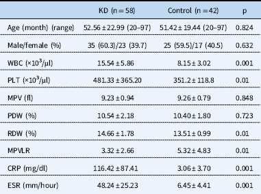
The characteristics of the patients and control groups are shown in Table 1. There was no significant difference between the patient and control groups in terms of gender, age, mean platelet volume, and platelet distribution width (p=0.632, p=0.824, p=0.848, and p=0.723, respectively). Compared with healthy controls, the mean levels of white blood cells, platelets, red blood cell distribution width, erythrocyte sedimentation rate, and C-reactive protein were significantly higher and mean platelet volume-to-lymphocyte ratio was significantly lower in children with Kawasaki disease (p<0.01, Table 1).
Comparison of characteristics between patients with Kawasaki disease (KD) and controls.

CRP=C-reactive protein; ESR=erythrocyte sedimentation rate; MPV=mean platelet volume; MPVLR=mean platelet volume-to-lymphocyte ratio; PDW=platelet distribution width; PLT=platelet; RDW=red blood cell distribution width; WBC=white blood cell
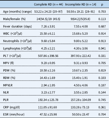
A total of 14 patients (24.1%) had incomplete Kawasaki disease and 15 (25.8%) children with Kawasaki disease had coronary involvement. There was no significant difference between fever duration, neutrophil-to-lymphocyte ratio, platelet-to-lymphocyte ratio, and the levels of white blood cells, platelets, red blood cell distribution width, erythrocyte sedimentation rate, and C-reactive protein in patients with complete and incomplete Kawasaki disease (p>0.05, Table 2). Children with complete Kawasaki disease had lower mean platelet volume-to-lymphocyte ratio levels than those with incomplete Kawasaki disease, but it was not statistically significant (p=0.187).
Comparison of characteristics between patients with complete and incomplete Kawasaki disease (KD).

CRP=C-reactive protein; ESR=erythrocyte sedimentation rate; MPV=mean platelet volume; MPVLR=mean platelet volume-to-lymphocyte ratio; NLR=neutrophil-to-lymphocyte ratio; PDW=platelet distribution width; PLR=platelet-to-lymphocyte ratio; PLT=platelet; RDW=red blood cell distribution width; WBC=white blood cell
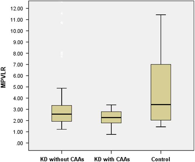
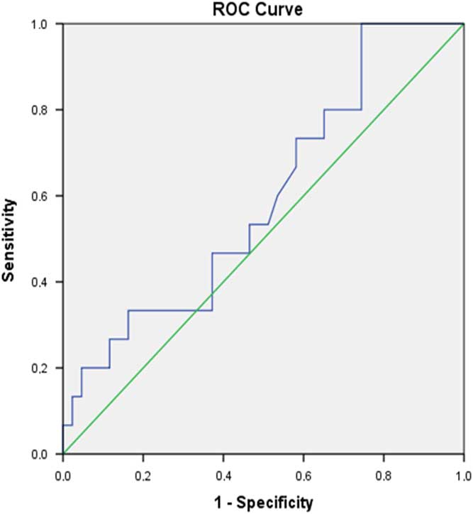
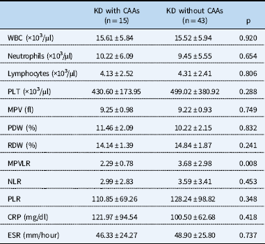
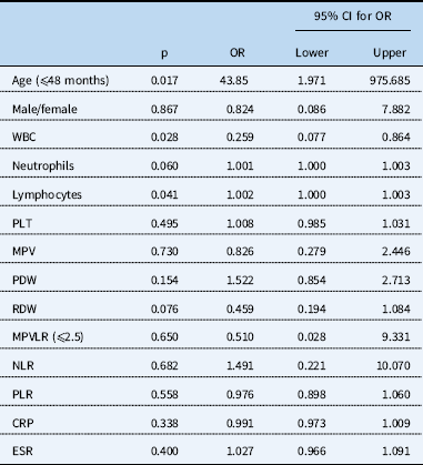
Mean ages of children with coronary artery abnormalities were significantly lower than those without coronary artery abnormalities – 41.06±18.12 months and 56.58±23.32 months, respectively, p=0.013. Laboratory results in Kawasaki disease patients with and without coronary artery abnormalities are shown in Table 3. The children with coronary artery abnormalities had significantly lower levels of mean platelet volume-to-lymphocyte ratio than did those without coronary artery abnormalities (p=0.008) (Fig 1). According to the receiver operating characteristic curve analysis, the best cut-off of mean platelet volume-to-lymphocyte ratio level on admission for predicting coronary artery abnormalities was 2.5 with a sensitivity of 53.3% and a specificity of 51.1% (area under curve=0.593, Fig 2). According to multivariable logistic regression analysis, age, white blood cells, and lymphocyte count were independent predictors of coronary artery abnormalities in Kawasaki disease (Table 4).
Mean platelet volume-to-lymphocyte ratio (MPVLR) levels in Kawasaki disease (KD) patients with and without coronary artery abnormalities (CAAs) and control subjects.

The receiver operating characteristic (ROC) curve analysis for mean platelet volume-to-lymphocyte ratio in the prediction of coronary artery abnormalities (area under curve=0.593, p=0.008).

Relationship between laboratory findings in Kawasaki disease (KD) patients with and without coronary artery abnormalities (CAAs).

CRP=C-reactive protein; ESR=erythrocyte sedimentation rate; MPV=mean platelet volume; MPVLR=mean platelet volume-to-lymphocyte ratio; NLR=neutrophil-to-lymphocyte ratio; PDW=platelet distribution width; PLR=platelet-to-lymphocyte ratio; PLT=platelet; RDW=red blood cell distribution width; WBC=white blood cell
Multivariate logistic regression analysis of patients with coronary artery abnormalities.

CI=confidence interval; CRP=C-reactive protein; ESR=erythrocyte sedimentation rate; MPV=mean platelet volume; MPVLR=mean platelet volume-to-lymphocyte ratio; NLR=neutrophil-to-lymphocyte ratio; OR=odds ratio; PDW=platelet distribution width; PLR=platelet-to-lymphocyte ratio; PLT=platelet; RDW=red blood cell distribution width; WBC=white blood cell
Discussion
To the best our knowledge, this is the first study that has investigated the role of mean platelet volume-to-lymphocyte ratio in children with Kawasaki disease. The results of our study indicated that children with Kawasaki disease had significantly lower mean platelet volume-to-lymphocyte ratio compared with the control subjects. We also showed that mean platelet volume-to-lymphocyte ratio level was significantly lower in Kawasaki disease children with coronary artery abnormalities than in those without coronary artery abnormalities. Although it was not statistically significant, we found that complete Kawasaki disease patients had lower mean platelet volume-to-lymphocyte ratio levels compared with those with incomplete Kawasaki disease.
Inflammation of the coronary arteries results in serious clinical outcomes of Kawasaki disease.Reference McCrindle, Rowley and Newburger 1 One of the most important problems in Kawasaki disease is cardiac morbidity, including coronary aneurysms, myocardial infarctions, and giant coronary aneurysms. Kawasaki disease is the leading cause of acquired heart disease among children in developed countries, and approximately 15–20% of the patients develop coronary artery abnormalities.Reference Makino, Nakamura and Yashiro 2 , Reference Al-Ammouri, Al-Wahsh and Khuri-Bulos 12 There are several biomarkers of inflammation and risk-scoring systems that use routine laboratory data to evaluate the outcomes of Kawasaki disease.Reference Sleeper, Minich and McCrindle 8 , Reference Tremoulet, Dutkowski, Sato, Kanegaye, Ling and Burns 13 Recent studies have been reported that complete blood tests including neutrophils, lymphocytes, and platelets, and their combinations, such as neutrophil-to-lymphocyte ratio and platelet-to-lymphocyte ratio, play a central role in the inflammation associated with acute Kawasaki disease and coronary artery abnormalities.Reference Ha, Lee and Jang 14 , Reference Kawamura, Takeshita, Kanai, Yoshida and Nonoyama 15
Mean platelet volume is a marker of platelet function and activation, and it can be easily measured in routine complete blood count test cycle. There has been rapidly growing interest in the association between mean platelet volume and inflammation, and the risk of cardiovascular events.Reference Gasparyan, Ayvazyan, Mikhailidis and Kitas 16 For this purpose, a study conducted by Liu et alReference Liu, Gao, Huo and Yi 17 evaluated the role of mean platelet volume in children with Kawasaki disease. They found that Kawasaki disease patients have lower mean platelet volume, but it was not a useful marker for predicting coronary artery abnormalities. In the current study, we found that there was no statistically significant difference in mean platelet volume between children with Kawasaki disease and controls. However, we have shown that mean platelet volume was not valuable for predicting coronary artery abnormalities.
In recent years, mean platelet volume-to-lymphocyte ratio has emerged as a novel marker that can be a prognostic indicator of adverse cardiovascular events in adult patients.Reference Hudzik, Szkodziński, Lekston, Gierlotka, Poloński and Gąsior 9 – Reference Ornek and Kurtul 11 Hudzik et alReference Hudzik, Szkodziński, Lekston, Gierlotka, Poloński and Gąsior 9 first defined mean platelet volume-to-lymphocyte ratio in 2016, and they reported that mean platelet volume-to-lymphocyte ratio plays a povital role in developing intravascular thrombus in ST elevation myocardial infarction. The findings supported with the study conducted by Kurtul et alReference Kurtul and Acikgoz 10 and they demonstrated that mean platelet volume-to-lymphocyte ratio was a strong independent predictor for angiographic no-reflow and short-term mortality in adult patients with ST elevation myocardial infarction. In patients with coronary artery disease, coronary collateral circulation develops as an adaptation to ischaemia and reduces the cardiovascular events. More recently, Ornek et alReference Ornek and Kurtul 11 found that mean platelet volume-to-lymphocyte ratio was associated with impaired collateral circulation in patients with stable angina pectoris.
As the role of mean platelet volume-to-lymphocyte ratio has not been defined in Kawasaki disease previously, the studies mentioned above encourage us to assess whether mean platelet volume-to-lymphocyte ratio may have a value in children with Kawasaki disease. In contrast to the previous studies in adults, we found that the levels of mean platelet volume-to-lymphocyte ratio was significantly lower in children with Kawasaki disease and those with coronary artery abnormalities. The real mechanism of the decrease in mean platelet volume-to-lymphocyte ratio in children with Kawasaki disease is not clearly understood. The evidence suggests that that platelet volume is correlated with platelet function and may be a more sensitive index than platelet number as a marker of clinical interest in various disorders including systemic infection and inflammatory conditions.Reference Hudzik, Szkodziński, Lekston, Gierlotka, Poloński and Gąsior 9 Mean platelet volume was decreased in active rheumatological diseases.Reference Gasparyan, Ayvazyan, Mikhailidis and Kitas 16 It has been speculated that the reduced platelet volume could be because of the consumption or sequestration of the large activated platelets in the vasculature.Reference Liu, Gao, Huo and Yi 17
Mean platelet volume-to-lymphocyte ratio has two parameters, and it is calculated by dividing the mean platelet volume value by the lymphocyte count. There is strong evidence that high-grade inflammatory diseases, such as active rheumatoid arthritis or attacks of familial Mediterranean fever, present with low levels of mean platelet volume.Reference Gasparyan, Ayvazyan, Mikhailidis and Kitas 16 , Reference Makay, Turkyilmaz and Unsal 18 During the acute stage of Kawasaki disease, lymphocyte predominance may be seen. Igarashi et alReference Igarashi, Hatake, Tomizuka, Yamada, Gunji and Momoi 19 found that macrophage colony-stimulating factor, granulocyte colony-stimulating factor, and interleukin-6, derived from monocytes as monokines or derived from vascular endothelial cells, might play an important role in the acute phase of Kawasaki disease. They have suggested that these markers increase during the acute phase of Kawasaki disease, which might contribute to the decreased platelet volume in Kawasaki disease. On the other hand, there were no statistically significant differences in mean platelet volume and lymphocyte counts between Kawasaki disease patients with and without coronary artery abnormalities in our study. Further studies are needed to clarify the exact mechanism of low levels of mean platelet volume-to-lymphocyte ratio in Kawasaki disease patients with coronary artery abnormalities.
Our study had several limitations. First, this study was designed as retrospective in nature. Second, the sample size was relatively small. Third, mean platelet volume-to-lymphocyte ratio was evaluated only on admission, and we did not evaluate fluctuations in mean platelet volume-to-lymphocyte ratio levels. As most of the children with Kawasaki disease had elevated inflammatory markers in the current study, we did not evaluate the role of mean platelet volume-to-lymphocyte ratio in patients with normal levels of white blood cells, platelets, red blood cell distribution width, erythrocyte sedimentation rate, and C-reactive protein. Hence, our findings may need to be confirmed in prospective studies. On the other hand, our study has several strengths as well. To our knowledge, this is the first study to examine the value of mean platelet volume-to-lymphocyte ratio in children with Kawasaki disease. The level of mean platelet volume-to-lymphocyte ratio may be affected with several conditions such as haematological diseases, allergic diseases, malignant and inflammatory diseases, and medications. In the current study, children with these conditions were excluded. We did not provide information regarding the value of mean platelet volume-to-lymphocyte ratio in children with fever and no Kawasaki disease. This issue may require further evaluation.
To date, there has been relatively little research on mean platelet volume-to-lymphocyte ratio. No study has assessed the role of mean platelet volume-to-lymphocyte ratio in children with Kawasaki disease. The results of this study show that mean platelet volume-to-lymphocyte ratio may be a novel marker to predict coronary artery abnormalities in children with Kawasaki disease. We believe that further studies with larger numbers of patients are needed to clarify the relationship between mean platelet volume-to-lymphocyte ratio and Kawasaki disease.
Acknowledgements
None.
Financial Support
This research received no specific grant from any funding agency or from commercial or not-for-profit sectors.
Conflicts of Interest
None.
Ethical Standards
The authors assert that all procedures contributing to this work comply with the ethical standards of the relevant national guidelines on human experimentation (Mersin University) and with the Helsinki Declaration of 1975, as revised in 2008, and has been approved by the institutional committees of Mersin University.