

Ian Hacking’s (Reference Hacking1983) “little” book is best remembered for his defense of “scientific entity realism,” encapsulated in the slogan, “it’s real if you can spray it.” Of more interest to me are lessons in the book’s second part, on experiments (“interventions”). Hacking was hopeful that the book’s second part “might initiate a Back-to-Bacon movement,” in which philosophers “attend more seriously to experimental science” (150). Numerous philosophers heeded Hacking’s call, including “new experimentalists” Alan Franklin, Deborah Mayo, and others, and more recently philosophers of neuroscience (Bickle Reference Bickle2003; Craver Reference Craver2007; Sullivan Reference Sullivan2010; Silva, Landreth, and Bickle Reference Silva, Landreth and Bickle2014; Atanosova Reference Atanosova2015).Footnote 1 One of Hacking’s (Reference Hacking1983) arguments stemmed from some scientific details about microscopes and their historical development. It is useful to reexamine Hacking’s “microscope” argument for the relative independence of experiment from theory, because exactly his premises are illustrated by a newly developed experimental tool in recent neurobiology: optogenetics and DREADDs (designer receptors exclusively activated by designer drugs). These comparisons hold despite the very different background sciences and fields of engineering that yielded microscopes and optogenetics/DREADDs. As radical as Hacking’s “contra-theory” conclusion still seems to some philosophers, he appears to have latched onto an important general point about tool development that holds across vastly different sciences.
Optogenetics and DREADDs: Some Scientific Details
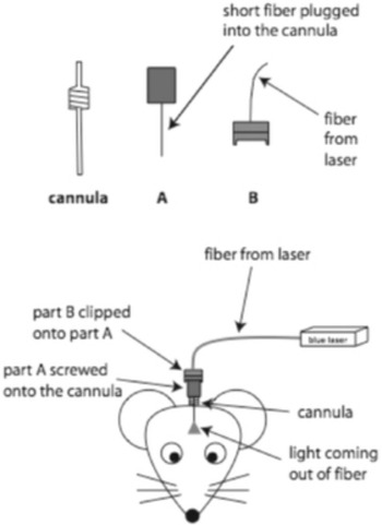
Now in existence for only 1 decade, the use of optogenetics and DREADDs in neurobiological experiments has increased dramatically since Nature Methods dubbed optogenetics “Method of the Year” in 2011 (Deisseroth Reference Deisseroth2015, fig. 7). These tools offer experimenters unprecedented control over the activation or silencing of specific neurons in living, behaving animals. For optogenetics, light-responsive protein receptors, most prominently the microbial opsins from algae, serve as membrane-bound ion channels for cation or anion influx, thereby depolarizing or hyperpolarizing the cell when these receptors are activated by light. The algae DNA coding for these opsin proteins is engineered to be expressed in mammalian (typically rodent) DNA. It is inserted into mammalian neurons via a virus vector, nowadays typically an engineered adeno-associated virus whose own genetic material has been removed to permit the insertion of the engineered opsin transgene. With stereotactic surgery, the virus vector is microinjected into precise locations in the animals’ brains. The virus infects neurons nearby the injection site, which take up the engineered opsin DNA by the usual mechanisms of virus replication. These infected neurons begin expressing the engineered gene and synthesizing the new protein, under the control of the promoter region of the engineered transgene. These synthesized opsin proteins embed in the infected neurons’ membranes. Depending on the specific opsin chosen, they can be activated by a light stimulus to pass a specific cation (sodium, Na+) or anion (chloride, Cl−) into the neuron. These neurons are thereby depolarized (activated) or hyperpolarized (inactivated) literally at the flip of a light switch—by turning on or off a laser or LED light source embedded in the brain, in range of the infected neurons. Activating (or inactivating) specific neurons can have downstream effects of activating (or inactivating) entire neuronal circuits. The stimulating light source can be inserted deep into the brain through chronically implanted cannulas cemented permanently into the animals’ skulls.Footnote 2 The DNA for an imaging protein (mCherry, green florescent protein, yellow fluorescent protein) typically is also coupled to the engineered DNA for the opsin protein in the virus vector. All neurons expressing the opsin protein will also express and synthesize the imaging protein and glow a particular color (red, green, yellow) under standard light microscopy of brain tissue slices. Experimenters can thereby verify exactly which neurons expressed the opsin protein and were activated or inactivated by the light stimulus.
That is the basic science. Additional refinements typically depend on the specific experimental questions optogenetics is employed to address. These refinements are usually variants drawn from the standard toolbox of genetic engineering, often the insertion of specific promoter regions into the virus vector to limit transgene expression to specific types of infected neurons. Judicious choices of promoters can control both neuron-specific and temporal expression of the opsin transgene, so that only neurons infected with the transgene and possessing the promoter binding proteins in sufficient quantity and in the required activating background state express the transgene and synthesize the protein.Footnote 3
DREADDs, a kind of chemogenetics, is a related technique that dispenses with optogenetics’ inserted light stimulus. Instead of an engineered microbial opsin gene, the gene for an entire receptor for a designer drug—a compound that nowhere occurs naturally in biological tissue—is microinjected via the virus vector. These engineered receptors are metabotropic and coupled to an intracellular G-protein, to induce either bursting (Gq-type protein) or silencing (Gi-type protein) in infected neurons.Footnote 4 The designer drug is administered systemically, through either peritoneal injections or diet. It binds only to the engineered receptors in infected neurons, activates the attached G-protein complex, and induces bursting or silencing in those neurons for the duration of the designer drug’s pharmacological activity.
Neurobiologists are furiously exploring the relative advantages and disadvantages of optogenetics versus DREADDs. No need for an implanted light source is one technological advantage of DREADDs. So can be the number of neurons activated by the drug, since systemic administration of the designer drug means that virtually all neurons expressing the transgene for the designer receptor will be activated, no matter where they are located in the brain. With optogenetics, only those neurons infected (in which the transgene gets expressed) and in the range of the stimulating light source are activated. This extent of the drug’s effect is a two-edged sword, however. It puts a premium on surgical placement of the DREADD-containing virus and subsequent verification of this placement. Leakage of the virus into other nontarget neurons, and subsequent expression of the designer receptor in them, can confound behavioral results. DREADDs’ temporal control is tied to the pharmacokinetics and dynamics of the designer drug: its absorption, distribution, metabolism into inactive metabolites, and excretion. This slow and imprecise temporal effect contrasts sharply with optogenetics’ temporally much more tightly controlled laser or LED on/off light switch. Practical laboratory concerns are also important. Optogenetics requires expensive, specialized equipment and specific experimental expertise to succeed. DREADDs can be up and running quickly in most competent neurophysiologists’ labs.
Toward a Metascience of Tool Development in Neurobiology
In an initial attempt to explicate a metascience of tool development in neurobiology, Bickle (Reference Bickle2016) investigated the motivating problem and initial successful and second phase hook experiments for optogenetics and DREADDs. The motivating problem for optogenetics stemmed from the causal-mechanistic explanatory goals of contemporary neurobiology. To test causal hypotheses that relate specific neurons’ activities directly to behaviors operationalizing specific cognitive phenomena, experimenters need the capacity reliably to intervene into the hypothesized neuronal mechanisms: to activate and inhibit specific neurons. They then can measure the effects of these interventions on those behaviors in tightly controlled experimental protocols using animal, preferably mammal, model organisms. These experimental interventions can be difficult because of often sparse anatomical distributions of the hypothesized neuronal mechanisms. Pharmacological interventions easily can be confounded by the spread of the drug to nearby nontarget neurons, and nature does not always cooperate to clump target neurons together into discrete cortical columns or microcolumns amenable to direct microelectrode stimulation. The intriguing suggestion of using light stimuli to activate or inhibit target neurons was already on offer (Crick Reference Crick1999). The “obvious” solution was to develop some way to induce the target neurons themselves to synthesize light-sensitive ion channels. But that problem space explodes quickly. For speed of activation or inhibition, the expressed ion channels should be ionotropic and simply open direct channels for cation or anion influx. Expression and synthesis of the channels must be at levels both safe, so as not to kill the cells, and in sufficient numbers to produce enduring effects on neuron membrane potentials. The activating light stimulus had to be deliverable safely, often deep into brains, yet also with sufficient strength to induce activity in enough of the target neurons to affect circuit activity. And all of this had to be implemented in vivo. Tough problems. And yet the prospects of having such an experimental tool beckoned. Like most motivating problems that generate revolutionary new tools in neuroscience, this one was a composite of numerous technical problems with a daunting interactive dynamic.
The microbial opsins prominent in algae were alluring possible candidate receptors. But the predicted currents they would generate individually were very small, and so high gene expression levels and light stimuli seemed necessary to induce sufficient activity in these receptors to affect mammalian neuron activity. Yet neurons were well known to be detrimentally sensitive to membrane protein overexpression and to damaging side effects of light and heat. High specificity of expression and synthesis would be needed for microbial opsins to be useful in tightly controlled in vivo experiments. Finally, attempts were already underway to engineer metazoan rhodopsin genes and channel proteins (which mediate photon transduction in vertebrate retinas) toward this same end. These ongoing attempts further reduced initial enthusiasm for trying to solve these technical problems using a family of genes and proteins from algae, so foreign to, and evolutionarily distant from, the mammalian genome.
This combination of features of its motivating problem is why the initial successful hook experiments using microbial optogenetics were so surprising. Even scientists “in the know” about early attempts to develop this tool assigned a low probability to these experiments succeeding. Despite this, the Adamantidis et al. (Reference Adamantidis, Zhang, Aravanis, Deisseroth and de Lecea2007) study, out of the Deisseroth and de Lecea labs at Stanford, successfully engineered microbial opsin DNA in a lentivirus and attached it to a promoter region that limited transgene expression to orexin-producing neurons and injected it specifically into mouse lateral hypothalamus. The inserted opsin transgene, channelrhodopsin2 (ChR2), expresses a membrane-bound protein that responds to blue light to open a channel for Na+ influx, to depolarize (activate) infected neurons. Electrophysiological studies in vitro and c-fos gene expression studies in vivo showed that these ChR2-expressing neurons reliably responded to light stimuli with volleys of action potentials (Adamantidis et al. Reference Adamantidis, Zhang, Aravanis, Deisseroth and de Lecea2007, fig. 1). Behaviorally, photostimulation to lateral hippocampus in vivo showed that direct activation of these neurons in experimental animals significantly decreased latency (time) to a wakeful state, from both slow wave sleep (SWS) and rapid eye movement (REM) sleep, compared to both baseline (nonphotostimulated) latency in these same optogenetically treated mice and photostimulation in control mice (who received the virus vector injections minus the ChR2 transgene; Reference Adamantidis, Zhang, Aravanis, Deisseroth and de Lecea2007, fig. 3). The mechanism producing these photostimulation-induced effects influenced the sleep-wake circuitry by affecting transmitter release in the infected neurons, which was demonstrated by administering a specific receptor antagonist that blocked this photostimulation effect in the optogenetically treated animals (Reference Adamantidis, Zhang, Aravanis, Deisseroth and de Lecea2007, fig. 4).
The initial hook experiments that demonstrate successful application of a new tool to professional scientists are important, but genuinely revolutionary new tools generate wider appeal, sometimes capturing the public’s attention. This comes from second-phase hook experiments. With optogenetics, the Tonegawa lab’s (at MIT) creation of false memories in a mouse model was one clear example of this second kind of result. Using a hippocampus-dependent memory task, Liu et al. (Reference Liu, Ramirez, Pang, Puryear, Govindarajan, Deisseroth and Tonegawa2012) activated ChR2 opsin transgene expression and synthesis in hippocampus dentate gyrus neurons only when mice were undergoing training to associate a foot shock with a novel context. They accomplished this temporal restriction by tying the activation of the ChR2 transgene to a tetracycline transduction activator system, so expression only occurred in infected neurons when experimental animals were taken off doxycycline in their diets. They limited expression of the transgene only to the most active hippocampal dentate gyrus neurons at the time of context-shock training, by tying the activator system to a c-fos activator system, so transgene expression was limited to only the most active neurons at the time of training, that is, those whose activity generated sufficient c-fos expression and synthesis to drive the activator system. These neurons constitute the hypothesized engram for the context-shock association. The experimenters then introduced the mice into a second novel context in which no foot shock was delivered, but the experimenters activated the neurons in the experimental animals that expressed the transgene during the previous context-shock training in the different context by turning on the hippocampus-embedded blue light. They then tested for fear conditioning in the second, never-shocked context. As expected, nonoptogenetic control animals displayed no fear conditioning to the second context. But optogenetic-treated animals whose infected hippocampus neurons that encoded the fear memory in the first, shocked context were activated while they explored the second, never-shocked context did demonstrate stereotypical rodent fear behavior (freezing) when placed back into the second, never-shocked context.
Ramirez et al. (Reference Ramirez, Liu, Lin, Suh, Pignatelli, Redondo, Ryan and Tonegawa2013) did the reverse manipulation. They activated the transgene-infected dentate gyrus neurons only while the experimental animals explored a first novel context, in which no shock was delivered. They then shocked the animals in a second novel context while activating the neurons coding for the memory of the first, never-shocked content in the optogenetic-treated experimental animals. When placed back into the first, never-shocked context, as expected control animals displayed no fear behavior. But the optogenetic-treated experimental mice displayed fear in the first, never-shocked context.
These experimenters interpreted their results straightforwardly: “we created a false memory in mice by optogenetically manipulating memory engram-bearing cells in the hippocampus” (Ramirez et al. Reference Ramirez, Liu, Lin, Suh, Pignatelli, Redondo, Ryan and Tonegawa2013, 387).Footnote 5 The Tonegawa lab’s false memory second-phase hook experiments using optogenetics brought this new tool wider scientific and public attention. Two years after Liu et al. (Reference Liu, Ramirez, Pang, Puryear, Govindarajan, Deisseroth and Tonegawa2012) first appeared, a review article appropriately titled “The Optogenetic Revolution in Memory Research” was published in Trends in Neurosciences (Goshen Reference Goshen2014). It referenced 100 publications already employing the tool for this single cognitive phenomenon. The Tonegawa lab’s results also attracted huge public interest. Search for “false memory optogenetics” on the Internet to get a quick feel for the range of popular coverage.
Hacking on Microscopes: Sounds Familiar
With optogenetics/DREADDs’s basic scientific details now explained, and the beginnings of a metascience of tool development in neurobiology applied to them, I turn next to Hacking’s (Reference Hacking1983) “microscope argument.” He starts with a seemingly obvious claim: “Some theory of light is of course essential for building a new kind of microscope, and is usually important for improving an old kind.” But in the next sentence he warns: “We should not, however, underestimate the pretheoretical role of invention and fooling around” (199; my emphasis). He defends his warning with some history of the development of microscopes. Here it is illuminating to view a page from the Deisseroth lab notebooks, published in a recent historical overview of the development of optogenetics (see fig. 1). This was the lab’s “initial engineering sketch” of the fiber-optic neural interface (Deisseroth Reference Deisseroth2015, 1215). It is a beautiful illustration of Hacking’s appeal to “invention and fooling around,” especially the lab’s cartoon bewildered mouse. The lab members literally were playing around, in an effort to figure out how to make these gadgets work.
“Initial engineering sketch of the fiber-optic neural interface for spatially registering viral transduction with focal high-intensity illumination (drawing courtesy F. Zhang, Stanford)” (Deisseroth Reference Deisseroth2015, 1215, fig. 2).

Remarking on the “initial trials” using the electron microscope—hook experiments—Hacking continues: “When the idea of the electron microscope was put into practice, it was a long shot, because people were convinced, on theoretical grounds, that the specimen would almost instantly be fried and then burnt out” (Reference Hacking1983, 199; my emphasis). This premise of Hacking’s microscope argument should be familiar from the discussion above of the initial development of optogenetics. This same scientific judgment (vastly improbable), from the same scientific ground (‘theory’) attached to the proposed use of microbial opsin genes and proteins in mammal models in vivo. Algae DNA was too foreign to be engineered to be expressible in metazoan DNA. The currents through the synthesized membrane-bound microbial opsins would be too weak, neurons would have to express so many of these foreign ion channels to generate a current sufficient to drive action potentials that their cell membranes would rupture, the heat from the light source that would be needed to drive enough of these engineered receptors would destroy the neurons expressing them, and so on. But theory be damned. It worked.
Hacking’s (Reference Hacking1983) microscope argument emphasizes another point relating theoretical possibility and practical engineering reality. He writes (more than 30 years ago), “The X-ray microscope had been a theoretical possibility for ages, but can effectively be built only in the next few years using high-quality beams that can be bought from a linear accelerator. Likewise the acoustic microscope … only in the last 10 years has one had the fast electronics to produce good high frequency sound and quality scanners” (199; my emphasis). Identical points hold for optogenetics. Recall from my discussion above Crick’s ‘theoretical’ speculations about tricking neurons into synthesizing their own light-responding receptors and then activating or inhibiting those neurons in behavioral neuroscience experiments using brain-embedded light sources. He knew that light-responsive ion-channel proteins existed, both in mammal nervous systems and in algae. “Gene targeting” was already an increasingly influential tool of basic neurobiology. But ‘theoretical possibility’ had to wait for engineering ingenuity and specific implementations. Optogenetics’ dependence on engineering problem solving, not theory, persists to the present day. Would-be users face critical engineering problems to get optogenetics techniques to work. One example is successfully delivering the light source through the optic fiber deep into brains, without significant power loss. In fact, it is technical engineering problems like this one that lead many labs to embrace DREADDs/chemogenetics, when speed of specific neuronal activation or silencing turns out not to be critical to experimental interventions. These are make-it-work, experiment-in-practice considerations, not theoretical ones.
The conclusion Hacking urges from his microscope argument is still radical for many philosophers of science: “Theory has only a modest amount to do with building these ingenious devices. The theory involved is mostly of the sort we learn in Physics I at college. It is engineering that counts” (Reference Hacking1983, 199). But since the basic premises of his microscope argument carry over so straightforwardly to optogenetics and DREADDs, it should surprise no one that this conclusion does, too. The background sciences are different: basic cell physiology instead of Physics I. The fields of engineering are different: bio- and genetic engineering instead of optical. But Hacking’s conclusion about the relative independence of the development of these revolutionary new experiment tools from theory holds for optogenetics/DREADDs just as well as it does for microscopes.
Hacking ends his microscope argument for the relative independence of experimental tool development from theory by reflecting on the changes in theoretical explanation that accompanied the continuing use of the light microscope. Initially the images were thought to result from absorption. Eventually they were understood to result from diffraction. Yet this theory change “made no whit of difference. People kept on regarding the micrographs of the molecules as genuinely correct representations.” He asks, “Is this all mumbo-jumbo, a kind of confidence trick?” and answers, “Only a theory dominated philosophy could make one say so. The experimental life of microscopy uses non-theory to sort out artifice from the real thing” (Hacking Reference Hacking1983, 200; my emphasis). The tie-in between optogenetics/DREADDs and behavioral neuroscience, which has existed since these experimental tools’ initial hook experiments, renders Hacking’s last point especially applicable. The scientific confidence in optogenetics and DREADDs rests on the integration of the behavioral results these experimental tools have generated with those gathered previously using less precise methods for intervening into hypothesized cellular and molecular mechanisms. The behavioral results using optogenetics- and DREADDs-treated animal models elide smoothly with previously published studies using pharmacological, ablation, microstimulation, and more standard gene targeting interventional techniques. This integration with already-existing behavioral results, not theory, is what gave experimenters confidence that these techniques actually were a more direct experimental path into the hypothesized cellular mechanisms of cognitive functions.
Which Came First? (The Theory or the Tool?)
A common criticism of my application of Hacking’s microscope argument to optogenetics/DREADDs points out the many theoretical advances that had to be made before optogenetics/DREADDs were conceivable as tools to develop.Footnote 6 This list includes the theory of protein ion channels as a mechanism for controlling selective ion movements across membranes, thereby depolarizing or hyperpolarizing neurons; the theory of opsin structure and function, whose configuration is changed by photon absorption to open a pore for specific ions to flow; and the theory of DNA coding for specific proteins (opsins, designer receptors), its insertability into viruses, and the virus replication cycle by which those engineered DNA segments come to be embedded and expressible in the mammalian genome. A lot of biochemical, biophysical, molecular-genetic, and virological theory predated, and made possible, these engineering marvels.
Without question this is true. But what this objection misses is the tool development dependence of every one of these prior theoretical advances. I will develop this response by working with one of these examples, although I insist that a similar story about other experimental tools can be told for the others. Consider the theory of protein ion channels. Although they had no verifiable account of the mechanism for the sodium and potassium currents that generated the neuron action potential that their famous mathematical model characterizes,Footnote 7 Hodgkin and Huxley (Reference Hodgkin and Huxley1952) speculated about some key features these mechanisms must possess, whatever they turned out to be. These mechanisms had to (1) permit both sodium and potassium ions to move across the membrane at high rates, (2) make use of electrochemical gradients (positively charged sodium ions, at a much higher concentration outside the cell, flow rapidly into the negatively charged neuron; a millisecond later, positively charged potassium ions, at a much higher concentration inside the cell, flow out of the depolarized, now positively charged neuron), (3) discriminate between sodium ions and potassium ions and allow the flow of each independently of the other (the inward sodium current occurs first, sodium does not flow out of the depolarized neuron, the outward potassium current begins only after the sodium current has ceased), and (4) sense voltage changes across the neuron and be differentially sensitive to different voltages (the sodium and potassium currents begin at different specific membrane voltages). Membrane-bound proteins provided one intriguing model for these “ion channels.”
It was another quarter century before evidence confirmed that these “ion channels” were two classes of membrane-bound proteins that were ion selective and whose configurations changed with membrane voltage. It was not theory that confirmed this. Hodgkin and Huxley had stated their basic functional properties all the way back in one of their original publications. What confirmed their physical structure and mechanics was the joint use of two tools. One tool, x-ray crystallography, had been in use of three-quarters of a century (at that time). The other, the patch clamp, was new. It was developed in the late 1970s and early 1980s by numerous labs, but two scientists, Erwin Neher and Bert Sakmann, received the lion’s share of credit.Footnote 8 The patch clamp was hardly deep theory. At bottom it is a miniaturization of Cole’s voltage clamp. The experimenter brings a glass pipette with a tiny opening (typically in the micron range) into tight contact with a tiny area of cell membrane, or “patch,” and applies suction to the back of the pipette. This creates a seal between the pipette and membrane tight enough so that no ions can pass. (The tightness of the seal is confirmable empirically, by an ultrasensitive electronic amplifier connected to the pipette.) Thus, all of the ionic current that flows out of the cell when a single ion channel opens flows into the pipette. Varying ion concentrations of, or other experimental manipulations to, the nutrient bath containing the “patched” neuron enable measurements of microscopic currents of specific ions through the patch (e.g., sodium, potassium). Armed with this new tool, experimenters quickly showed that the average of hundreds of summed microcurrent measurements revealed the same shape and time dimensions of the macroscopic ion currents (measured via the voltage clamp). Even the probability of a single ion channel opening was shown to depend on the specific membrane voltage “clamped” at different millivolt potentials.Footnote 9
With single ion channels and their functional properties now isolable experimentally, elucidating their physical structure was a straightforward matter of applying x-ray crystallography. This tool had existed since the second decade of the twentieth century and was most famously used in biology by Rosalind Franklin, Francis Crick, James Watson, and Maurice Wilkins to elucidate the double-helical structure of the DNA molecule. This technique quickly revealed these “channels” to be configured proteins. The entire sequence of charged amino acids composing specific channels could be determined, and their functional properties thereby explained. These included the ion selectivity of the pore and their membrane-voltage responsiveness, based on the specific location of differently charged amino acids on the configured protein. These details have been worked out most completely for a bacterial potassium channel (Tao et al. Reference Tao, Lee, Limapichat, Dougherty and MacKinnon2010). It was chosen as an experimental model because the large number of channel proteins required for x-ray crystallography could be obtained most easily by growing large numbers of bacteria. In the cellular and molecular mainstream of contemporary biology, experiments and their concerns are primary. Innovation and revolution here depend first and foremost on tools, new or newly applied (Bickle Reference Bickle2016). These tools are the result of engineering ingenuity and laboratory tinkering, in developmental chains often stretching back decades. Deep theory lags behind.
Fundamentally different background sciences and fields of engineering built microscopes and optogenetics/DREADDs. Hacking’s conclusion about the relative independence of experiment from theory seems truly to be a correct general point about tool development across science. Is, perhaps, theoretical physics one of science’s exceptions, rather than its exemplar?