INTRODUCTION
Babesia bovis is an intraerythrocytic apicomplexan parasite responsible for babesiosis in bovines, ruminants that are the natural hosts of this parasitosis. This tick-borne disease, prevalent in tropical and subtropical areas worldwide, is poorly controlled by the existing chemotherapy and immunoprophylaxis, remaining as an important constraint for the development of cattle industries (Bock et al. Reference Bock, Jackson, de Vos and Jorgensen2004). Babesia bovis-infected erythrocytes undergo sequestration by attachment to capillary endothelium, resulting in organ damage, cerebral dysfunction and pulmonary oedema, similar to the most severe form of human malaria caused by another apicomplexa, Plasmodium falciparum (Wright et al. Reference Wright, Goodger and Clark1988). In B. bovis infection, innate immune mechanisms have been recognized to have an important role in the limitation of acute infection, whereas acquired immunity is likely to be more important for resistance to parasite challenge (Fell and Smith, Reference Fell and Smith1998; Brown and Palmer, Reference Brown and Palmer1999). In this sense, it has been demonstrated that innate immune molecules derived from B. bovis-activated macrophages, such as NO and inflammatory cytokines, contribute to that limitation (Shoda et al. Reference Shoda, Palmer, Florin-Christensen, Florin-Christensen, Godson and Brown2000). Although pro-inflammatory mediators function as elements of early protective immunity against haemoprotozoan parasites, as infection progresses in severity, overproduction of these molecules has been implicated in the pathological sequelae of disease (Krause et al. Reference Krause, Daily, Telford, Vannier, Lantos and Spielman2007).
As concerns lipids from microorganisms, these compounds have an important modulatory role in the immune system (Schofield and Hackett, Reference Schofield and Hackett1993; Roach and Schorey, Reference Roach and Schorey2002; Quesniaux et al. Reference Quesniaux, Nicolle, Torres, Kremer, Guerardel, Nigou, Puzo, Erard and Ryffel2004). In B. bovis, it has been reported that this parasite possesses a high rate of phosphatidylcholine (PC) and phosphatidic acid (PA) biosynthesis (Florin-Christensen et al. Reference Florin-Christensen, Suarez, Florin-Christensen, Hines, McElwain and Palmer2000). Furthermore, we have demonstrated that the lipid composition of 2 B. bovis strains with polar behaviour, attenuated R1A and virulent S2P, present quantitative differences that contribute to the distinct inflammatory responses observed in a murine experimental model (peritoneal macrophages) (Gimenez et al. Reference Gimenez, Magalhaes, Belaunzarán, Poncini, Lammel, González Cappa, Bozza and Isola2010). It is well known that macrophage stimulation leads to the activation of transcription factors related to inflammatory cytokines and NO production, and that the mitogen activated-protein kinase (MAPK) and the nuclear factor-κB (NF-κB) pathways are crucially involved in this process (Roach and Shorey, Reference Roach and Schorey2002; Hayden and Ghosh, Reference Hayden and Ghosh2008). In this regard, we have previously determined in murine macrophages that lipids of B. bovis R1A strain activate extracellular signal-regulated kinases 1/2 (ERK1/2) as well as the involvement of this pathway in pro-inflammatory cytokine release (Gimenez et al. Reference Gimenez, Magalhaes, Belaunzarán, Poncini, Lammel, González Cappa, Bozza and Isola2010).
In the present work the immunomodulatory effect of lipids from these B. bovis strains in cells from its natural host was studied. With this aim, we analysed in bovine macrophages the induction of NO and the cytokines TNFα, IL-12 and IL-10, the activation of MAPKs and NF-κB signalling pathways and investigated the effect of these lipids in the proliferation of bovine peripheral blood mononuclear cells. Moreover, as we previously demonstrated in non-infected bovines the presence of natural IgM anti-PC and anti-phosphatidylethanolamine (PE) (Gimenez et al. Reference Gimenez, Florin-Christensen, Belaunzarán, Isola, Suarez and Florin-Christensen2007), we also here analysed the anti-phospholipid antibody response in bovines experimentally infected with each strain.
MATERIALS AND METHODS
Reagents and antibodies
Silica gel 60 thin layer chromatography plates and organic solvents were purchased from Merck (Darmstadt, Germany). Egg L-α-phosphatidylcholine (PC), egg sphingomyelin (SM) and egg L-α-phosphatidylethanolamine (PE) were purchased from Avanti Polar Lipids (Alabaster, Alabama, USA). Lipid standards, concanavalin A (Con A), protease inhibitors cocktail, Histopaque-1077, LPS from Escherichia coli (serotype O127:b8), sulphanilamide and naphthylethylendiamine dihydrochloride were acquired from Sigma Chemical Co. (St Louis, MO, USA). The Roche blocking reagent® was purchased from Roche (Mannheim, Germany). Peroxidase-labelled (HRP) anti-bovine IgM and anti-bovine IgG antibodies were obtained from KPL (Gaithersburg, Maryland, USA). O-phenylendyamine was acquired from DakoCytomation (Denmark). Recombinant bovine IFNγ (Ciba-Geigy) was kindly provided by Dr Carlos Suárez (WSU, Pullman, USA). Rabbit polyclonal anti-mouse β-actin, goat anti-rabbit IgG-HRP, anti-mouse IgG-HRP, anti-phospho p38 and anti-IκBα antibodies were purchased from Santa Cruz Biotechnology Inc (Santa Cruz, CA, USA). Rabbit anti-phospho ERK and anti-total ERK antibodies were obtained from Cell Signaling Technology, Inc (Danvers, MA, USA). Nitrocellulose membrane and autoradiographic films were from GE Healthcare (Piscataway, NJ, USA). Supersignal® West Pico Chemiluminescent substrate was purchased from Pierce (Rockford, IL, USA). Protein and DNA molecular weight markers, random primers, SuperScript II Reverse Transcriptase, Taq Polimerase, DNAse and oligonucleotides were obtained from Invitrogen Corporation (CA, USA). Maxisorp 96-well ELISA plates, 6- and 96-well culture plates and 100-mm polystyrene Petri dishes were purchased from Nunc (Roskilde, Denmark). Hanks balanced salt solution, TRIzol, RPMI 1640 medium were obtained from GIBCO BRL (Grand Island, New York, USA). The RNA extraction kit and PCR SYBR green QuantiTec were purchased from Qiagen Sciences Inc. (Venlo, Holland).
Parasites
Merozoites from 2 strains of B. bovis with polar behaviour were employed: R1A, attenuated and S2P, virulent. R1A is a vaccinal strain derived from a virulent isolate responsible for a babesiosis outbreak in Santa Fe, Argentina, and attenuated by a series of 22 passages of parasitized blood in splenectomized calves (Anziani et al. Reference Anziani, Guglielmone, Abdala, Aguirre and Mangold1993). The virulent S2P strain was isolated from a bovine during a babesiosis outbreak in Salta, Argentina and has been adapted to and maintained in continuous in vitro culture (Mangold et al. Reference Mangold, Aguirre, Cafrune, de Echaide and Guglielmone1993; Baravalle et al. Reference Baravalle, Thompson, Valentini, Ferreira, Torioni de Echaide, Florin-Christensen and Echaide2012). Continuous in vitro multiplication does not modify the attenuated or virulent phenotypes of B. bovis strains (Baravalle et al. Reference Baravalle, Thompson, Valentini, Ferreira, Torioni de Echaide, Florin-Christensen and Echaide2012). Parasites from both strains were cultured in bovine erythrocytes under micro-aerophilic conditions (Levy and Ristic, Reference Levy and Ristic1980). To isolate R1A- and S2P-free merozoites, cultures were left overnight at 4 °C to favour parasite release from bovine erythrocytes to the extracellular media. Cultures were then centrifuged at 400 g for 10 min at 4 °C and parasites contained in the resulting supernatants were further separated by centrifugation at 12800 g for 20 min at 4 °C. Pellets were stored at −20 °C until used for lipid extraction.
Experimental cattle
Six Holstein calves (6 months old), from INTA Experimental Station, Rafaela, Santa Fe, Argentina, were infected with R1A or S2P B. bovis-parasitized erythrocytes. Holstein calves, located at INTA-Castelar, Argentina, serologically negative to B. bovis were used to obtain cells from whole blood samples for in vitro studies. First, 107 (R1A) or 105 (S2P) B. bovis-parasitized erythrocytes suspended in 2 ml of PBS, were packed into 2·5 ml individual sterile syringes and inoculated into 3 calves each via the jugular vein within 30 min after harvest from the tissue culture. The concentration of parasitized erythrocytes was determined in a Neubauer haematocytometer after staining the DNA with fluorochrome acridine orange. The calves were provided with water ad libitum, standard hay and commercial cattle concentrate. Calves were observed daily throughout the experimental period. All animal usage was according to protocols from the Animal Ethics Committee at INTA, Argentina, under the guidelines of the European Union (EU directive 86/609/EEC).
Serum samples
Blood samples were collected from B. bovis-inoculated calves following experimental infection protocols. Calves were bled weekly from the jugular vein, starting 1 week before the experimental infection (day 0) and ending during week 10 (day 66). Serum samples were stored at −20 °C until used for anti-phospholipid antibodies analysis.
Detection of anti-phospholipid antibodies in the sera of B. bovis experimentally infected bovines
The serum samples previously obtained were analysed by ELISA (Gimenez et al. Reference Gimenez, Florin-Christensen, Belaunzarán, Isola, Suarez and Florin-Christensen2007). Briefly, 100 μl aliquots of PC, PE or SM in ethanol at 50 μg/ml were loaded into test wells of microtitre plates. The solvent was evaporated and blocking was performed with a commercial blocking system (Roche Blocking Reagent®) for 2 h at 37 °C. The plates were then washed with Tris-buffered saline (TBS), incubated with 60 μl of the different serum samples (1:100, v/v) for 2 h at 37 °C, washed again and incubated with 60 μl of the conjugates anti-bovine IgM – HRP or anti-bovine IgG – HRP (1:500, v/v) for 2 h at 37 °C. Plates were then washed and incubated with H2O2 + O-phenylendiamine (OPD). After 4 min the absorbance was measured in a microplate reader Model 450 (Bio-Rad) at 490 nm. Results are expressed as the ratio of A490 of each day post-inoculation (p.i.)/0 days p.i. (basal level).
Preparation of total lipid extracts from R1A (LA) and S2P (LV) B. bovis merozoites
The method of Bligh and Dyer (Reference Bligh and Dyer1959) was used for lipid extraction. The solvents were evaporated under nitrogen to a constant weight and lipids from different batches of R1A (LA) and S2P (LV) B. bovis merozoites were re-suspended individually in ethanol for macrophage stimulation assays or in chloroform for preparation of lipid fractions. Lipid extracts were stored at −20 °C until used.
Preparation of lipid fractions from LA and LV
Each chloroform lipid extract previously obtained was separated by thin-layer chromatography (TLC) in silica gel plates, as previously described (Gimenez et al. Reference Gimenez, Magalhaes, Belaunzarán, Poncini, Lammel, González Cappa, Bozza and Isola2010). Briefly, a double solvent system was employed: chloroform/methanol/acetic acid/water (65:35:6:2, v/v) up to the middle of the plate to separate phospholipids and hexane/diethylether/acetic acid (90:10:1, v/v) up to the top to separate neutral lipids (Florin-Christensen et al. Reference Florin-Christensen, Suarez, Florin-Christensen, Hines, McElwain and Palmer2000). For mass determination, plates were sprayed with 10% CuSO4 in 8% H3PO4 and charred by exposure to 150 °C for 13 min (Baron et al. Reference Baron, Cunningham, Strauss and Coburn1984). Lipids were identified by comparison with authentic standards and the following fractions: sphingomyelin + lysophosphatidylcholine (SM + LPC), PC, phosphatidylserine + phosphatidylinositol (PS + PI), PE, PA, free fatty acids (FFA) and cholesterol + diacylglycerol (CHO + DG) were eluted from the TLC plates with 2 × 2 ml portions of chloroform/methanol (1:1, v/v). The solvents were evaporated under nitrogen to a constant weight and lipids were re-suspended in ethanol for macrophage stimulation assays.
Culture of bovine monocyte-derived macrophages
Monocyte-derived macrophages were isolated from peripheral blood mononuclear cells (PBMC) by a modification of a method previously described by Stich et al. (Reference Stich, Shoda, Dreewes, Adler, Jungi and Brown1998). Briefly, bovine peripheral blood was collected from the jugular vein into heparinized 60-ml syringes and centrifuged at 1200 g for 30 min. Buffy coats were collected and diluted approximately to 30 ml with 2 volumes of PBS, under layered with 15 ml of Histopaque-1077, and centrifuged at 1500 g for 20 min. The PBMC were then removed from the interface between the plasma and Ficoll solution, pooled, diluted at least 1:3 with RPMI medium, and centrifuged at 500 g for 15 min. The pellets containing PBMC were washed repeatedly at 250 g until the supernatants were clear, re-suspended in complete RPMI medium (with 10% bovine autologous plasma), placed in 100-mm polystyrene Petri dishes at approximately 1–2 × 10⁸ PBMC/ml (10 ml/dish) and incubated overnight at 37 °C in 5% CO2. Non-adherent cells were removed by washing the plates 3 times with complete RPMI 1640 medium, and adherent cells were cultured in 10 ml of complete RPMI at 37 °C for an additional 6–8 days to allow maturation into macrophages. Cell morphology was controlled by optic microscopy and the culture medium was changed every 2–3 days. After approximately 1 week, adherent macrophages were washed once with complete RPMI and once with Mg2+- and Ca2+-containing Hanks balanced salt solution (HBSS), and removed by vortexing and incubated at 37 °C for 1 h in 0·5 mm EDTA in Mg2+- and Ca2+-free HBSS. Macrophages were harvested and used for nitric oxide (NO) and cytokine induction assays, as well as for the study of p38, ERK1/2 and NF-κB signalling pathways.
Nitric oxide detection
NO production was determined by measurement of nitrite (NO2−) levels, a stable derivative of NO by the Griess assay, according to Shoda et al. (Reference Shoda, Palmer, Florin-Christensen, Florin-Christensen, Godson and Brown2000). Briefly, macrophages were cultured for 48 h at a concentration of 105 cells/well (100 μl/well) in 96-well flat-bottom plates with complete RPMI medium, vehicle (0·5% ethanol) +50 U/ml bovine IFNγ (control), 10 μg/ml LPS +50 U/ml bovine IFN-γ (positive control) or the different batches of LA or LV (50 μg/ml) +50 U/ml bovine IFN-γ. In all the assays vehicle was added in the equivalent amount as present in each of the lipid mixtures at each concentration. Culture supernatants were transferred (50 μl/well) to new 96-well flat-bottom plates, and 100 μl of 1% sulfanilamide in 2·5% H3PO4/well followed by 100 μl of 0·1% naphthylethylenediamine dihydrochloride in 2·5% H3PO4/well were added to the supernatants. Absorbance at 540 nm was compared to a NaNO2 standard curve.
Cytokine mRNA analysis
Macrophages (1 × 10⁷ cells/well) were plated into 6-well plates and left to adhere for 2 h at 37 °C in 5% CO2. Cells were incubated overnight with 50 U/ml bovine IFNγ and then stimulated with the different batches of LA or LV (50 μg/ml) or vehicle (0·5% ethanol) for 0, 3, 6 and 16 h, at 37 °C in 5% CO2. Total cellular RNA was isolated from macrophage cultures using the TRIzol reagent RNA isolation method as specified by the manufacturer (GIBCO) and the sediment was cleaned up following the protocol of a commercial kit for RNA extraction (Qiagen). Quality and quantity of total RNA were estimated by UV spectrophotometry (Nanodrop, Wilmington, DE, USA) and electrophoresis was performed on 0·8% agarose gels. DNA-free RNA (1 μg) was mixed with 50 ng of random primers (Invitrogen) in 20 μl of final volume and reverse transcribed to total cDNA with SuperScript II reverse transcriptase (Invitrogen) according to the manufacturer's instructions. One μl of the cDNA was used as template for each real-time quantitative PCR (qPCR) reaction. The primers for bovine: TNFα (F: CCCCCAGAGGGAAGAGTCC, R: GGGCTACCGGCTTGTTACTTG, melting temperature (Tm) 84 °C), IL-12p35 (F: TAGCCACGAATGAGAGTTGCC, R: TTTCCAGAAGCCAGACAATGC, Tm 78 °C), IL-10 (F: GGAAGAGGTGATGCCACAGG, R: AGGGCAGAAAGCGATGACAG, Tm 84 °C) and pol II genes (F: GGACCCGTGTGGACAAGAAT, R: ACAAGCCCCAGGTAATCATCC, Tm 76 °C) were those previously described (Blanco et al. Reference Blanco, Schierloh, Bianco, Caimi, Meikle, Alito, Cataldi, Sasiain M del and Bigi2009). qPCR reactions were performed as previously described (Coussens et al. Reference Coussens, Verman, Coussens, Elftman and McNulty2004). Briefly, SYBR green QuantiTec Mastermix (Qiagen) was employed and reactions were made on Applied Biosystem 7000 SDS using standard cycling conditions. All reactions were performed in duplicate and qPCR data were analysed using the 2–ddCT with efficiency correction according to Pfaffl et al. (Reference Pfaffl, Horgan and Dempfle2002). REST beta 9 software (http://www.gene-quantification.info/, [rest-mcs-beta-9august2006]) was used for final calculations and statistical analysis. The value for each treatment was compared with the value of the control by using a Pair Wise Fixed Reallocation Randomization test attached to software REST beta 9. Results were presented as the relative expression ratio of each target gene (TNFα, IL-12p35 or IL-10), which considers the response of the target gene of the sample versus control (cells incubated overnight with IFNγ + vehicle), and also compared to a reference non-regulated gene (Pol II).
Signalling pathway analysis by immunoblot
Macrophages (4 × 10⁶ cells/well) were plated into 6-well plates and left to adhere 2 h at 37 °C in 5% CO2. Cells were incubated overnight with 50 U/ml IFN-γ and then stimulated with the different batches of LA or LV (50 μg/ml) or vehicle (0·5% ethanol) for 5, 15 and 30 min at 37 °C in 5% CO2. Cells were washed thrice with PBS and treated for 30 min with cold lysis buffer (50 mm Tris, pH 7, 1 mm EDTA, 1% Triton X-100, NaF 0·1 mm, protease inhibitor cocktail). Proteins from cells adhered to the 6-well plates were re-suspended in Laemmli´s sample buffer, boiled and stored at −20 °C until used for phospho-ERK1/2, phospho-p38 and IκB immunoblot analysis. Samples were separated in a 10% SDS-PAGE, transferred to nitrocellulose membranes using a Trans-blot semi-dry transfer cell (Bio-Rad, California, USA) and blocked with TBS +0·05% Tween 20 (TBST) +5% skimmed milk for 2 h at room temperature. After 3 washes with TBST, the membranes were incubated with the following antibodies: anti-phospho-ERK1/2 (1:1000, v/v), anti-phospho-p38 (1:1000, v/v) or anti-IκBα (1:1000, v/v) in TBST overnight at 4 °C. Membranes were then washed thrice with TBST, incubated with goat polyclonal anti-rabbit IgG-HRP (1:2000, v/v) or anti-mouse IgG-HRP (1:2000, v/v) for 1 h at room temperature and washed thrice before detection. HRP-labelled conjugates were detected using Supersignal® West Pico chemiluminescent substrate; membranes were exposed to autoradiographic films and scanned using an HP Scanjet 2400 apparatus.
For the loading control, detection of total ERK1/2 or β-actin was performed. The same membranes were blocked as described above and incubated with rabbit anti-total ERK1/2 antibody (1:1000, v/v) or rabbit polyclonal anti-mouse β-actin (1:5000, v/v) overnight at 4 °C. After 3 washes membranes were incubated with goat polyclonal anti-rabbit - HRP (1:2000, v/v) for 1 h at room temperature and washed thrice; HRP - labelled conjugates were detected as described above. The intensity of the signal of phospho-ERK1/2, phospho-p38 and IκBα bands was quantified by densitometry using Gel-Pro® Analyzer 4·0 software and normalized to the intensity of the corresponding ERK or β-actin bands.
Bovine PBMC proliferation assays
Cells (2 × 10⁵ cells/well) were incubated for 48 h at 37 °C in 5% CO2, in 96-well flat-bottom plates with culture medium, vehicle (0·5% ethanol) or 2 μg/ml concanavalin A (Con A) alone or together with: the different batches of LA or LV (0·5, 5 and 50 μg/ml) and the lipid fractions SM + LPC, PC, PS + PI, PE, PA, FFA or CHO + DG of each B. bovis strain (0·5 or 5 μg/ml). The assays were conducted in triplicate and the mixtures were radio-isotope labelled with 1 μCi/well [3H]-thymidine during the final 18 h of culture, harvested and counted in a liquid scintillation counter (Rackbeta, Liquid Scintillation counter, Amersham-Pharmacia).
Statistical analysis
Results were expressed as mean±s.e.m. or S.D. Unpaired Student´s t-test was used to compare between groups, using GraphPad Prism 4 software for Windows. One- or two- way analysis of variance (ANOVA) was used for multi-group analysis depending on the assay.
RESULTS
LA induced a significant release of NO in bovine macrophages
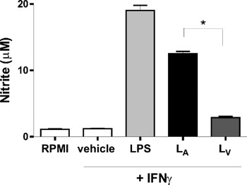
Considering that B. bovis lipids are relevant antigens (Goodger et al. Reference Goodger, Commins, Waltisbuhl, Wright and Rode-Bramanis1990; Shoda et al. Reference Shoda, Palmer, Florin-Christensen, Florin-Christensen, Godson and Brown2000) that can be recognized by innate immune receptors such as TLR2 (Gimenez et al. Reference Gimenez, Magalhaes, Belaunzarán, Poncini, Lammel, González Cappa, Bozza and Isola2010), we here analysed the production of NO, a macrophage secretory product with inhibitory effect in in vitro growth of intracellular B. bovis (Montealegre et al. Reference Montealegre, Levy, Ristic and James1985; Shoda et al. Reference Shoda, Palmer, Florin-Christensen, Florin-Christensen, Godson and Brown2000). First, we assessed the induction of NO in bovine macrophages stimulated with the lipid extracts of B. bovis merozoites from attenuated R1A (LA) or virulent S2P (LV) strains, in the presence of IFNγ. Even though the lipids from both strains induced significant levels of NO compared to control cells, LA induced 4-fold higher levels than LV, indicating that the lipids of the attenuated strain generated a stronger inflammatory response (Fig. 1).
LA induced a higher release of NO than LV. Results are presented as the mean±s.e.m. of triplicate determinations and are representative of 3 independent experiments performed with different LA or LV batches and macrophages from different cattle. *Statistically significant for macrophages stimulated with LA compared to LV (P < 0·05). Lipids from both strains induced significant levels of nitrite compared to control cells (P < 0·05). LA: total lipids of R1A Babesia bovis attenuated strain; LV: total lipids of S2P B. bovis virulent strain.

LA produced an earlier/stronger induction of inflammatory cytokine mRNA in bovine macrophages compared to LV
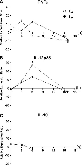
Cytokines IL-12 and TNFα, produced by macrophages and other antigen-presenting cells, are critical for generating and regulating innate and acquired immune responses against many pathogens (Trinchieri, Reference Trinchieri1995). On the other hand, IL-10 has been shown to suppress a broad range of inflammatory responses (Sabat et al. Reference Sabat, Grütz, Warszawska, Kirsch, Witte, Wolk and Geginat2010). Herein, we analysed the induction of TNFα, IL-12 and IL-10 by real-time qPCR in bovine macrophages stimulated with LA or LV at different times. Control cells did not display variations in cytokine mRNA levels in the times assayed (data not shown) and all results were presented as the relative expression ratio of each target gene which considers the response of the sample versus control. Figure 2A shows that lipids from both strains induced a TNFα mRNA peak of similar intensity but LV-stimulated macrophages reached the peak 3 h later than those treated with LA. As regards IL-12p35, LA and LV induced significant levels of this cytokine mRNA at 3 h post-stimulation, reaching their maximum levels at 6 h post-stimulation (Fig. 2B). Noteworthy, at this time the expression ratio of IL-12p35 mRNA was 2-fold higher in macrophages stimulated with LA with respect to those stimulated with LV (Fig. 2B). As concerns IL-10, neither LA nor LV induced significant mRNA levels in the times assayed (Fig. 2C). Collectively, these results indicate that lipids of the virulent strain induced a reduced/retarded inflammatory cytokine response with respect to that induced by lipids of the attenuated strain.
LA produced an earlier/stronger induction of inflammatory cytokine mRNA compared to LV. Results are expressed as the relative expression ratio of each cytokine of each sample versus control and are representative of 3 independent experiments performed with different LA or LV batches and macrophages from different cattle. *Statistically significant for IL-12p35 mRNA expressed in macrophages at 6 h post-LA stimulation compared to LV (P < 0·005). LA: total lipids of R1A Babesia bovis attenuated strain; LV: total lipids of S2P B. bovis virulent strain.

LA and LV induced a differential activation of p38 and ERK1/2 MAPKs and NF-κB signalling pathways in bovine macrophages
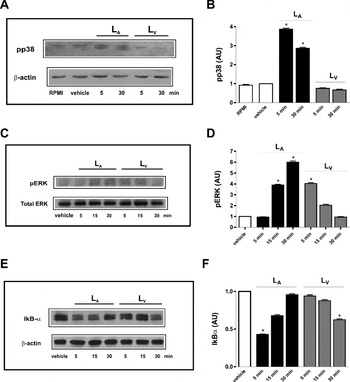
Immunoblot analysis showed that LA induced a transient phosphorylation of p38 (pp38) with a maximum at 5 min, decreasing at 30 min post-stimulation compared with control cells; in contrast, cells stimulated with LV did not display phosphorylation signals (Fig. 3A). No changes were observed at the different times assayed in control cells stimulated with IFNγ + vehicle (basal phosphorylation pattern) (data not shown). The densitometry analysis of these results is shown in Fig. 3B.
LA and LV induced a differential activation of p38 and ERK1/2 MAPKs and NF-κB signalling pathways. Immunoblot analysis of (A) p38 phosphorylation, (B) ERK1/2 phosphorylation, (C) IκB degradation, was performed. Blots were re-probed for β-actin or total ERK detection, as the loading control. Cells incubated overnight with IFNγ + vehicle (IFNγ) show the basal phosphorylation profile (control). The blots are representative of 3 independent assays with different cattle. The intensity of the signals of (D) pp38, (E) ERK and (F) IκB were quantified by densitometry using Gel-Pro® analyser 4·0 software, normalized to the intensity of the corresponding β-actin or total ERK bands and expressed as arbitrary units (AU) with respect to control cells. The values are the mean±s.d. of duplicate determinations and are representative of 3 independent experiments performed with different LA or LV batches and macrophages from different cattle. *Statistically significant with respect to control cells (P < 0·05). LA: total lipids of R1A Babesia bovis attenuated strain; LV: total lipids of S2P B. bovis virulent strain; pp38: phospho-p38; pERK: phospho-ERK.

In the case of ERK1/2, LA induced an increasing and sustained phosphorylation with the maximum levels at 30 min post-stimulation (Fig. 3C). At variance, macrophages stimulated with LV rapidly reached (5 min) the maximum levels of pERK but with lower values than those obtained with LA, recovering the basal levels at 30 min (Fig. 3C). No changes were observed in control cells (basal phosphorylation pattern) at the different times assayed (data not shown). The densitometry analysis of these results is shown in Fig. 3D.
NF-κB is an inducible transcription factor whose activation is regulated by the cytoplasmatic inhibitor IκB; herein we evaluated the degradation of IκB as a hallmark of the activation of the NF-κB pathway (Pahlevan et al. Reference Pahlevan, Wright, Andrews, George, Small and Foxwell1999; Chen et al. Reference Chen, Sun, Chen and Chiu2000; Eligini et al. Reference Eligini, Brambilla, Banfi, Camera, Sironi, Barbieri, Auwerx, Tremoli and Colli2002). Macrophages stimulated with LA showed at 5 min an important decrease of IκB compared to control cells, recovering the basal levels at 30 min post-stimulation (Fig. 3E); whereas, in cells stimulated with LV a significant degradation of IκB was observed but at 30 min post-stimulation (Fig. 3E). No changes were observed in IκB levels at the different times assayed in control cells (data not shown). The densitometry analysis of these results is shown in Fig. 3F. Taken together, these results indicate that LA lipids promote a rapid/strong activation of p38, ERK1/2 and NF-κB pathways in contrast to LV that generated a delayed/reduced stimulation of these signalling cascades.
LV produced a strong inhibition of bovine PBMC proliferation
Having demonstrated that LA and LV were able to induce different bovine macrophage activation patterns, we studied the effect of both lipid extracts on PBMC proliferation as an early indicator of lymphocyte activation. As shown in Fig. 4A, both LA and LV inhibited ConA-induced PBMC proliferation in a dose-dependent manner. Interestingly, this effect was significantly higher in cells stimulated with LV with respect to those stimulated with LA, indicating that the former might have a stronger inhibitory effect over lymphocyte response. Next, we analysed the effect of the lipid fractions: SM + LPC, PC, PS + PI, PE, PA, CHO + DG and FFA from R1A and S2P strains, at 2 concentrations (0·5 or 5 μg/ml). With respect to R1A lipid fractions, only FFA at the higher concentration produced a significant inhibitory effect in Con A-induced PBMC proliferation (Fig. 4B). In the case of S2P lipid fractions, most of them exerted an inhibitory effect, except for SM + LPC and PA in both concentrations and PC in the lower concentration (Fig. 4C). These results are in accordance with those previously obtained with the whole lipid extracts (LA and LV), corroborating that the marked inhibitory effect of LV in the lymphocyte proliferative response is a consequence of the individual inhibitory activities that the majority of the lipid fractions exert.
S2P lipids produced a higher inhibitory effect in ConA-induced PBMC proliferation compared R1A lipids. Bovine PBMC were stimulated for 48 h with ConA alone and in the presence of: (A) LA or LV (0·5, 5 or 50 μg/ml), (B) lipid fractions of R1A (0·5 or 5 μg/ml) or (C) lipid fractions of S2P (0·5 or 5 μg/ml). Control cells were incubated with culture medium (C) or vehicle. The values are the mean±s.e.m. of triplicate determinations and are representative of 3 independent experiments performed with different LA or LV batches and PBMC from different cattle. The results obtained with LA, LV or the different lipid fractions were statistically significant with respect to those obtained with ConA alone (*P <0·05, **P <0·001). LA: total lipids of R1A B. bovis attenuated strain; LV: total lipids of S2P Babesia bovis virulent strain; ConA: Concanavalin A, SM: sphingomyelin, LPC: lysophosphatidylcholine, PC: phosphatidylcholine, PS: phosphatidylserine, PI: phosphatidylinositol, PE: phosphatidylethanolamine, PA: phosphatidic acid, CHO: cholesterol, DG: diacylglycerol, FFA: free fatty acids.

Anti-phospholipid antibodies increased in bovines infected with R1A or S2P strains
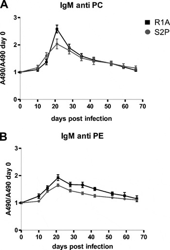
Previously, we determined in non-infected bovines the presence of natural IgM anti-PE and anti-PC antibodies (approximately 5-fold higher levels against PE than PC) and no detectable activity of anti-SM antibodies (Gimenez et al. Reference Gimenez, Florin-Christensen, Belaunzarán, Isola, Suarez and Florin-Christensen2007). Herein, we analysed anti-phospholipid antibody levels during the course of R1A or S2P B. bovis infection. For R1A the parasitaemia peak (approx. 0·02%) appeared at 7–8 days p.i. and lasted 1–2 days and for S2P, appeared at 8–10 days p.i. (approx. 2%) and lasted for more than 3 days (data not shown). Figure 5 shows that the increase in anti-phospholipids antibodies against PC and PE (IgM isotype) was significantly higher in animals infected with each B. bovis strain with respect to their corresponding basal levels (0 days p.i.), reaching a peak at 21 days p.i. At 21 days p.i., the R1A strain produced a significantly higher increase in IgM anti-PC levels than that of IgM anti-PE, whereas S2P generated a similar increase for both antibodies. In addition, bovines infected with the R1A strain developed at 21 days p.i. a significantly higher increase in IgM anti-PC levels than those inoculated with S2P strain (Fig. 5A), whereas IgM anti-PE did not show significant differences between animals inoculated with each strain (Fig. 5B). No IgM anti-SM antibodies were detected, as expected (data not shown). Regarding the IgG isotype, no reactivity was demonstrated against the phospholipids here studied (data not shown).
Anti-phospholipid antibodies in Babesia bovis-infected bovines. IgM anti-PC (A) or anti-PE (B) antibodies in bovines infected with R1A-attenuated (black square) or S2P virulent (grey circle) B. bovis strains were detected by ELISA. The graphics show the ratio between absorbance at 490 nm (A490) of each day post-inoculation (days p.i.) and 0 days p.i. Results represent the mean±s.e.m. of triplicate measurements of 3 independent animals for each strain. At 21 days p.i., the values for the IgM anti-PC increase in animals inoculated with R1A were significantly higher with respect to those obtained in animals inoculated with S2P (P < 0·03). On the same day, the values for the IgM anti-PC increase were significantly higher than IgM anti-PE in animals inoculated with R1A (P < 0·02). PC: phosphatidylcholine, PE: phosphatidylethanolamine.

DISCUSSION
Macrophages, crucial cells of innate immunity, can inhibit early Babesia bovis replication in acute infection through the generation of soluble factors such as cytokines and inflammatory mediators (Brown et al. Reference Brown, Norimine, Knowles and Goff2006). This is the first study that demonstrates in bovine macrophages the induction of NO by lipids purified from merozoites of 2 B. bovis strains with polar behaviour: attenuated R1A or virulent S2P (LA or LV). It has been reported that NO has a relevant role in B. bovis growth inhibition (Johnson et al. Reference Johnson, Cluff, Goff and Wyatt1996; Shoda et al. Reference Shoda, Palmer, Florin-Christensen, Florin-Christensen, Godson and Brown2000), therefore the present finding that, in the early stage of parasite lipids-host cell interaction LA induced the highest levels of NO, could be related to the attenuated virulence of R1A (vaccinal strain). At variance, the low NO induction by LV could contribute to the establishment of this virulent strain in host cells. However, in long-term infections, the increase in the concentration of inflammatory mediators may correlate with the increase in the number and severity of clinical manifestations such as anaemia, coagulopathy, respiratory distress syndrome, encephalopathy and even death (Krause et al. Reference Krause, Daily, Telford, Vannier, Lantos and Spielman2007).
Regarding cytokine induction, LV generated in bovine macrophages lower levels of IL-12p35 (Th1 polarizing cytokine) and a delayed induction of the inflammatory cytokine TNFα compared to LA. This could correlate with the virulent behaviour of S2P through the generation of a lower inflammatory response and consequent evasion of host immune mechanisms. These results are in line with those previously described in murine peritoneal macrophages, another experimental model (Gimenez et al. Reference Gimenez, Magalhaes, Belaunzarán, Poncini, Lammel, González Cappa, Bozza and Isola2010). Other authors have reported that lipids from erythrocytes infected with B. bovis Mexico strain did not induce TNFα or IL-12p35 or p40 by bovine macrophages (Shoda et al. Reference Shoda, Palmer, Florin-Christensen, Florin-Christensen, Godson and Brown2000). These differences could be mainly attributed to the fact that they used a lipid mixture of erythrocytes plus B. bovis merozoites, a different strain and lipid quantities, as well as to variations in the kinetics of cytokine induction and determination of cytokine mRNA. On the other hand, the lipid structure and the relative quantity of each lipid in the total extracts of the B. bovis strains used, might account for the differential bovine macrophage activation here observed. In this respect, it has been demonstrated that lipoarabinomannans isolated from non-pathogenic mycobacterias stimulated a stronger cytokine response in macrophages, compared to those from pathogenic mycobacterias, demonstrating that slight variations in these molecules have a relevant impact in immunogenicity (Roach et al. Reference Roach, Barton, Chatterjee and Blackwell1993; Riedel and Kaufmann, Reference Riedel and Kaufmann2000). Concerning IL-10, neither LA nor LV induced this anti-inflammatory cytokine in bovine macrophages. In the case of LA, this was in accordance with the pro-inflammatory profile here described for this lipid extract, whereas the results obtained with LV were unexpected. In this sense, similar results have been reported for a lipid fraction of a pathogenic strain of M. ulcerans that inhibits the release of anti-inflammatory IL-10 as well as pro-inflammatory TNFα in human monocytes (Pahlevan et al. Reference Pahlevan, Wright, Andrews, George, Small and Foxwell1999).
Mitogen activated-protein kinases (MAPK) including ERK1/2 and p38, and the NF-κB pathways are important cellular targets of diverse pathogens (Ruckdeschel et al. Reference Ruckdeschel, Machold, Roggenkamp, Schubert, Pierre, Zumbihl, Liautard, Heesemann and Rouot1997; Feng et al. Reference Feng, Goodridge, Harnett, Wei, Nikolaev, Higson and Liew1999; Reiling et al. Reference Reiling, Blumenthal, Flad, Ernst and Ehlers2001; Roach and Schorey, Reference Roach and Schorey2002; Surewicz et al. Reference Surewicz, Aung, Kanost, Jones, Hejal and Toossi2004; Yadav et al. Reference Yadav, Roach and Schorey2004; Souza et al. Reference Souza, Evanson and Weiss2007; Hayden and Ghosh, Reference Hayden and Ghosh2008). The inhibition of the p38 pathway and the transient and lower activation of ERK 1/2 cascades by LV compared to LA here observed, are in agreement with the results described above. In addition, other studies have reported decreased MAPK activity only for pathogenic mycobacteria-infected cells (Roach and Schorey, Reference Roach and Schorey2002). Moreover, the high ERK1/2 activation induced by LA is in accordance with the high release of TNFα since it has been reported that this signalling pathway is involved in the expression of this cytokine in human and murine macrophages stimulated with different pathogens or derived molecules (Ruckdeschel et al. Reference Ruckdeschel, Machold, Roggenkamp, Schubert, Pierre, Zumbihl, Liautard, Heesemann and Rouot1997; Surewicz et al. Reference Surewicz, Aung, Kanost, Jones, Hejal and Toossi2004; Gimenez et al. Reference Gimenez, Magalhaes, Belaunzarán, Poncini, Lammel, González Cappa, Bozza and Isola2010). As TNFα is one of the target genes regulated by NF-κB and also an activator of this signalling pathway (Verma et al. Reference Verma, Stevenson, Schwarz, Van Antwerp and Miyamoto1995), the reduced and delayed activation of NF-κB cascade produced by LV with respect to LA, is congruent with the retarded induction of TNFα detected in bovine macrophages stimulated by LV. Altogether, the distinct activation patterns of these cascades induced by the lipids of each B. bovis strain could account for the contrasting effects observed in the inflammatory response and consequently with the outcome of infection.
As regards bovine PBMC proliferation assays, our results indicated that B. bovis lipids could modulate the LT response in a dose-dependent manner, being the lipids of the virulent S2P strain (LV) able to exert a higher inhibitory effect in the adaptive immune response with respect to LA. Accordingly, it has been reported that a lipid fraction from a pathogenic strain of M. ulcerans, but not from a non-pathogenic strain, had an inhibitory effect in the proliferation of human PBMC/LT (Pahlevan et al. Reference Pahlevan, Wright, Andrews, George, Small and Foxwell1999). In addition, M. leprae and M. tuberculosis lipoarabinomannans suppress in vitro proliferation of LT from lepromatous patients in response to mitogens and antigens (Kaplan et al. Reference Kaplan, Gandhi, Weinstein, Levis, Patarroyo, Brennan and Cohn1987). In fact, we also observed different responses between the lipid fractions from LA and LV that could be attributed to qualitative differences in the structure of bioactive molecules, since they were tested in the same concentrations.
Finally, the significant increase in IgM anti-PC antibodies detected in bovines infected with R1A or S2P B. bovis strains with respect to non-infected animals could have resulted from the high rate of PC synthesis in B. bovis merozoites (Florin-Christensen et al. Reference Florin-Christensen, Suarez, Florin-Christensen, Hines, McElwain and Palmer2000) and the recognition of this antigenic molecule by the immune system. The finding that R1A generated a higher increase in anti-PC antibody response than S2P, despite having the former lower and short-term parasitaemias than the latter, might contribute in the better control of infection that R1A establishes with less or null clinical manifestations. In this sense, it has been described that the pre-immunization of bovines with a lipid extract of B. bovis-infected erythrocytes, developed lower and delayed parasitaemias after the challenge with a virulent strain of the same parasite (Goodger et al. Reference Goodger, Commins, Waltisbuhl, Wright and Rode-Bramanis1990). As we only detected IgM anti-phospholipid antibodies that decreased with time, we propose that they could partially restrict the early phase of B. bovis infection. On the other hand, despite the fact that animals inoculated with S2P displayed higher parasitaemias than R1A-infected animals, the lower humoral response here observed could be attributed to the higher antigen offer, thus interfering with the efficiency of this immune response, as reported for malaria (Hisaeda et al. Reference Hisaeda, Yasutomo and Himeno2005; Wykes et al. Reference Wykes, Keighley, Pinzon-Charry and Good2007).
In summary, the strong inflammatory response generated by lipids from attenuated R1A strain could contribute to limit B. bovis acute infection and promote host survival, whereas the reduced inflammatory response induced by lipids from virulent S2P might collaborate in an early stage to evade the innate immune system, thus allowing B. bovis establishment in the host. Further studies should be performed using a larger set of strains of polar phenotypes in order to confirm and generalize the conclusions of this study. Despite the discovery of B. bovis over a century ago, there are still no safe and effective vaccines that protect cattle against this pathogen, therefore the ability of R1A lipids to stimulate protective innate immune responses here described, opens up the possibility that they might be a key factor in designing vaccine adjuvants for bovine babesiosis prophylaxis.
ACKNOWLEDGEMENTS
We thank Drs Silvina Wilkowsky for helpful discussions and Fabiana Bigi for allowing the performance of qPCR assays in her laboratory (INTA-Castelar, Argentina).
FINANCIAL SUPPORT
This work was supported by a grant from Agencia Nacional de Promoción Científica y Tecnológica (FONCYT) (PICT 08-12074).