Introduction
Coblation (also known as electrodissociation) and flexible fibre CO2 laser are novel technologies with potential uses in functional endoscopic sinus surgery (FESS) and endoscopic skull base surgery. Temperature-limited tissue ablation with minimisation of collateral thermal injury has been advocated as the key advantage of coblation.Reference Chinpairoj, Feldman, Saunders and Thaler1 Recent studies on healing of coblation wounds in rabbit and sheep sinus mucosa indicate appropriate mucosal healing after coblation injury.Reference Swibel Rosenthal, Benninger, Stone and Zacharek2, Reference Swibel-Rosenthal, Benninger, Stone and Zacharek3 Importantly, longer duration of coblation application does not result in deeper injury. In tandem with studies on safety, the use of coblation in FESS and endoscopic skull base surgery has been increasing, with consistent reports of low intra-operative blood loss, and one report of shorter operative time in reducing encephalocoeles compared with bipolar cautery.Reference Kostrzewa, Sunde, Riley and Woodworth4–Reference Smith, Riley and Woodworth7
There are fewer studies on the use of CO2 laser in the sinonasal cavity, as flexible fibre lasers and hand-pieces (which facilitate endonasal manoeuvrability, improving ease of delivery) have been introduced only recently.Reference Jayarao, Devaiah and Chin8, Reference Agarwal, Kupferman, Holsinger and Hanna9 However, precise cutting and coagulation have been reported, especially when creating a transsphenoidal window for sellar lesions.Reference Jayarao, Devaiah and Chin8
Patterns of thermal injury differ between the two devices. In animal studies of coblation, lesions applied to the lamina papyracea were not associated with any histological changes in the adjacent orbit.Reference Swibel Rosenthal, Benninger, Stone and Zacharek2, Reference Swibel-Rosenthal, Benninger, Stone and Zacharek3 The ethmoid skull base is another site of extremely thin bone at risk of inadvertent intracranial thermal injury. There are currently no reports on associated dural changes following either coblation or CO2 laser use at the ethmoid skull base.
The aim of this study was to describe the depth of injury resulting from coblation and CO2 laser use at the ethmoid skull base in fresh cadaveric specimens.
Materials and methods
The study was performed on fresh frozen cadaveric heads. In each specimen side, wide sphenoidotomy, removal of all ethmoid partitions and Draf 2a frontal sinusotomy was performed so as to obtain an unobstructed view of the entire medial orbital wall and ethmoid roof. Mucosal removal was performed over an area bounded by the anterior ethmoidal artery anteriorly, attachments of the middle and superior turbinates medially, junction of the lamina papyracea and the fovea ethmoidalis laterally, and the plane of the face of the sphenoid posteriorly.
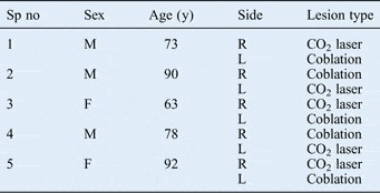
In each cadaveric head, the mucosa removal described above was performed using laser on one side and coblation on the other. While method and side allocation was not randomised, pairing of the mucosa and skull base was performed to minimise the effect of intrinsic differences for each specimen side. The characteristics of the cadavers in each group are shown in Table I.
Specimen and lesion characteristics

Sp no = specimen number; y = years; M = male; R = right; L = left; F = female
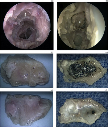
Each lesion was created using either CO2 laser (AcuPulse WaveGuide CO2 Laser; Lumenis, Santa Clara, California, USA) or coblation (Coblator PROcise XP Plasma Wand; Arthrocare, Austin, Texas, USA). Carbon dioxide laser was delivered using the AcuPulse WaveGuide laser fibre system (super pulse; repeat mode; 4.0 W; spot size, 320 µm; time on, 0.20 seconds; time off, 0.10 seconds). A power of 4.0 W was chosen as this was the lower end of the power range used at the pituitary sella in Jayarao and colleagues' report.Reference Jayarao, Devaiah and Chin8 For the Coblator, the standard settings (i.e. ablation, 7; coagulation, 3) were used (Figure 1). The CO2 laser or Coblator was applied within the defined anatomical limits until no further mucosa was observed endoscopically.
Endoscopic views and pathological photographs presenting the application of coblation and flexible fibre CO2 laser to the ethmoid skull base, showing: (a) mucosal ablation after coblation (arrow); (b) precise area of charring due to laser application (asterisk); (c) & (d) inferior surface of the ethmoid skull base after coblation and laser application, respectively; and (e) & (f) superior surface of the ethmoid skull base after coblation and laser application, respectively.

For coblation specimens, the ethmoid roof mucosa changed colour to grey-white as coblation was applied with light, brushing strokes. At the end-point of application (i.e. no further change noted), complete mucosal removal was achieved (Figure 1a and 1c).
The CO2 laser fibre delivery system was able to deliver point lesions with submillimetre precision, using the HeNe aiming beam. Each lesion resulted in gradual charring with shrinkage of the tissue, accompanied by a small plume. At the end-point of application, there was no observable change to the remaining layer of char on the ethmoid roof (Figure 1b and 1d).
After the lesions were created, each ethmoid roof was resected by dissecting around the perimeter of the ethmoid skull base, using a combination of cutting endoscopic instruments and a 4 mm diamond burr where required. Dural cuts were made with endoscopic scissors.
High definition digital photographs of the sinus and dural aspects of each dissected specimen side were obtained. The photographs, under ×10 magnification, were presented in a randomised fashion to an assessor (the second author) who was blinded to the dissection technology used. The assessor was asked to determine whether macroscopically visible thermal injury was present or absent.
Histological assessment of the specimens was not performed, as the aim of this study was to ascertain macroscopic changes. Furthermore, any microscopic changes would have been difficult to interpret in a fresh cadaveric specimen.
Results
Ten cadaveric head specimen sides were obtained; five received CO2 laser and five coblation. Both the Coblator PROcise XP and the AcuPulse WaveGuide were easily manoeuvred within the sinonasal cavity under endoscopic vision.
On macroscopic examination, no visible tissue remnants were observed on the inferior surface of any coblation specimen side. However, all five CO2 laser specimens had a thin layer of char tissue on the inferior surface. The dural (superior) surface of all coblation specimen sides appeared normal (Figure 1e), while foci of black discolouration were observed on the dura in two of the five (40 per cent) CO2 laser specimen sides (Figure 1f).
Discussion
Coblation and CO2 fibre laser represent technological innovations with potential applications in FESS and endoscopic skull base surgery. Each offers unique advantages. Coblation technology enables controlled tissue removal within a 40–70°C range, with minimal circumferential and limited deep tissue injury.Reference Chinpairoj, Feldman, Saunders and Thaler1 Combined irrigation and suction enables the surgical bed to be kept clear. Additionally, the integrated bipolar diathermy facilitates haemostasis, reducing the need to change instruments. While the shaft of the Coblator PROcise XP wand is malleable, it is 3.9 mm in width and thus substantially larger than the laser delivery fibres. Additionally, the point of dissection is also 3.9 mm wide, compared with the 0.6 mm beam width of the CO2 fibre laser.
Histological specimens from animal studies have demonstrated that coblation of inferior turbinate mucosa results in immediate loss of respiratory epithelium and thermal injury to underlying seromucinous glands; the depth of injury was dependent on soft tissue type rather than duration of coblation application.Reference Swibel-Rosenthal, Benninger, Stone and Zacharek3 Underlying bone demonstrated reactive, regenerative changes; however, a study of rabbit specimens observed only one case of histological changes beyond the maxillary sinus bony wall, even with prolonged application.Reference Swibel Rosenthal, Benninger, Stone and Zacharek2 Therefore, it is important that the characteristics at each potential site of application are well understood. In the present study of human cadaveric specimens, the macroscopic finding of complete removal of mucosa at the ethmoid roof, along with the lack of dural injury and low working temperature range, suggest that coblation is ideally suited to, and safe for, bulky soft tissue removal at the skull base.
• Coblation enables reduced thermal mucosal injury and less operative blood loss
• Endonasal fibre CO2 laser enables precise, haemostatic dissection
• This study found macroscopic dural injury in 40 per cent of specimens after ethmoid skull base CO2 laser
• No dural injury was seen after coblation
• Coblation may be more suitable for periorbital and peridural surgery
The main advantages of the CO2 laser have been listed as (1) haemostatic dissection of scar tissue, adhesions and tumour capsules, and (2) the ability to precisely dissect lesions from the surrounding soft tissue.Reference Agarwal, Kupferman, Holsinger and Hanna9 Unlike its non-fibre predecessors, the flexible fibre system does not require ‘line of sight’ delivery, and the hand-piece allows the tip to be manipulated like a straight endoscopic instrument. Because CO2 laser is absorbed to a large degree by water within cells, it has a shallow depth of penetration which confers relative safety, compared with other lasers. Submillimetre spot sizes enable lesions to be precisely positioned, minimising circumferential damage around the target. Multiple parameters can be adjusted to vary performance characteristics, in order to reduce collateral thermal injury at the target site. The superpulse mode used in the present study provides alternating laser time on and off and allows regular dissipation of thermal energy between applications. Studies on the use of flexible CO2 laser in FESS are emerging and will provide more information on appropriate applications.
Conclusion
Both coblation and flexible fibre CO2 laser have their advantages in FESS. However, in the present, experimental study of thermal injury sustained during endoscopic skull base surgery using either CO2 laser or coblation, dural injury was observed for CO2 laser but not for coblation. The lower thermal working power of coblation may be more suitable for periorbital and peridural surgery.