Introduction
Kerria chinensis (Mahdihassan) (Hemiptera: Kerriidae) is one of the most economically important beneficial insects of Thailand (Chen et al. Reference Chen, Chen, Feng, He and Yang2011). Female K. chinensis secrete protective substances that are used to produce valuable natural products such as dye, shellac, and wax used in varnishes, textiles, electronics, medicines, and cosmetics (Lagowska and Golan Reference Lagowska and Golan2009). Kerria chinensis are members of the Kerriidae, one of 17 families of Neococcoidea (Cook et al. Reference Cook, Gullan and Trueman2002; Gullan and Cook Reference Gullan and Cook2007; Hardy Reference Hardy2013; Hodgson and Hardy Reference Hodgson and Hardy2013). Kerria chinensis are sexually dimorphic. The adult females are sac-like and immobile, ~1 cm long, whereas the adult males have two forms, either winged or wingless, and are about 1 mm long. After emerging from their cells (cages), the mature males live for only three to four days during which time they seek out females to mate. The full lifespan of male and female K. chinensis is about 60 days and 180 days, respectively (Miller and Kosztarab Reference Miller and Kosztarab1979; Sharma Reference Sharma1991; Morotta Reference Morotta1997).
Kerriidae and Pseudococcidae (Hemiptera) spermatogenesis are known to be quite different from the typical process in insects since their spermatids are obtained via inverted meiosis (Brown Reference Brown1959; Dikshith Reference Dikshith1965; Bongiorni et al. Reference Bongiorni, Fiorenzo, Pippoletti and Prantera2004). In typical meiosis, the chromosome number is reduced at the first meiosis (meiosis I) as homologous chromosomes segregate and at the second meiosis (meiosis II) sister chromatids segregate so the number of chromosomes is equal. By contrast, in inverted meiosis, the first meiosis is equal, sister chromatids segregating, resulting in two identical diploid cells. The reductional division occurs in the second division where homologous chromosomes segregate and four haploid spermatids are obtained. In Kerriidae and Pseudococcidae, instead of producing four haploid spermatids, one spermatid with four haploid nuclei is obtained (Dikshith Reference Dikshith1965; Bongiorni et al. Reference Bongiorni, Fiorenzo, Pippoletti and Prantera2004).
In Neococcoidea, males and females of each species have the same chromosome number. Their chromosomes are holocentric and they have no sex chromosome. To determine sex, one haploid chromosome set in male-fated embryos becomes heterochromatic (Brown Reference Brown1959; Jaipuriar et al. Reference Jaipuriar, Teotia, Lakhotia and Chauhan1984; Khosla et al. Reference Khosla, Mendiratta and Brahmachari2006). Studies in Pseudococcidae revealed that the heterochromatic set is the paternally derived chromosome set (Brown and Nelson-Rees Reference Brown and Nelson-Rees1961). During spermatogenesis in the second meiosis, the paternal chromosome set is separated from the maternal set and then becomes heterochromatic. The spermatids with the paternal chromosome set then degenerate; only the spermatids with the maternal chromosome set differentiate to be functional spermatozoa (Brown Reference Brown1959; Brown and Nur Reference Brown and Nur1964; Prantera and Bongiorni Reference Prantera and Bongiorni2012). This became known as the lecanoid system of paternal genome elimination (Ross et al. Reference Ross, Pen and Shuker2010). In Kerria lacca (Kerr), spermatogenesis was shown to follow a pattern typical of the lecanoid system, where the paternal genome set is lost during spermatogenesis. Each primary spermatocyte cyst is composed of 16 primary spermatocytes that divide to produce spermatids via inverted meiosis. In meiosis II, cytokinesis is suppressed so two binucleated spermatids result. Later, the two binucleated spermatids fuse together to form a quadrinucleated spermatid with two heterochromatic and two euchromatic nuclei and only two spermatozoa develop from a spermatid that has two euchromatic nuclei (Jaipuriar et al. Reference Jaipuriar, Teotia, Lakhotia and Chauhan1984). Thus, each spermatid cyst produces 32 spermatozoa that are divided into two bundles with 16 spermatozoa per bundle (Dikshith Reference Dikshith1965).
The lecanoid system of spermatogenesis also produces unusual sperm morphology. In most insects, a typical spermatozoon possesses an acrosome, a nucleus, mitochondrial derivatives and an axoneme within a long flagellum (Baccetti Reference Baccetti1972; Jamieson et al. Reference Jamieson, Dallai and Afzelius1999). In Neococcoidea, the spermatozoa are filamentous entities that lack the mitochondria, the axoneme and an acrosome (Robinson Reference Robinson1966, Reference Robinson1972; Ross and Robinson Reference Ross and Robinson1969; Dallai et al. Reference Dallai, Lupetti and Mencarelli2006, Reference Dallai, Gottardo and Beutel2016; Paoli et al. Reference Paoli, Roversi, Mercati, Marziali, Cocco and Dallai2015). The spermatozoa are arranged in bundles that resemble the individual spermatozoa of most insects, initially leading to the misunderstanding that sperm bundles were in fact spermatozoa (Dikshith Reference Dikshith1965). A light microscope study of the morphology of K. lacca sperm revealed the bundled arrangement, with each bundle being composed of 16 spermatozoa that intertwine in a helical pattern within a membrane that shows a corkscrew-like head region (Dikshith Reference Dikshith1965).
In Kerriidae, spermatogenesis has been studied only in the species, K. lacca. Since that time, no further comprehensive light microscopic or ultrastructural study has been carried out on sperm morphology in lac insects. In this study, we describe morphology of the testes, sperm bundles sperm and spermiogenesis of the lac insect, K. chinensis, using light and electron microscopy. The knowledge gained from examination of K. chinensis spermatozoa, as a representative of the family Kerriidae, may be useful for ongoing research on taxonomy, phylogeny and reproductive biology of the scale insects.
Materials and methods
Insect samples
Kerria chinensis were obtained from Sukhothai Province Forest Products Research Station, Royal Forest Department, Sukhothai Province, Thailand during February to April, 2011–2013. The insects are commonly found on twigs of Samanea saman (Jacquin) Merrill (Fabaceae).
Light microscopy
Mature males (around 60 days old) and late-stage male nymphs (40–55 days old) were dissected in phosphate buffered saline and their testes were removed and observed using a BX41 Olympus light microscope (Olympus, Tokyo, Japan). Photomicrographs were taken using an Olympus DP72 digital camera (Shinjuku-ku, Tokyo, Japan). Some males were fixed in Carnoy’s solution (three parts ethanol: one part acetic acid) overnight then transferred to 70% alcohol and stored at 4 °C until used. The testes were dissected from fixed males and the testicular cells were dissociated in a few drops of 60% acetic acid for 5–10 minutes and spread on slides, then placed on a slide warmer at 50 °C and kept at room temperature for further staining. Some slides were stained with 4% Giemsa (Merck, Darmstadt, Germany) in phosphate buffer, pH 6.8, for 30 minutes then rinsed thoroughly with distilled water and air dried. For silver nitrate staining (Howell and Black Reference Howell and Black1980), two solutions were prepared; one was colloidal developer solution (2 g powder gelatin, 1 mL formic acid in 100 mL distilled water) and the other, silver nitrate solution (4 g AgNO3 dissolved in 8 mL distilled water). For staining, 200 μL of colloidal developer solution was mixed with 400 μL of silver nitrate solution and then dropped on the slides, covered with coverslips and placed on a slide warmer at 70 °C for two minutes. Distilled water was used to remove the coverslips and excess stain. The slides were air-dried and examined. All slides stained with Giemsa and silver nitrate were observed under a BX41 Olympus microscope using an immersion oil objective (100×) and photographed with an Olympus DP72 digital camera. For 4′,6-diamidino-2-phenylindole (DAPI) staining, 200 μL of DAPI solution (1 μg/mL of DAPI in phosphate buffered saline, pH 6.8) was dropped on the slides and cover-slipped. After storage in the dark for 30 minutes, the slides were observed under a Zeiss fluorescence microscope (Obetkochen, Germany) and images were captured using the Isis Fish imaging system (version 5.2, MetaSystems Hard and Software, Altlussheim, Germany).
Electron microscopy
Adult males were fixed with 2% glutaraldehyde at 4 °C overnight and washed with phosphate buffered saline for one hour. After washing, the males were post-fixed in 1% osmium tetroxide for two hours, then washed with phosphate buffered saline for one hour and dehydrated using an ethanol series. The fixed males were then embedded in epon resin before sectioning. Semi-thin sections (350 µm) were stained with 1% toluidine blue and observed with BX41 Olympus microscope. Thin sections (70 nm) were obtained using the Leica EM UC7 ultramicrotome (Leica Microsystem, Wien, Austria) and stained with saturated uranyl acetate in 70% methanol and 0.1% lead citrate and observed on a JEOL 1230 transmission electron microscope (JEOL, Peabody, Massachusetts, United States of America). For scanning electron microscopy, the testes were dissected in Carnoy’s solution and fixed males were mounted, sputter-coated, and examined using an FEI Quanta 450 scanning electron microscope (FEI, Tokyo, Japan).
Results
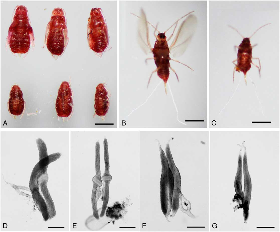
Kerria chinensis has two male forms, one winged and the other wingless. Winged males are larger (Fig. 1A–C) and are the more common form but their testes are smaller than those of wingless males (Fig. 1D–G). In this study, the testes, sperm bundles, spermatozoa, and spermiogenesis in wingless males are described.
Kerria chinensis nymphs and testis morphology. A, Representatives of late-stage nymphs, the upper row developing wings and the lower three wingless. B, An adult winged male. C, An adult wingless male. D, F, Testes of wingless males. E, G, Testes of winged males. D–E, Testes of late-stage nymphs. F–G, Testes of adult males. Scale bars: A–C=500 μm, D–G=200 μm.
Structure of testes and sperm bundles
A mature male lac insect (about 60 days old) has a pair of ovoid testes (~700 μm long and 150 μm diameter). Each testis tapers into a 200 μm long, narrow anterior thread and a shorter, about 60 μm long, thread posteriorly. The vas deferens exits the testes from the central region (Fig. 2A). The mature sperm bundles have a needle-like appearance (Fig. 2B) with a screw-like structure at the head and are approximately 460 μm long and about 2.5 μm in diameter. The screw-like region shows six or seven helical turns from the anterior, tapered end (16 μm long) transitioning into an inflated region with more compact helical turns (40 μm long and 3 μm diameter) (Fig. 2C–E). The wall of the rest of the filamentous bundle is smooth with no apparent helical turns. Posteriorly, the bundle body tapers to a fine thread (Fig. 2I). The heads of living sperm bundles dissected from males at the last nymphal stage and viewed under the light microscope have a smooth appearance (Fig. 2F); however, the helical arrangement was visible after staining with silver nitrate (Fig. 2G) and the anterior region did not stain with Giemsa (Fig. 2H). The sperm bundles stained with DAPI showed brightly stained filamentous regions in the posterior portion next to the centre (Fig. 8).
Testes and sperm bundles of Kerria chinensis. A, Paired ovoid testes. The vas deferens connects with the central widest portion of each testis (arrow). B, Needle-like sperm bundles. C–E, Screw-like heads of mature sperm bundles. C, The head of a single bundle (unstained). D, Scanning electron micrograph of a single bundle head showing the helical turns of the bundle membrane. E, Silver nitrate-stained bundle head. F–H, Heads of immature sperm bundles. F, The bundle membrane has not developed the helical turns seen in adult bundles (unstained). G, Silver nitrate staining of a nymphal bundle showing the developing gyres. H, Geimsa staining of an immature bundle. I, The sperm bundle stained with silver nitrate, the anterior region is on the right and the posterior is on the left. Scale bars: A–B=100 μm, C, E, G–I=10 μm, D=50 μm, H=20 μm.

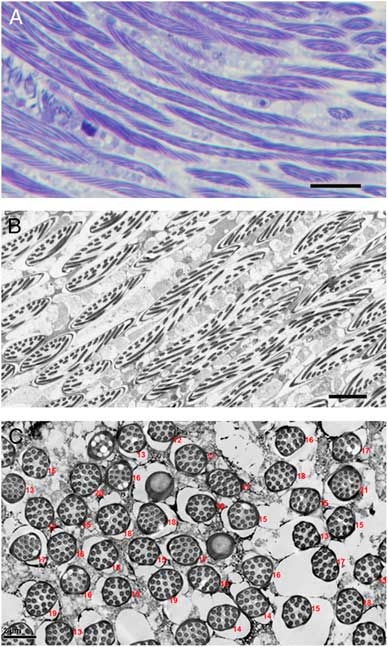
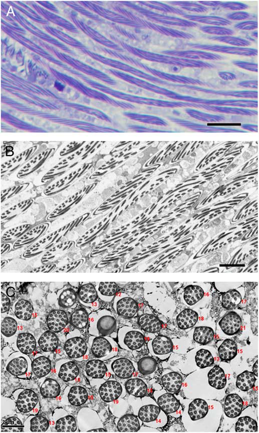
Each sperm bundle contained filamentous spermatozoa arranged in a spiral starting from the base of the apical inflated region and ending approximately 40 μm before the end of the bundle (Figs. 3A–B, 10A). The number of spermatozoa per bundle was counted in 270 bundles; most contain 16 spermatozoa, varying from 12 to 21 (e.g., Fig. 3C).
Configuration and number of spermatozoa in sperm bundles. A, Light microscope section showing spiral arrangement of spermatozoa within bundles. Scale bar=10 μm. B, Transmission electron microscope of a section similarly oriented to A. Scale bar=5 μm. C, Transverse section of sperm bundles show variation in the number of spermatozoa within bundles. Numerals indicate numbers of spermatozoa within bundles. Scale bar=2 μm.
Spermiogenesis
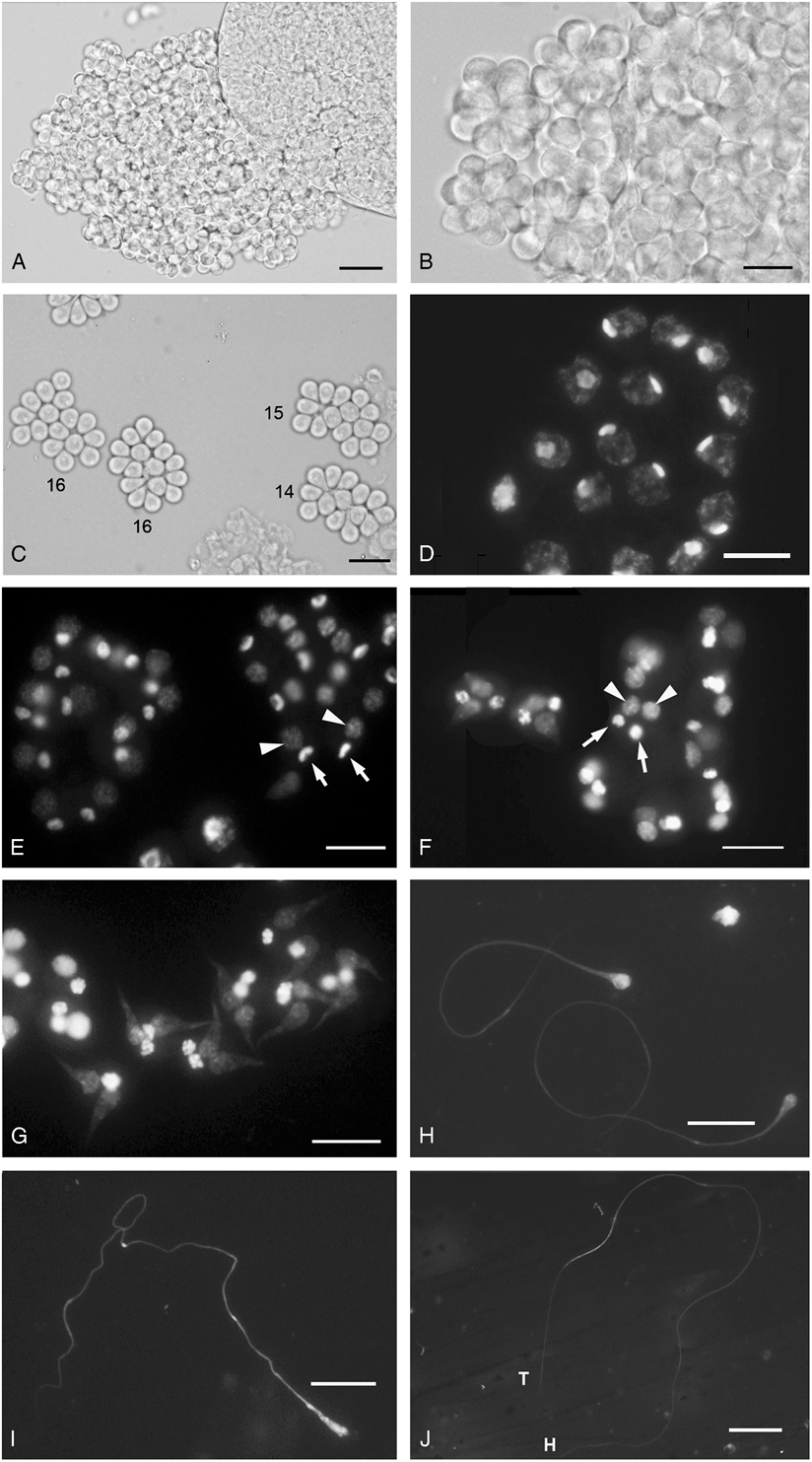
The developing testes dissected from 40-day-old male nymphs contain numerous spermatocyte cysts (Fig. 4A). Most cysts consist of 16 primary spermatocytes loosely tied together to form a rosette-like structure (Fig. 4B) but some had fewer than 16 spermatocytes (Fig. 4C). At the end of meiosis, each primary spermatocyte produces only two binucleate spermatids, rather than four haploid spermatids. Each spermatid contains two nuclei, one euchromatic and the other heterochromatic (Fig. 4D). Later, the two spermatids fuse together, yielding one quadrinucleate spermatid (Fig. 4E), with two heterochromatic and two euchromatic nuclei. Ultimately, each cyst contains 32 heterochromatic and 32 euchromatic nuclei. The euchromatic nuclei are larger in size and lightly stained compared with heterochromatic nuclei. Spermiogenesis initiates with the development of two sperm tails from each spermatid (Fig. 4F). DAPI staining indicated that only the euchromatic nuclei progressively elongated along with the protruding sperm tails until the spermatozoa reached maturity (Fig. 4F–J).
Spermatogenesis of Kerria chinensis. A, Spermatocyte cysts in a developing testis. Scale bar=20 μm. B, Spermatocyte cysts at higher magnification. C, The number of spermatocytes within each cyst is shown for four cysts. D, Primary spermatocytes stained with 4′,6-diamidino-2-phenylindole. E, Binucleate spermatids. Arrows indicate heterochromatic nuclei and arrowheads indicate euchromatic nuclei. F, Quadrinucleate spermatids, each containing two euchromatic (arrowheads) and two heterochromatic nuclei (arrows). G–I, Sperm tails in the process of elongating from euchromatic nuclei. J, Mature sperm. Scale bars: B–J=10 μm.
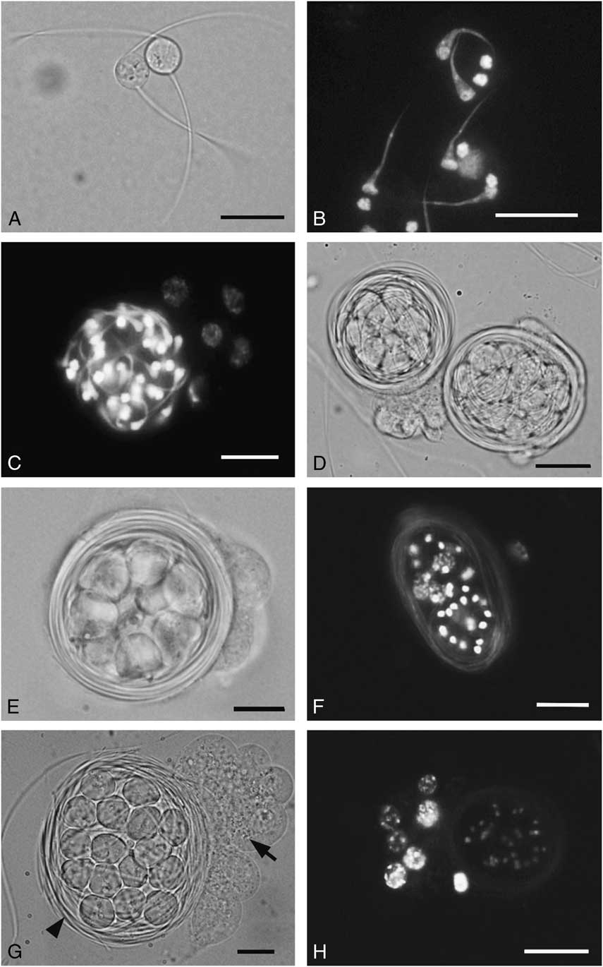
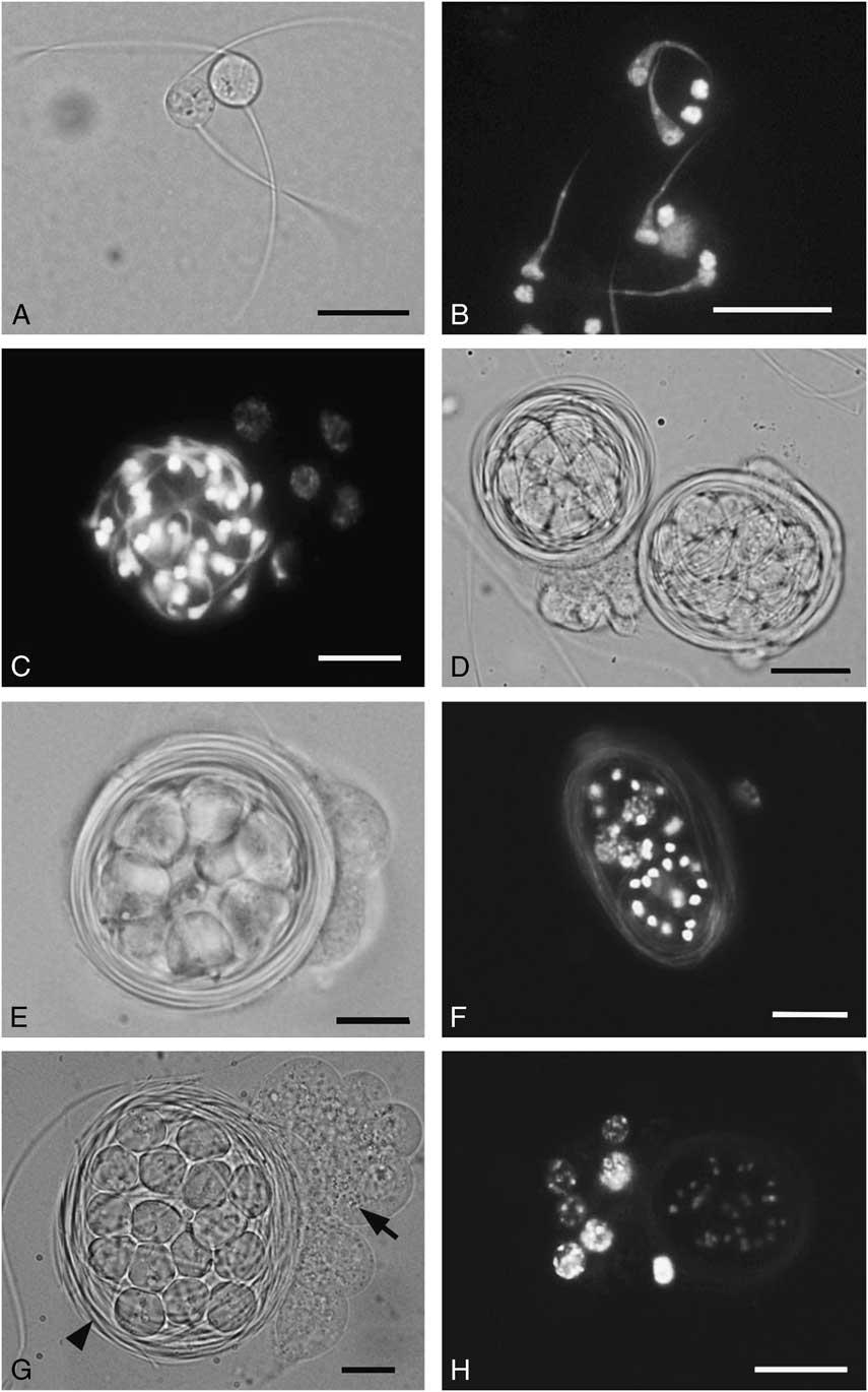
The tails of the two spermatozoa originating from the same spermatid elongate in opposite directions, and wrap around their spermatid cyst, one rotating clockwise and the other counter-clockwise (Fig. 5A). Thus, in each cyst, one group of 16 spermatozoa grow clockwise and the other 16 counter-clockwise. At an early stage, the wrapping of all spermatozoa causes the cyst take on the appearance of a ball of yarn. Spermatozoa then form a ring-like structure, the cyst taking the appearance of a bird nest with spermatid cells in the middle (Fig. 5B–D). The 32 heterochromatin nuclei and cell bodies lie in the middle of the nest (Fig. 5F) and each cyst is surrounded by seven to eight cyst wall cells (Fig. 5E–F).
Sperm development. A, D–E, G, Unstained sperm and spermatid cysts. B–C, F, H, Sperm and spermatid cysts stained with 4′,6-diamidino-2-phenylindole (DAPI). A, Two sperm tails growing from each spermatid in opposite orientations, one clockwise and the other counter-clockwise. B, Two sperm tails elongating from euchromatic nuclei of each spermatid. C, A spermatid cyst containing elongating spermatozoa and heterochromatic nuclei. Four large nuclei of cyst wall cells are located near the cyst. D, The elongating sperm tails enwrap the cyst to create a ball-like structure. E, The sperm tails wrap to produce a ring configuration. F, DAPI staining shows heterochromatic nuclei located centrally within the ring configuration. G, A spermatid cyst containing 16 spermatids surrounded by a ring of sperm tails (arrowhead) with cyst wall cells (arrow) located on one side of the cyst. H, DAPI stain reveals the nuclei of cyst wall cells located to one side of the cyst. Scale bars: A–D=20 μm, E–H=10 μm.
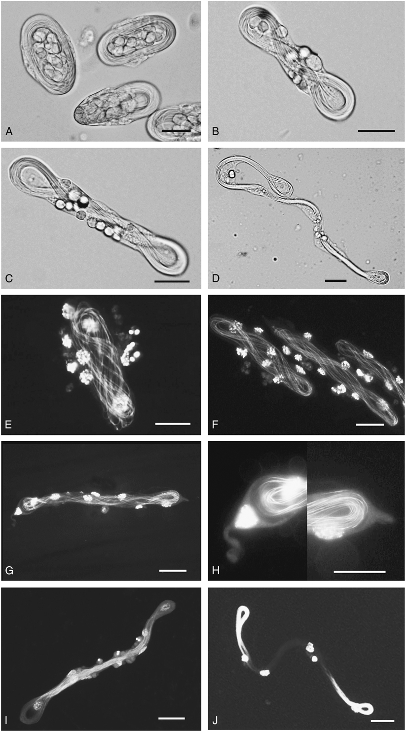
Later, the cyst becomes ovoid in shape and then twists (Fig. 6A–B). While twisting, all sperm heads became detached from their cells and the cyst extends longitudinally, taking on the appearance of a skein (Fig. 6C–D). DAPI staining showed that several twisted spermatozoa are present in a skein (Fig. 6E–F). The skein lies embedded in the cytoplasm sac with the cyst wall cells at either end having a pointed appearance (Fig. 6G–H). The seven to eight nuclei of cyst wall cells attach at intervals along the skein (Fig. 6E–G). The skein then elongates further and nuclear cores are present in the middle of the skein (Fig. 6I). As the skein continues to extend, the nuclear cores move forward to the loops at the opposite end of the skein (Fig. 6J). The intense DAPI stain at either end indicates the location of core nuclei of developing spermatozoa that divide into two groups depending on their wrapping orientation, clockwise or counter-clockwise.
Sperm bundle formation. A, The spermatid cyst in oval configuration. B–D, The twisting and elongating spermatid cysts taking on a skein-like appearance. E–J, Spermatid cysts stained with 4′,6-diamidino-2-phenylindole. E–G, Sperm tails in a cyst and nuclei of cyst wall cells adhering along the sperm tails. H, Higher magnification view of G showing the endpoints of the cyst wall cytoplasm containing a skein of spermatozoa embedded within. I, A skein with nuclear cores located along the central region. J, Elongate skein with two bright nuclear cores located at the opposite loops. Scale bars=20 μm.

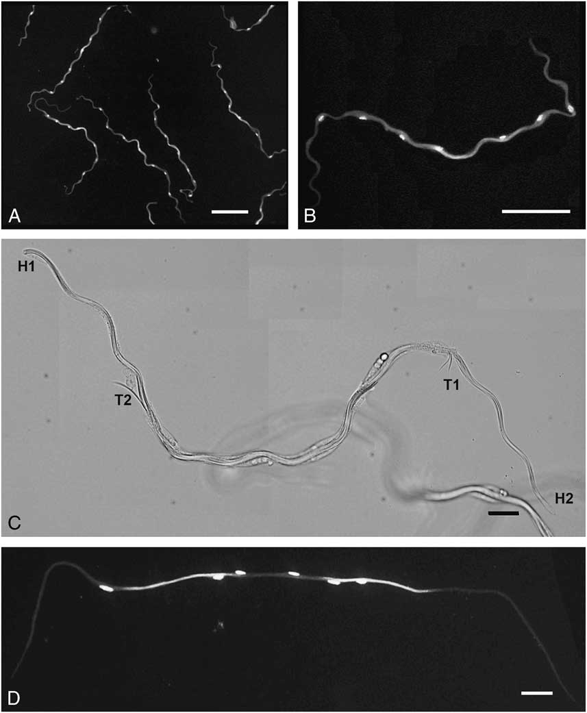
Later, the two loops straighten and develop further into two sperm bundles, each composed of a like-oriented group. At this stage, the bundles have a wavy appearance (Fig. 7A–B) and about three posterior parts of the four parts of their bundles overlap (Fig. 7C). In particular, the nuclear cores of the two bundles lay together (Fig. 7A–B). Moreover, the seven to eight nuclei of cyst wall cells are attached at intervals along the overlapping bundles (Fig. 8A–B). Subsequently, the wavy bundles elongate and become linear. The two overlapping bundles move apart and separate from each other to opposite sides, indicated by the two bright nuclear cores in the overlapping bundles (Fig. 7D). While stretching to complete a linear bundle, all cyst wall cells and other cytoplasm move down to the posterior end and eventually exit the bundle (Fig. 8). Finally, the two bundles separate from each other so the head or the anterior region of the two bundles point to different poles of the testis. Thus, half of the sperm bundles are oriented with their heads to the posterior end while the other half orient to the anterior end of the testis; the posterior ends of both bundles lie in the middle of the testes (Fig. 9). At the final stage of sperm bundle formation, all cyst wall cells and discarded cytoplasm move along the bundles to their posterior ends, producing a swelling in the middle region of the testes (Figs. 1D–E, 9).
Sperm bundle development. A, Wavy bundles stained with 4′,6-diamidino-2-phenylindole. B, A high magnification view of a wavy bundle showing the bright nuclear core in the middle and nuclei of cyst wall cells adhering at intervals along the bundle. C, The unstained wavy bundle consisting of two overlapping sperm bundles; H=head, T=tail. D, Two brightly stained nuclear cores indicating the two overlapping sperm bundles as they straighten and separate from each other. Scale bars: A–B=100 μm, C–D=20 μm.

Sperm bundle development. A–D, Sperm bundles with nuclei of cyst wall cells and cytoplasm gradually moving posteriorly and exiting (arrows). E, The mature sperm bundle. In all, anterior ends are to the left. Scale bars=20 μm.
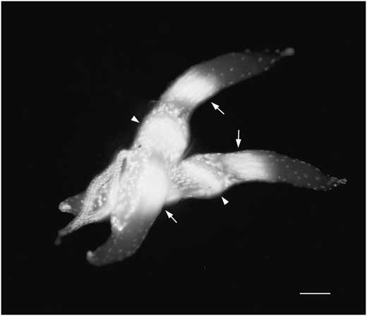
4′,6-diamidino-2-phenylindole-stained testes dissected from Kerria chinensis at the late nymphal stage. Each testis has two bright regions (arrows) that are nuclear cores of two sperm bundle groups orienting their heads toward opposite ends of testes. Arrowheads point at inflated regions in the middle of testes that are the posterior end of sperm bundles with extruding nuclei and cytoplasm of cyst wall cells and other spermatid debris. Scale bar=100 μm.

Sperm morphology
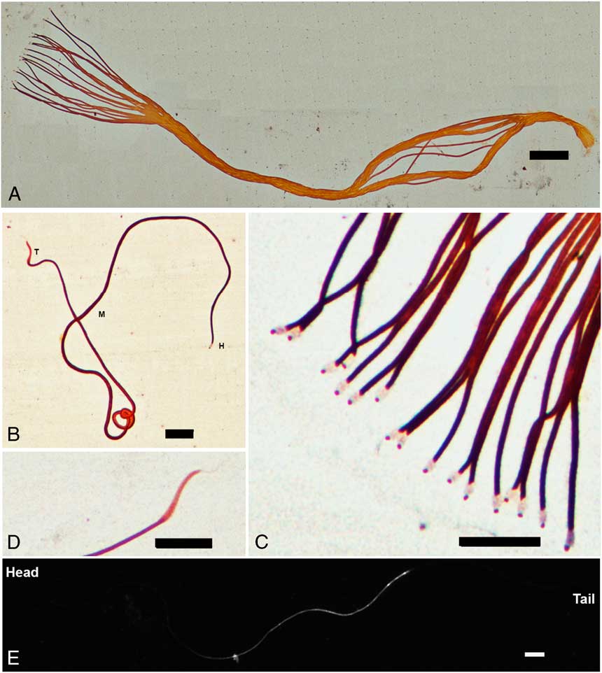
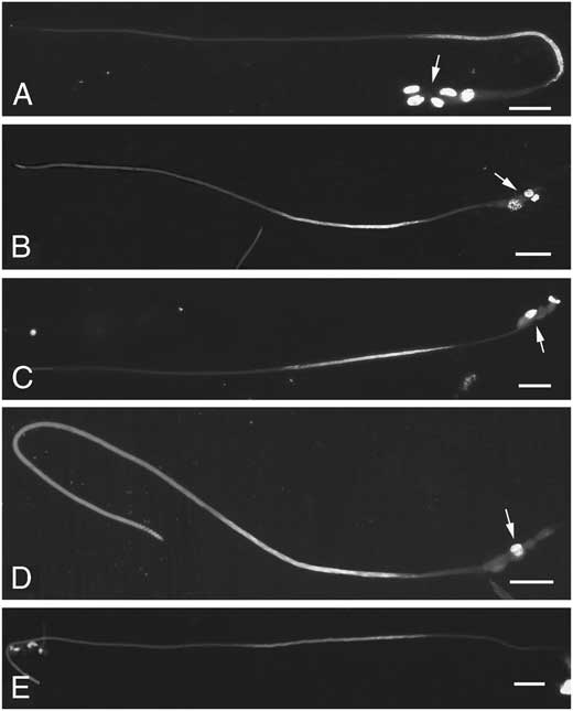
The spermatozoa are filamentous, ~ 0.6 μm in diameter and 420 μm long (Fig. 10A–B). Three main regions can be recognised: the anterior region, the main body, and the tail (Fig. 10D). Upon silver nitrate staining, the apical end of each spermatozoon shows a translucent lantern-like structure, about 2 μm long and 0.75 μm wide, with a small, dark-stained apex (Fig. 10C). From its location, this structure could be the acrosome. Below the pale-staining region, the main body is long and uniformly filamentous (Fig. 10E–F). The tail region, ~17 μm long, stains more lightly than the main body. The beginning of the tail region is slightly wider in diameter than the main body and then tapers into a fine thread (Fig. 10D). Spermatozoa nuclei are located in the third quarter from the head as indicated by DAPI staining (Fig. 10E). In each spermatozoon, the single long thread-like nuclear core was approximately 100 μm long and 0.05 μm in diameter.
Spermatozoa of Kerria chinensis. A, A single bundle showing spiral arrangement of 17 spermatozoa. B, Silver nitrate stains of a single spermatozoon, showing the head (H), mid-region (M), and tail (T). C, The heads of individual spermatozoa with a putative acrosome show a dark-staining apical spot distal to a clear, expanded region. D, The tail region of a single spermatozoon is slightly expanded and tapers to a point. E, Spermatozoon stained with 4′,6-diamidino-2-phenylindole shows a nuclear core. Scale bars: A=50 μm, B–E=10 μm.

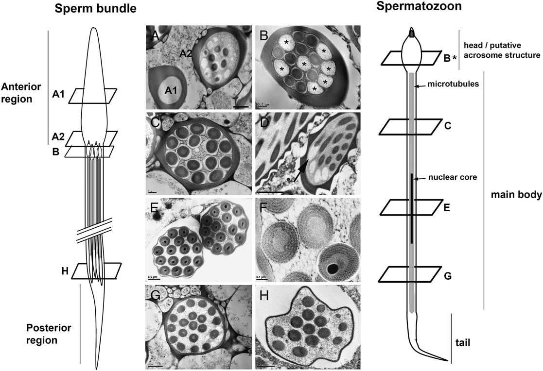
Using transmission electron microscopy, transverse sections across the anterior region of sperm bundles show that the bundle sheath is thickest anteriorly, gradually becoming thinner from anterior to the posterior (Fig. 11A–H). In cross-section, its profile is uneven, reflecting the thickening of the sheath where the plane of section crosses a helical turn of the bundle profile (Fig. 11A–B). The inflated region of the bundle head with the most concentrated helical turns is sectioned longitudinally in Figure 11D. It is apparent that the helical turns of the sheath are more compact than the helical turns of the spermatozoa. The sheath is uniformly thin at the basal region of the bundles (Fig. 11E–H). The number of spermatozoa visible in cross-section at the posterior end of sperm bundles gradually reduces more basally (Fig. 11G–H), indicating that their arrangement is staggered or that all spermatozoa are not of equal length. In sections through the anterior region of a bundle, some spermatozoa were sectioned through the anterior regions, indicated by their circular electron-lucent profiles, while others were sectioned across their main bodies, as indicated by the presence of microtubules (Figs. 11A–C, E–F). A longitudinal section through this region also confirms that the putative acrosome structure is apical to the beginning of the main body (Fig. 11D). The main bodies of spermatozoa contain only microtubules arranged in concentric rings (Fig. 11F) and if cut across the nuclear cores in the third region of their filamentous bodies, the round nuclei are present in middle of their bodies (Fig. 11E–F).
Transverse sections of sperm bundles and spermatozoa of Kerria chinensis, arranged from anterior to posterior regions. A, A section through two sperm bundles. In A1, the section is through the anterior-most region of sperm bundle. In A2, the section cuts across the anterior-most part of spermatozoa. B, Spermatozoa (n=17) within the bundle are sectioned at either the electron-lucent acrosome region (asterisks) or through the anterior region with peripheral arrays of microtubules. C, A section through a bundle cutting through spermatozoon at the anterior region of the main body, showing a central core of microtubules and electron-lucent outer ring. The bundle sheath is thinner than in the apical region; however, the irregular profile indicates the section is through the screw-like region. D, An oblique longitudinal section through the anterior regions of a bundle showing the ridged profile of the bundle membrane and the transition between the electron-lucid head region and the microtubule-filled anterior region of one spermatozoon (arrow). E, Sections through the posterior region of two bundles showing the nuclear core of (16 and 14) spermatozoa. F, The microtubules arranging in concentric rings within spermatozoa. G, Section through the region of spermatozoa beyond the nuclear core region to tail. H, The posterior most of sperm bundles showing the reduce number of spermatozoa. Diagram of a spermatozoon (right) and a sperm bundle (left), indicating positions of cross-sections. Scale bars: A, E, G–H=0.5 μm, B–C=0.2 μm, F=0.1 μm, D=1 μm.

Discussion
The morphology of testes, sperm bundles, spermatozoa, and the process of spermatogenesis of wingless male lac insects, K. chinensis, are described. It is not clear why most K. chinensis embryos develop as wingless males rather than as winged males. From our observations, winged males take longer to become adults so at the time of their emergence, all females nearby had already been fertilised by wingless males. The winged form might be the dispersive form due to their ability to fly to find females. Moreover, their smaller testes might enable dispersion due to their reduced weight.
Kerria chinensis show the unique characteristic of coccoid insects that the mature testes contain syncytial sperm bundles and the bundles contain syncytial spermatozoa only, with no premature stages (Nur Reference Nur1962). The presence of mature sperm at eclosion is characteristic of insects whose males are short-lived, being immediately prepared for mating and fertilisation (Phillips Reference Phillips1970). Within the testes, the sperm bundles separate into two groups arranged in antiparallel directions to different end of the testes.
Overall, spermatogenesis in K. chinensis, is similar to that described in other scale insects, particularly in the closely related species, K. lacca (Dikshith Reference Dikshith1965; Jaipuriar et al. Reference Jaipuriar, Teotia, Lakhotia and Chauhan1984; Paoli et al. Reference Paoli, Roversi, Mercati, Marziali, Cocco and Dallai2015). In K. lacca, 16 primary spermatocytes were reported to be present per cyst. In K. chinensis, the number of primary spermatocytes is the same overall except that a small proportion contains fewer than 16 primary spermatocytes. In insects, the number of spermatocytes per cyst is expressed as 2 n with “n” referring to the successive mitotic division number of the spermatogonia in a cyst (Phillips Reference Phillips1970). In the case of K. chinensis, the 16 primary spermatocytes state is likely to be derived from the four successive mitotic divisions (24) of a spermatogonium. It is possible that the reduced number of spermatocytes seen in some cases could be due to failure of one or two of the eight cells from the third round of mitotic division failing to divide again in the fourth round.
In Neococcoidea, the primary spermatocytes have been shown to produce spermatids via inverted meiosis (Hughes-Schrader Reference Hughes-Schrader1948; Paoli et al. Reference Paoli, Roversi, Mercati, Marziali, Cocco and Dallai2015). Also, each primary spermatocyte produces only one spermatid with four nuclei (quadrinucleated spermatids), in contrast to most insects where each primary spermatocyte produces four haploid spermatids. The same occurs in K. lacca, where two binucleated spermatids fuse to form quadrinucleated spermatids. The spermatids then undergo morphological change to become spermatozoa (Dikshith Reference Dikshith1965). In K. chinensis, most cysts harbor 16 quadrinucleate spermatids and in each quadrinucleate spermatid, two sperm tails grow out at the peripheral of spermatid next to the euchromatic nuclei in opposite directions, clockwise and counter-clockwise. Then, each round euchromatic nucleus becomes cylindrical and moves into the spermatozoon as a nuclear core. Thus, each cyst has 16 clockwise spermatozoa that give rise to one sperm bundle and 16 counter-clockwise spermatozoa that form the other bundle. Similarly, a study in Pseudococcus obscurus revealed that each cyst produced 32 spermatozoa of which 16 gave rise to a clockwise-turning bundle and to a counter-clockwise bundle (Nur Reference Nur1962).
The number of spermatozoa per bundle is generally given as an average because of variability in number within individuals of a species (Jamieson et al. Reference Jamieson, Dallai and Afzelius1999; Paoli et al. Reference Paoli, Roversi, Mercati, Marziali, Cocco and Dallai2015). It has been suggested that intraspecific variation was due to variation in the level of sectioning of bundles and the sectioning of immature cysts (Swiderski Reference Swiderski1980). Taking care to count spermatozoa in bundles sectioned at the sub-apical or mid-regions, the number of K. chinensis sperm per bundle ranged from 12 to 21; however, most bundles contain 16 spermatozoa per bundle and 15–17 spermatozoa per bundle was the next most common. The variation in number seen in K. chinensis could be due to variation in the number of primary spermatocytes per cyst due to abnormal mitotic division. Another possibility is that inconsistent orientation of sperm tails in each spermatid affects their allocation. For example, in a spermatocyte cyst with 16 spermatids, one of the 16 spermatids could produce two spermatozoa with their tails abnormally moving in the same direction and those of the remaining 15 spermatids move normally, resulting in two sperm bundles, one with 15 spermatozoa and the other one with 17 spermatozoa.
The formation of two sperm bundles per cyst is common in coccids (Moses and Wilson Reference Moses and Wilson1970). However, in some coccids such as Phenacoccus acericola King (Hughes-Schrader Reference Hughes-Schrader1935) and Steatococcus tuberculatus Morrison (Moses and Wilson Reference Moses and Wilson1970), one spermatid cyst produced one sperm bundle. Tenebrionidae (Coleoptera) have been reported to produce two sperm bundles per cyst but the mechanism is different to that of Neococcoidea. In Neococcoidea, the two bundles separated at the end of spermiogenesis while in Tenebrionidae, the two sperm bundles formed at the beginning of spermiogenesis (Dias et al. Reference Dias, Yotoko, Gomes and Lino-Neto2012).
We found that the spermatid cyst of K. chinensis was wrapped with seven to eight cyst cells. In K. lacca and some Pseudococcidae, the number of cyst cells was not reported (Nur Reference Nur1962; Dikshith Reference Dikshith1965; Moses and Wilson Reference Moses and Wilson1970). At the final stage of sperm bundle formation, these cyst cells and the cytoplasm of spermatids along with the heterochromatic nuclei are discarded in a process whereby they move along the bundle from the anterior to the posterior end and are then eliminated from the bundle. In Drosophila melanogaster Meigen (Diptera: Drosophilidae), each mature spermatid cyst contains 64 spermatozoa and is covered with two cyst cells. The two cyst cells were not eliminated at the same time as elimination of all spermatid cytoplasm. At the final stage of spermatogenesis, the 64 spermatozoa became individualised in a process by which cytoplasm, membranes, and organelles of 64 spermatozoa formed an individualisation complex at the nuclear end of the cyst. The individualisation complex moves down the length of the spermatozoa and is eliminated from the posterior end of the cyst. After individualisation, the individual 64 spermatozoa remained covered by two cyst cells and they were liberated from the cyst cells when they moved out the testis into the seminal vesicle (Tokuyasu et al. Reference Tokuyasu, Peacock and Hardy1972; Cross and Shellenbarger Reference Cross and Shellenbarger1979; Fabrizio et al. Reference Fabrizio, Hime, Lemmon and Bazinet1998; Fabian and Brill Reference Fabian and Brill2012). In K. chinensis, spermatozoa remain congregated in a bundle after excess cytoplasm is eliminated. During mating, the sperm bundles are transferred into the spermatheca of a young female (about 60 days old) whose ovaries have just started developing (Ngernsiri et al. Reference Ngernsiri, Piyajaraprasert, Wisoram and Merritt2015). Dissection of the ovaries of older, developing females (about 75 days old) revealed that spermatozoa were present in their lateral oviducts (image not shown). Therefore, to fertilise the eggs, the bundles are expelled from the spermatheca to the lateral oviducts within which the spermatozoa become individualised.
Insect spermatozoa are typically comprised of a head and a tail, with the head containing the nucleus and acrosome and the tail containing the axoneme and mitochondrial derivatives (Baccetti Reference Baccetti1972; Swiderski Reference Swiderski1980). However, scale insect spermatozoa are atypical with no distinctive head, mid-piece or tail, and lacking the acrosome, axoneme, centrioles, and mitochondrial derivatives (Ross and Robinson Reference Ross and Robinson1969; Moses and Wilson Reference Moses and Wilson1970; Robinson Reference Robinson1977; Jamieson et al. Reference Jamieson, Dallai and Afzelius1999; Dallai et al. Reference Dallai, Gottardo and Beutel2016). We found that K. chinensis spermatozoa lack most of these components as well. However, K. chinensis spermatozoa have small lantern-like structures with small dark tips at their anterior ends. In most animal spermatozoa, an acrosome is located at the apical end of the sperm head. Therefore, from its location, it is possible that the small lantern-like structure may be the putative acrosome of K. chinensis spermatozoa. The only existing report of a possible acrosome-like structure in Neococcoidea spermatozoa is an apical structure of medium electron-density seen at the tip of Parlatoria oleae (Colvée) (Hemiptera: Diaspididae) spermatozoa in some electron micrographs (Robinson Reference Robinson1966).
In scale insects, nuclear cores are typically thread-like and extremely elongate, comprising more than 50% of spermatozoon length and most often located in the anterior to middle region (Robinson Reference Robinson1966; Ross and Robinson Reference Ross and Robinson1969; Swiderski Reference Swiderski1980; Dallai et al. Reference Dallai, Gottardo and Beutel2016). In the K. chinensis sperm, the nuclear material is shorter, comprising ~25% of spermatozoon length and is located in the posterior half. In Pseudococcus viburni Signoret (Hemiptera: Pseudococcidae), it has been reported that the entire sperm entered the egg for fertilisation (Nur Reference Nur1962). Therefore, the location of nuclear material in the posterior region of K. chinensis spermatozoa may not affect fertilisation.
In conclusion, we describe the more processes of lac insect spermiogenesis in more detail than the process presented for the related species, K. lacca by Dikshith (Reference Dikshith1965). We provide a complete description of sperm bundle formation, using light microscopy, fluorescence microscopy, and transmission electron microscopy. The novel findings include a description of variation in number of spermatozoa per bundle, and the distinct morphology of K. chinensis spermatozoa, including a putative acrosome in the apical region.
Acknowledgements
The authors thank Mr. Wichian Piyajaraprasert and Mr. Nutthawuth Thanbaramee at Sukhothai Province Forest Products Research Station, Royal Forest Department, Sukhothai, Thailand, for collecting the insect samples used in this study. The authors also like to thank Southeast Asian Regional Center for Graduate Study and Research in Agriculture (SEARCA) travel grant for a travel grant for L.N. to visit the University of Queensland and for D.M. to visit the Faculty of Science, Kasetsart University. This work was supported by a grant from Kasetsart University Research and Development Institute and The Centre of Advanced Studied in Tropical Natural Resources, Kasetsart University.