Introduction
The most appropriate technique for superior canal dehiscence syndromeReference Minor, Solomon, Zinreich and Zee 1 has been a topic of discussion over the past decade. Transmastoid plugging seems more reliable than middle fossa approach resurfacing, except for the risk of inner-ear damage. Consequently, transmastoid resurfacing, endoscopic resurfacing and endaural approaches have been developed.Reference Shaia and Diaz 2
Here we propose a method of underwater endoscopic ear surgery that provides a clear operative view for dissection and which can prevent inner-ear damage in the treatment of superior canal dehiscence, as has been found for closure of labyrinthine fistulas.Reference Yamauchi, Yamazaki, Ohta, Kadowaki, Nomura and Hidaka 3 Underwater endoscopic ear surgery does not require suction and so protects the inner ear from unexpected aeration that may damage its function. Superior canal dehiscence plugging by underwater endoscopic ear surgery can be performed through the transmastoid approach instead of the more invasive middle fossa approach; the latter may cause relatively severe complications, including cerebrospinal fluid leak and intracranial bleeding.
Materials and methods
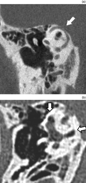
A 36-year-old male had suffered from superior canal dehiscence in the left ear. He developed hyperacusis at the age of 34 years and Tullio's phenomenon. High-resolution computed tomography (CT) revealed dehiscence of the top of the superior semicircular canal on the left (Figure 1a).
High-resolution computed tomography scans (Pöschl plane) of the left ear. (a) Pre-operative scan showing superior semicircular canal dehiscence (white arrow). (b) Post-operative scan after one year showing the superior semicircular canal plugged completely (white arrows).

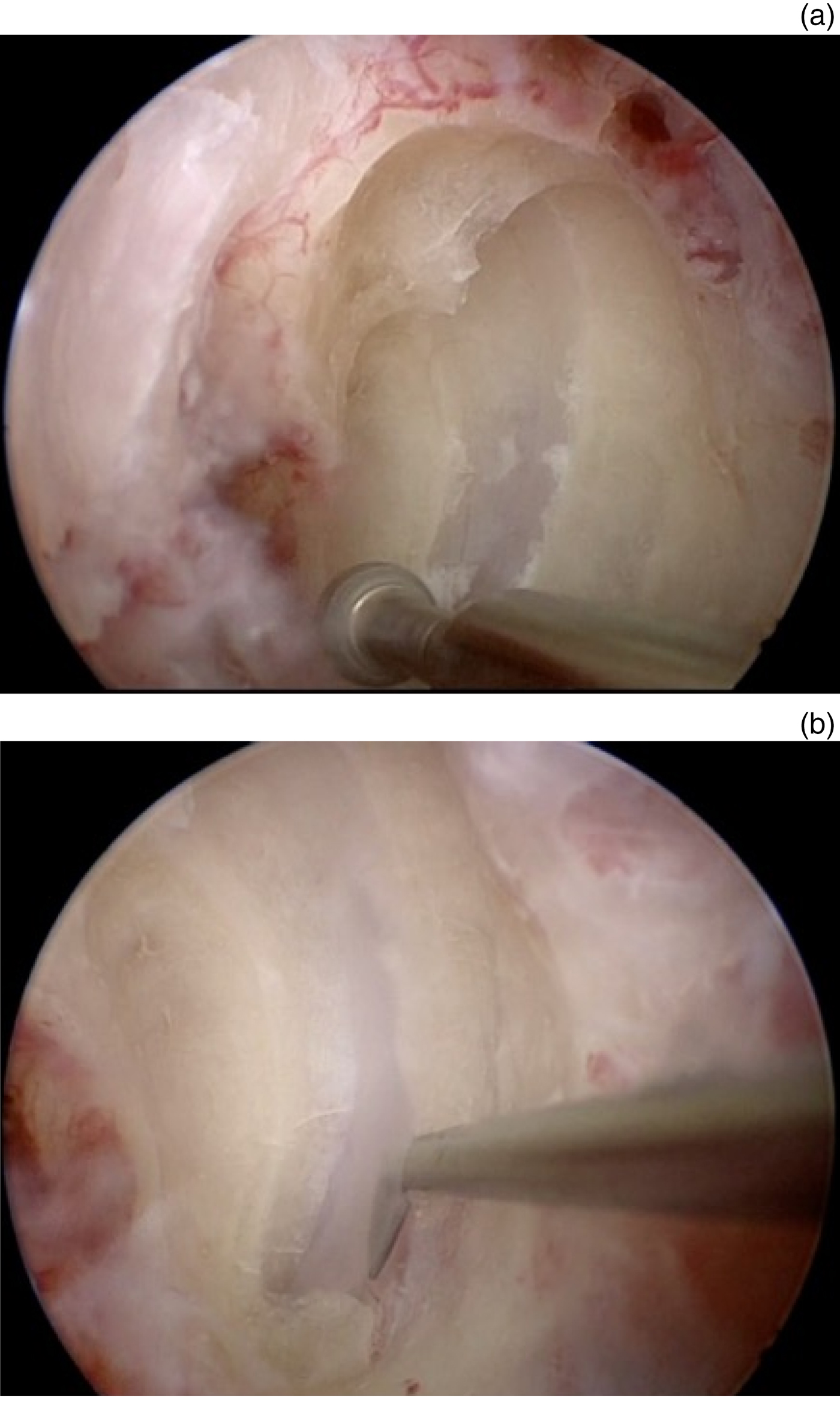
Underwater endoscopic ear surgery was performed for plugging of the superior semicircular canal through the transmastoid approach. Retroauricular incision and mastoidectomy were performed under a surgical microscope. Subsequently, saline solution was infused into the mastoid cavity through an Endo-Scrub Lens Cleaning Sheath (Medtronic ENT, Jacksonville, Florida, USA) mounted on a 0-degree, 2.7 mm diameter, high-definition endoscope (Karl Storz, Tuttlingen, Germany) coupled to a high-definition camera and video system (Karl Storz). Then, the tip of the inserted endoscope was filled completely with saline water to prevent refraction effects, resulting in a clear surgical field. Saline was supplied via an Integrated Power Console (IPC System; Medtronic ENT) so that the surgeon could directly control the amount of perfusion without instructing an assistant. Using this underwater endoscopic view, the superior semicircular canal was identified via the blue line, and opened by drilling with a 2 mm coarse diamond burr, and diamond curved burrs with diameters of 1.5 mm and/or 1 mm (IPC System) (Figure 2a). The canal was dissected with a 45-degree hook to expose the semicircular canal membranous labyrinth and dehiscence area (Figure 2b). The area corresponding to the semicircular canal membranous labyrinth was cut out, and the edges of the canal were completely plugged with muscle fascia and bone wax. This area was reinforced with bone paste and temporal muscle fascia (as indicated in a short video, available on The Journal of Laryngology & Otology website (Appendix 1)).
(a) The semicircular canal on the left was dissected by drilling with a 2 mm coarse diamond burr, and 1.5 mm and/or 1 mm diamond curved burrs (IPC System, Medtronic ENT). (b) The canal was dissected with a 45-degree hook to expose the semicircular canal membranous labyrinth and dehiscence area. The right side of the photograph is the head side. (Images taken from the supplementary video (Appendix 1).)

Results
Our novel underwater endoscopic ear surgery technique for the treatment of superior canal dehiscence syndrome provided a clearer operative field of view with an Endo-Scrub Lens Cleaning Sheath and IPC System than via a simple syringe operated by an assistant. The membranous labyrinth of the semicircular canal was not collapsed and was identified easily. The dehiscence area was clearly identified behind the membranous labyrinth (Appendix 1). No particular complication occurred during the surgical procedure, and post-operatively the patient did not experience vertigo, only slight dizziness without nystagmus.
The absence of post-operative tinnitus and lack of deterioration of bone conduction thresholds in the patient's left ear confirmed that cochlear function was successfully preserved. Pre-operative audiograms showed bone conduction thresholds of 10, 5, 0, 15 and −5 dB HL at 0.25, 0.5, 1, 2 and 4 kHz, respectively (Figure 3a). However, post-operative audiograms showed no deterioration, with bone conduction thresholds of 10, 5, 0, 10 and −5 dB HL (obtained one month after surgery) at 0.25, 0.5, 1, 2 and 4 kHz, respectively (Figure 3b).
(a) Pre-operative and (b) post-operative (one month after surgery) pure-tone audiometry. ○ = air conduction (unmasked) right ear; × = air conduction (unmasked) left ear; [ = bone conduction (masked) right ear; ] = bone conduction (masked) left ear

Pre-operative air-conducted vestibular-evoked myogenic potential responses demonstrated lower thresholds in the left ear than the right, but post-operatively these differences disappeared. One year after surgery, follow-up CT revealed that the fistula was completely covered with bone paste and soft tissues, and no aeration was observed within the labyrinth (Figure 1b). After the surgery, hyperacusis and Tullio's phenomenon had diminished. The patient has been completely free of the pre-operative symptoms, and without complications.
Discussion
Superior canal dehiscence syndrome is a relatively recent disease concept, and diagnosis depends on high-resolution CT detection of a superior semicircular canal anatomical defect. Therefore, superior canal dehiscence syndrome has been considered as a ‘third window disease’ that causes symptoms such as vertigo, conductive hearing loss, pulsatile tinnitus, autophony, migraine headaches and hyperacusis. Tullio's phenomenon and Hennebert's sign of vestibular symptoms are also consistent with superior canal dehiscence, because the ‘third window’ acts as an additional window for the vestibular system, allowing pressure and noise changes to induce vestibular activity.Reference Shaia and Diaz 2
Various surgical treatments for superior canal dehiscence have been proposed since the surfacing technique with the middle cranial fossa approach was first reported.Reference Minor 4 The plugging technique with the transmastoid approach is more direct and effective than the surfacing technique,Reference Kirtane, Sharma and Satwalekar 5 , Reference Beyea, Agrawal and Parnes 6 as the surfacing technique through the middle fossa approach may damage the temporal lobe because of the large craniotomy required. A modified surfacing technique with the transmastoid approach was described as less invasive, although direct visualisation is limited.Reference Hahn and Zappia 7 – Reference Teixido, Seymour, Kung and Sabra 9 Endoscopic resurfacing through the middle cranial fossa approach involves a smaller craniotomy, but still carries the risk of temporal lobe retraction.Reference Carter, Lookabaugh and Lee 10 More recently, reinforcement of the oval and round windows through a transcanal or endaural approach was proposed.Reference Silverstein, Kartush, Parnes, Poe, Babu and Levenson 11 This approach is consistent with the ‘third window theory’, and is much less invasive than the other techniques, but soft failure has been observed as early as six months after surgery.Reference Shaia and Diaz 2
The plugging technique seems anatomically compatible and effective. However, it may damage the inner ear, because even slight suction could cause unexpected aeration into the inner ear, resulting in unrecoverable damage.Reference Kobayashi, Sakurada, Ohyama and Takasaka 12 To overcome these problems, our novel technique of plugging by underwater endoscopic ear surgery offers a soft surgery approach for preventing inner-ear damage. We previously reported an underwater endoscopic ear surgery technique for closure of a labyrinthine fistula in cholesteatoma.Reference Yamauchi, Yamazaki, Ohta, Kadowaki, Nomura and Hidaka 3 Initially, saline was perfused with a syringe by an assistant, but using the Endo-Scrub Lens Cleaning Sheath and IPC System is more efficient because the surgeon can control the ideal supply of saline. The underwater endoscopic ear surgery technique can achieve: preservation of inner-ear function, clear operative visualisation with 1.33 times magnification, and a reduced thermal effect of the endoscope. Hence, it is an extremely effective technique, especially for inner-ear surgery.
Conclusion
Plugging of superior canal dehiscence was performed with a novel underwater endoscopic ear surgery technique. Saline perfusion with the Endo-Scrub Lens Cleaning Sheath and IPC System cleared the surgical field more efficiently than manual application with a syringe. Furthermore, the dehiscence was easily identified, with complete hearing preservation.
Appendix 1 Supplementary video material
A short video demonstrating the operative procedures of the underwater endoscopic ear surgery technique is available online at The Journal of Laryngology & Otology website, at https://doi.org/10.1017/S0022215117001104.