Introduction
The search for former, extant or recent life on other celestial objects of our Solar system will be one of the priority goals of future planetary missions (Reference SteeleSteele & Beaty). The answer to the question of the uniqueness of life on Earth will change mankind's view in all fields of science, philosophy and society and will be of the uttermost relevance for our perspective on bio-science. Since its characteristics and constraints are roughly unknown, the search for extraterrestrial life must be prepared very carefully. It is essential to know what to look for before flying to other planets, satellites and moons. Methodological diversity, objectiveness in interpretation and detailed background knowledge are crucial prerequisites for planning scientific experiments on future planetary missions in the Solar system. One astrobiological approach in reference to this question is to investigate the chemical, physiological and ecological characteristics of terrestrial extremotolerant and extremophile organisms with respect to their adaptations to predominant abiotic factors and harsh environmental conditions. Such studies will broaden our knowledge on the limits and limitations of terrestrial life and thus may help to focus our efforts on the detection of life.
Several spaceflight and ground based experiments were already performed with colonies of (cyano-)bacteria, fungi and lichens that survived to a certain extent real space exposure or simulated space conditions and were able to recover their activity afterwards (de Vera et al. Reference de Vera, Horneck, Rettberg and Ott2003, Reference de Vera, Horneck, Rettberg and Ott2004a, Reference de Vera, Horneck, Rettberg and Ottb, Reference de Vera, Möhlmann, Lorek, Wernecke and Ott2010; de la Torre et al. Reference de la Torre, Sancho, Pintado, Scherer, Facius, Deutschmann, Reina, Baglioni and Demets2004; de la Torre Noetzel et al. Reference de la Torre Noetzel, Sancho, Pintado, Rettberg, Rabbow, Panitz, Deutschmann, Reina and Horneck2007, Reference de la Torre Noetzel2010; Sancho et al. Reference Sancho, de la Torre, Horneck, Ascaso, de los Ríos, Pintado, Wierzchos and Schuster2007; Sancho Reference Sancho2009; Raggio et al. Reference Raggio, Pintado, Ascaso, de la Torre, de los Ríos, Wierzchos and Sancho2011; Onofri et al. Reference Onofri2012; Sánchez et al. Reference Sánchez, Mateo-Martí, Raggio, Meeßen, Martínez-Frías, Sancho, Ott and de la Torre2012). The Biology and Mars Experiment (BIOMEX) is an international and interdisciplinary Low Earth Orbit (LEO) exposure experiment which will be performed on EXPOSE-R2 – the ESA exposure facility attached to the Russian Module Zwesda on the International Space Station (ISS). This facility is designed for the exposure of samples to vacuum, temperature, solar and cosmic radiation as well as the insolation regimen of the LEO (Rabbow et al. Reference Rabbow2009, Reference Rabbow2012; Cottin Reference Cottin2011; de Vera et al. Reference de Vera2012). The launch is scheduled for April 2014. The exposure duration is scheduled to be 1.5 years. BIOMEX focuses on two main objectives: (1) The first objective of BIOMEX is to investigate the resistance and possible decomposition of biomolecules like pigments and cellular components under LEO-space and simulated Mars-like conditions. (2) The second objective is to analyse to what extent terrestrial extremophiles are able to survive LEO-space and Mars-like conditions, and to determine which interactions between biological samples and selected minerals (including original terrestrial substrata as well as Moon- and Mars analogues) could occur.
Biominerals – biogenically formed minerals – may serve as biomarkers as they may be remnant for geological time-scales even if the producing organisms itself vanished long before (Thomas-Keprta et al. Reference Thomas-Keprta, Bazylinski, Kirchvink, Clemett, McKay, Wentworth, Vali, Gibson and Romanek2000). As indicators of biological activity, these minerals are of high interest for the search for former life on other planetary objects, e.g. Mars. Several extremotolerant micro-organisms were identified to excrete oxalic acid into their surrounding where it disintegrates the rock and binds leaching calcium ions to form calcium oxalate deposits (Sohrabi Reference Sohrabi2012). This process allows that many organisms grow into the rock to shelter from extreme environmental conditions and facilitate bio-weathering, a potential biomarker itself. In the context of future planetary missions, the characteristics and detectability of biogenically formed oxalates need to be understood in detail, especially if extremotolerant organisms prove to produce such compounds as an adaptation to harsh environmental conditions. Among 13 other organisms, Circinaria gyrosa is chosen as a biosample in BIOMEX. C. gyrosa is a lichen, namely a symbiotic association composed of a fungal and a photosynthetic partner. After the investigation of this lichen species in space and in Martian-simulated environment (Sancho et al. Reference Sancho, de la Torre, Horneck, Ascaso, de los Ríos, Pintado, Wierzchos and Schuster2007; de la Torre Noetzel et al. Reference de la Torre Noetzel2010; de Vera Reference de Vera2012), and the positive results of tolerance and resistance obtained under these extreme conditions, it can be confirmed that it is a suitable model organism in Astrobiology. We will take a step further trying to understand the process of biomineralization under harsh environmental parameters.
In the present study, we will show that C. gyrosa is producing calcium oxalate monohydrate in restricted areas of its thallus. Thus, this lichen is suitable to investigate the products of biomineralization under extreme environmental conditions. We will present the Raman spectral characteristics of calcium oxalate monohydrate, its precise distribution in the lichen thallus, and suggest a hypothesis on its biological role. Moreover, we will demonstrate its role as a biomarker as well as the Raman spectral characteristics of additional biomarker compounds.
Raman spectroscopy is a non-destructive method to investigate the molecular and crystal structure of a sample. It is based on inelastic scattering of monochromatic light, i.e. a laser. The energetic shift of the reflected laser photons gives the Raman spectrum of the sample. The Raman spectrum is unique for the sample and thus allows its identification. As the Raman laser spectrometer (RLS) is scheduled to be part of the future planetary exploration mission ExoMars (Esa robotic exploration of mars), this technique is used in the present work to determine biominerals as remanent biomarkers. This will be demonstrated by investigating the characteristics and the distribution of calcium oxalate within the lichen C. gyrosa by Raman spectroscopy. The interpretation of Raman spectra requires considerable a priori information and background knowledge. We will contribute in the present study to the background knowledge of Raman spectra of C. gyrosa. Besides a valuable progress in understanding the internal structure and Raman properties of a lichen model organism, the results can be applied for future astrobiological experiments, especially in the context of the BIOMEX project and the possible changes that C. gyrosa might undergo during space exposure.
The following two steps have been applied to investigate and interpret the Raman characteristics of C. gyrosa: (1) thallus thin sections are used to determine the amount and distribution of calcium oxalate and other lichen substances within the lichen; (2) a fractured part of the thallus is examined to figure out which of the previous findings can be identified as well on a natural sample. To substantiate our findings, the distribution of inner medullary crystals was verified by scanning electron microscopy (according to Reference Meeßen, Sánchez, Brandt, Balzer, de la Torre, Sancho, de Vera and OttMeeßen et al., Reference Meeßen, Sánchez, Brandt, Balzer, de la Torre, Sancho, de Vera and Ott2013) and the identity of calcium oxalate was checked by a complementary specific reaction test (according to (Pizzolato Reference Pizzolato1964)).
The morphological and anatomical structures of C. gyrosa are briefly described in The lichen species C. gyrosa section. The sample preparation and the experimental setup for the Raman measurements are presented in the Methods section. The measurement results will be presented and discussed in the Results and discussion section with emphasis on the particular implications for C. gyrosa and the general role of calcium oxalates in lichens followed by summary and an outlook section.
The lichen species C. gyrosa
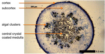
The subject of the present study is the lichen C. gyrosa (nom. provis. (Sohrabi Reference Sohrabi2012), recently renamed from Aspicilia fruticulosa). This lichen species naturally grows in steppe-like and continental deserts of the Northern hemisphere, and therefore, it is adapted to extreme environmental conditions such as heat, long-term drought and high levels of insolation (Sancho et al. Reference Sancho, Schroeter and del Prado2000). Our samples were collected at an open forest area of the steppic highlands of Guadalajara (Spain), near the locality of Zaorejas. They grow scattered in the clay-like soil of a region characterized by drastic diurnal and seasonal variations in terms of temperature and rainfall. It has also survived simulated Mars- and space conditions (Sancho et al. Reference Sancho, de la Torre, Horneck, Ascaso, de los Ríos, Pintado, Wierzchos and Schuster2007, Reference Sancho, de la Torre and Pintado2008; de la Torre Noetzel et al. Reference de la Torre Noetzel2010; Sánchez et al. Reference Sánchez, Mateo-Martí, Raggio, Meeßen, Martínez-Frías, Sancho, Ott and de la Torre2012). The coralloid to compact, vagrant thalli of C. gyrosa are frequently relocated by wind and water and are often found partially embedded in its clay-like substrate. Its fruticose thalli are brownish to ochre and measure up to 2.5 cm in diameter. At the branch tips, the pores (pseudocyphellae) could appear as whitish openings and play a crucial role in the lichen's gas exchange (Sancho et al. Reference Sancho, Schroeter and del Prado2000; Meeßen et al.). Compared with other lichen species used in astrobiological studies (as Xanthoria elegans, Rhizocarpon geographicum and Buellia frigida), C. gyrosa reveals intriguing morphological and anatomical differences that may help to explain its high resistance to harsh environmental but also to simulated Martian and to space conditions (Meeßen et al.): Thin sections of the lichen branchings (Fig. 1) reveal distinct structures and a complex internal stratification. The outer cortex of the thalli is composed of a brownish pigmented layer of dead cells followed by a layer of vital isodiametric cells. Below, the lichen forms a compact and extended subcortex consisting of tightly packed fungal hyphae which are agglutinated by high amounts of extracellular mucilage. That subcortex is supposed to contribute to the lichen's mechanical stability which is necessary due to its vagrant life-style. Nonetheless, naturally occurring thalli are more or less frequently fractured. In addition, the subcortex acts as a diffusion barrier for gas and water exchange, and as an irradiation-protective layer. Below, algal cell clusters are distributed evenly. Most inward, the central tube of each branching as well as the layer in which the algal clusters are embedded are filled with a medullary fungal tissue that is rich in inner aerial spaces and connected to the surrounding air by the tip-located pores.
Thin section of a thalline branch tip of Circinaria gyrosa below the distal pseudocyphellae. From the outside to the centre, the section depicts the dark pigmented outer cortex, followed by the extended, dense, and highly gelatinated subcortex and evenly distributed algal clusters. The centre is made of loose medullary hyphal tissue. The thin section measures 30 μm and is stained with 5% lactoglycerol-blue. A digital light microscope (Axio imager A1, Zeiss) was used for the analysis and recording of the micrographs.

Methods
Raman analysis of thin sections of C. gyrosa
Thin layer sections of 15–30 μm of C. gyrosa (Fig. 1) were obtained from the distal branches of a representative specimen by using a cryostatic microtome (Frigomobil 1206, Reichert-Jung, −20 to −30 °C). Some specimens of the sections were used to examine and document the morphological–anatomical characteristics under the microscope (Axio Microscope and Imager A1, Carl Zeiss AG) after staining with 5% lactoglycerol/cotton blue. In addition, thin sections were fixed on a glass slide with 1% gelatine, air-dried for 8 hours and investigated by Raman laser spectroscopy.
Raman measurements were performed with the confocal WITec Alpha 300 system with a Nikon 10× (NA 0.25) objective (WITec 2008). The Raman laser excitation wavelength of 532 nm was applied and a spectral resolution of about 4 cm−1 was achieved in a spectral range between 100 and 3800 cm−1. The ideal spot size on the sample is in focus at about 1.5 μm. The laser power was set to 1 mW for the thin section. The measurements were performed in air at room temperature and under ambient air pressure. Single spectra, line scans and image scans with up to 175 μm×175 μm and up to 22.500 image points were measured. The image scans were performed on flat thin sections to identify the distribution of the typical lichen substances within the different tissues of C. gyrosa.
Raman analysis of the complete thallus of C. gyrosa
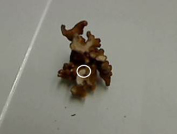
In a second step, Raman measurements were made on whole, natural lichen thalli (Fig. 2). Taking into account that these thalli are erratic and live freely in the ground, they are subjected to all kinds of environmental influence and so could be found fractured. Single spectra measurements and line scans were obtained from the natural lichen with rough, uneven and sometimes damaged surfaces. Here, it is of interest to find out which of the results from the thin section can be reproduced with the natural sample.
Fractured part of the thallus of Circinaria gyrosa; the circle encloses the area investigated with Raman microspectroscopy, including all internal stratification (depicted in Fig. 1) for ulterior reproduction of results.

Results and discussion
General characteristics of obtained Raman spectra of C. gyrosa
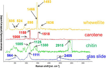
Detailed investigation performed by image scanning revealed differences in the distribution of Raman signatures for the various anatomical structures and hyphal tissues of C. gyrosa. Thus, physiologically different thalline layers were distinguishable by Raman imaging. Typical Raman spectra found in the thin section and representing basic compounds of the lichen are shown in Fig. 3. This investigation focuses on β-carotene as an accessory pigment of the photosynthetic apparatus of the algal symbiont and chitin as the main component of the cell wall of the fungal symbiont, both with known Raman signatures. The peak positions of the spectra representing the lichen compounds are assigned correspondingly.
Typical Raman spectra found in Circinaria gyrosa; the dotted rectangles describe the spectral range filtered for the discrimination of the various compounds. All spectra were baseline corrected applying the correction procedure provided by the Raman spectrometer software WITecProject.

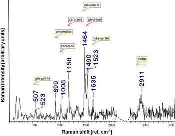
The spectrum of calcium oxalate monohydrate Ca2(C2O4)×H2O (Fig. 3), referred to as whewellite in mineralogical context, is characterized by the double peak at 1466 and 1493 cm−1, an additional peak at 1636 cm−1 of the CO symmetric stretching vibration, the C–C stretching mode peak at 898 cm−1, and the CO2 mode double peak at 505 and 524 cm−1 (Shippey Reference Shippey1980; Frost & Yang Reference Frost, Yang and Ding2003]. The double peak at 1466 and 1493 cm−1 is well pronounced and suitable for interpretation purposes. Nevertheless, polarization effects have to be considered due to the orientation of the whewellite crystals. The polarization effect influences the relative intensities of the two peaks. On the other hand, this effect can be used as an indication for the orientation of the whewellite crystals, e.g. in image scans. The same is true for the less pronounced double peak at 505 and 524 cm−1. The main, characterizing Raman shifts of β-carotene (red spectrum in Fig. 3) are typically about 1008, 1158 and 1518 cm−1. The 1518 and 1158 cm−1 bands are strong and characteristic for the C=C and C–C stretching vibrations (Edwards et al. Reference Edwards, Moody, Villar and Wynn-Williams2005). The smaller feature at 1008 cm−1 corresponds to the rocking modes of the CH3 groups (Vitek et al. Reference Vitek, Edwards, Jehlicka, Ascaso, De Los Ríos, Valea, Jorge-Villar, Davila and Wierzchos2010).
The spectrum assigned to chitin (green spectrum in Fig. 3) contains a large number of lines and bands. Only the main Raman shifts are mentioned here: the bands between 1085 and 1125 cm−1 represent C–O–C and C–O stretching, the band at 1380 cm−1 represents the rocking CH2 vibrations, and the 2915 cm−1 band is associated with the C–H x stretching mode (Ehrlich et al. Reference Ehrlich, Maldonado, Spindler, Eckert, Hanke, Born, Goebel, Simon, Heinemann and Worch2007). The lines between 1260 and 1360 cm−1 can be assigned to amide bands (Ehrlich et al. Reference Ehrlich, Maldonado, Spindler, Eckert, Hanke, Born, Goebel, Simon, Heinemann and Worch2007). It is also important to take into account the Raman spectrum of the background that is a glass slide on which the thin sections are mounted. The spectrum of the glass slide is characterized by a broad band around 1111 cm−1 and two smaller but also broad bands around 564 and 2405 cm−1. The main band of the background overlaps with the main band of chitin. To decide if there is a glassy background the band about 2405 cm−1 is used.
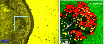
In the following investigation, only the main lines and bands are considered. Special care is taken for overlapping Raman bands and lines among the compounds and the background. The dotted squares around the specific Raman bands and lines in Fig. 3 mark the spectral ranges used for the integral filters for analysis and interpretation. These filters integrated over Raman shift ranges that contained characteristic lines for the lichen substances for calcium oxalate monohydrate (whewellite), carotene and chitin. The integral filter ranges were 1450–1482 cm−1 for calcium oxalate monohydrate (whewellite), 1130–1185 cm−1 for carotene and 2835–3000 cm−1 for chitin. As seen in Fig. 3, the chosen integral filter ranges do not overlap and thus are appropriate to be used for the representation of the substances in the colour-coded images mentioned below. The range between 2340 and 2480 cm−1 is unaffected by the Raman spectra of the lichen substances. To avoid false interpretation, this range is used to identify the areas of the underlying glass slide in the images. Combining these integral filter ranges, colour-coded images were derived as represented in Figs 4-right, 5-right and 6-right. To determine the type and distribution of lichen substances, several image scans were performed at specific areas of the thallus: outer cortex, subcortex, algal clusters and central medulla.
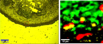
Outer cortex of Circinaria gyrosa: Left – microscopic image. Right – Raman image of the scan, described by the red square in the microscopic image (50 μm×50 μm with 50×50 measurement points). The colours correspond to the different matter (tissue) of the lichen – green – chitin, red – carotene, calcium monohydrate (whewellite) – yellow. Applied integral filters for substance identification are taken as shown in Fig. 3.

Algal clusters of Circinaria gyrosa: Left – microscopic image marked with a square for the colour-coded Raman image scan. Right – colour-coded Raman image scan, described by the red square in the microscopic image (175 μm×175 μm with 150×150 measurement points). The colours correspond to the different matter (tissue) of the lichen – green – chitin, red – carotene, calcium monohydrate (whewellite) – yellow. Applied integral filters for substance identification are taken as shown in Fig. 3.

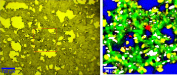
Central medulla of Circinaria gyrosa: Left – microscopic image marked with a square for the colour-coded Raman image scan. Right – colour-coded Raman image scan, described by the red square in the microscopic image (175 μm×175 μm with 150×150 measurement points). The colours correspond to green – chitin; red – carotene; yellow, white – calcium monohydrate (whewellite); blue – background (glass slide, fluorescence). Applied integral filters for substance identification are taken as shown in Fig. 3.

Raman spectra of the thin section of C. gyrosa
As mentioned in the Methods section the thin sections are investigated first. In the thin section of C. gyrosa, the layers of the thallus can be identified clearly (Fig. 1). Starting from the most external part the pigmented cortex is followed by the non-pigmented subcortex. Algal clusters are embedded partially within or below the subcortex and are ensheathed by fungal hyphae. This layer is followed by the inner medulla. The hyphae of the central medulla are coated with crystals. Raman image scans are performed for each of the layers.
The imaged area of the outer cortex (and subcortex) – the surface of the thallus – can be seen in Fig. 4. On the left, the microscopic image with the scanned area (red square) is shown. On the right, the colour composite of the Raman scan of the outer cortex could be seen. The different colours correspond to the lichen compounds: green – chitin, red – β-carotene, yellow – calcium monohydrate (whewellite). The spectra describing these substances are shown in Fig. 3 with the same colour code. Black areas indicate that no single substance can be assigned and/or the spectra are highly fluorescent. As expected the outer cortex consists of chitin (green) and some β-carotene (red) of uncertain origin is also present. The areal fraction of chitin is increasing from the edge of the cortex in the direction of the subcortex representing the extremely dense packed hyphae in the subcortex. The presence of calcium monohydrate (whewellite) is quite unexpected in the outer cortex. Deeper analysis of the spectra showed that the yellow areas in Fig. 4 are characterized by fluorescence and thus the assignment to whewellite is misleading. The spectra of the black areas are, as already mentioned above, characterized by high fluorescence and/or a non-resolvable mixture of different spectra. So the surface of the lichen C. gyrosa is highly fluorescent, which is important for measurements on the natural sample.
Algal clusters embedded in hyphal tissue are shown in Fig. 5. The square in the microscopic image (left) is marking the area scanned with Raman microspectroscopy and corresponds to the colour-coded image on the right. The spectra of whewellite (yellow), chitin (green) and β-carotene (red) (see Fig. 3) represent the main compounds of the algal cluster and attached hyphae. Chitin gives a very clear Raman signal and is surrounding the algal cluster. In the colour-coded image (Fig. 5 right) chitin of the subcortex is characterized by brighter green compared with the chitin in the inner medulla. The spectra of the latter area exhibit high fluorescence connected to chlorophyll in the algae. However, the β-carotene signal (red) is still visible on top of the fluorescence and the spectrum reflects the areal distribution of the photobionts and the shape of the algal cluster. Spectra of calcium monohydrate (whewellite) can be found apart from the subcortex and the algal cluster in the direction of the inner medulla. The yellow patches in the white-rimmed area in the Raman colour-coded image in Fig. 5 (right) are whewellite. The spectra of whewellite here show two strong lines about 1466 and 1493 cm−1 in contrast to the spectra of the outer cortex where only one strong line exists at 1466 cm−1.
The following layer, located in the centre of the thallus represents the inner medulla. In Fig. 6 (left), the microscopic image of the inner medulla with the red square marking the Raman scanned area is shown. The colour-coded image (right) derived from the Raman scan shows the distribution of chitin (green), the glass slide (blue) and whewellite (yellow), which is always attached to the chitin. β-carotene (red) is present as well, but only in a very small portion. The morphological–anatomical structure of the inner medulla (Fig. 6) is not as compact as the outer cortex and subcortex (Fig. 4). That's why the (blue) background is visible in the inner medulla. The hyphal chitin signal (green) is clearly observed forming a loose medullary tissue. Whewellite (yellow, white) is remarkably present in the inner medulla. Interestingly, calcium oxalate is always attached to the hyphae, which means that C. gyrosa forms extracellular whewellite in the inner medullary space. This is new and important to take into account for the investigation of the natural sample. The corollary of this peculiarity will be discussed in Calcium oxalate and its role in C. gyrosa section. The different colours for whewellite (yellow and white) correspond to the different ratios of the intensity of the main lines around 1466 and 1493 cm−1. For the yellow areas the line about 1466 cm−1 is much stronger than the 1493 cm−1 line. For the white areas an opposite ratio is observed. The change in the ratio cannot be explained by polarization and thus, orientation effects. Further investigation is needed to study this change, which is outside the scope of this paper and will be the aim of a new publication.
Raman spectra of natural thalli of C. gyrosa
In a second analysis, the question was answered which of the findings of Raman microspectroscopy can be confirmed for the fractured part of a complete thallus of the lichen C. gyrosa. Several single spectra and line scans were measured on various parts of the lichen thallus. Image scans were not possible to be performed because of the too rough structure of the surface.
Spectra measured on the brownish areas of C. gyrosa showed mainly fluorescence. In very few cases, some indication for Raman signatures of β-carotene and whewellite could be detected. This coincides with the results presented for the outer cortex of the thin section, which showed strong fluorescence.
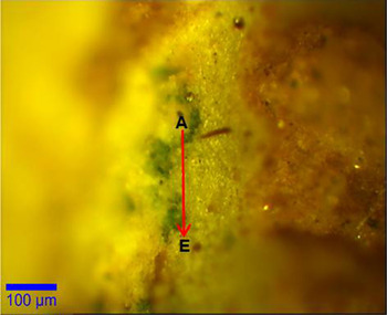
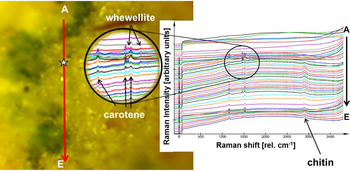
Whitish areas of the thallus of C. gyrosa that appear in Fig. 2 were identified either as pseudocyphellae or as branches layed bare by fracturing. In Fig. 7, the microscopic image of the subcortex with algal clusters and some parts of the inner medulla can be seen. This area is the surface of a fractured part and no additional manipulation was done with the lichen in order to perform Raman measurements. The red (A→E) line represents the line scan sweeping over the subcortex and the inner medulla and algal clusters. The corresponding raw Raman spectra are shown in Fig. 8. Raman spectra of whitish areas are characterized by fluorescence. In addition, Raman features of β-carotene, chitin and whewellite can be observed rather well. The interpretation of the Raman spectra with respect to the corresponding line scan positions shows that chitin is present in almost all positions, except at the first points of the scan. This can be derived from the Raman band of the C–H stretching mode around 2911 cm−1. Raman bands of 1008, 1158 and 1523 cm−1 can be found on positions with algal clusters indicating β-carotene, which is in agreement with the results of the thin section. Whewellite signatures are present in Raman spectra of the positions of the first part of the line scan near algal cluster positions and starts where chitin (of the inner medulla) arises in the spectrum as well (Fig. 8). The Raman spectra of whewellite contain also signatures of chitin and carotene (Fig. 9). One reason can be that the Raman signal comes from the surface as well as from layers below that can contain all compounds of the inner medulla. Another explanation can be that the border area between the medulla and the algal clusters is observed during the scan and all three signals exist in parallel.
Microscopic image of the subcortex with algal clusters and some parts of the subcortex and the inner medulla. The red arrow represents the line scan.


Raman spectrum of the position marked in Fig. 8 with an asterisk. The assignments represent whewellite, β-carotene and chitin.

Thus, the conclusions drawn from Raman measurements on the thin section can be applied to the natural sample: the outer cortex (brownish in the natural sample) is highly fluorescent and difficult to investigate with Raman spectroscopy. On parts with damaged surface, the underlying layers can be investigated and give decisive Raman signals. The algal clusters can be seen clearly by the β-carotene Raman signature. The subcortex and the inner medulla (whitish in the natural sample) are characterized by the spectrum of chitin, especially the C–H stretching mode around 2911 cm−1, and whewellite. In summary, whewellite can be measured by Raman spectroscopy mainly together with chitin and carotene and is located in the inner medulla and near the algal clusters. This co-occurrence of three biogenic substances within one lichen thallus and their peculiar spatial distribution could be seen in the Raman spectra for the thin section as well as for the pristine thallus sample. Such results give valuable information for astrobiological modelling and for the search and definition of bio-signatures.
Calcium oxalate and its role in C. gyrosa
Whewellite is discussed as a product of biological as well as of mineralogical processes. Its appearance in hydrothermal deposits indicates abiogenic origin, while the biogenic origin is supported by its appearance in coal and sedimentary nodules (Hoefs Reference Hoefs1969; Zák & Skála Reference Zák and Skála1993; Vassilev & Vassilev Reference Vassilev and Vassilev1996; Ward Reference Ward2002). In plants, calcium oxalate crystals are formed as virtually insoluble vacuolic deposits to compensate the surplus of calcium supply and act as herbivore defence (Sitte et al. Reference Sitte, Weiler, Kadereit, Bresinsky and Körner2002). Some extremotolerant lichens excrete oxalic acid and degrade their rock habitats by bioweathering (Edwards et al. Reference Edwards, Russell and Wynn-Williams1997), allowing the symbionts to colonize more sheltered endolithic habitats. As a result, calcium oxalates are formed by the reaction of oxalic acid with the rock. Thus, its abundance and multiple biogenic formation make calcium oxalate a valuable biomarker for astrobiological research.
For C. gyrosa an alternative function of calcium oxalate monohydrate is proposed. Its medullary location, its hyphal attachment as demonstrated in the above Raman investigation, together with its virtual insolubility and the gas exchange promoting function of the medulla (Sancho et al. Reference Sancho, Schroeter and del Prado2000; Sánchez et al. Reference Sánchez, Mateo-Martí, Raggio, Meeßen, Martínez-Frías, Sancho, Ott and de la Torre2012) indicate a role in water balance. As all lichens, C. gyrosa is a poikilohydric organism, i.e. their water content will tend to equilibrium with the water status of the environment, under wet conditions they become hydrated and active and under dry conditions they dry out and become dormant. It tolerates dehydration without physiological damage and is metabolically active under moist environmental conditions. Under wet conditions lichens take up water, leading to the soaking of interhyphal spaces. Thus, liquid water strongly decreases the diffusion rate of CO2 towards the algae and inhibits photosynthesis at the very moment when water is not a limiting factor (Lange et al. Reference Lange, Green, Reichenberger, Hesbacher and Proksch1997). Besides other strategies found in lichens, extracellulary deposited crystals on loosely connected medullary hyphae are discussed to prevent complete soaking of the medullary gaseous space (Lange et al. Reference Lange, Green, Reichenberger, Hesbacher and Proksch1997; Honegger Reference Honegger2009). In this context calcium oxalate is suggested to enhance the air/hyphae surface in C. gyrosa, to prevent increased diffusion resistance of CO2 (Sancho et al. Reference Sancho, Schroeter and del Prado2000) and thus facilitates efficient gas exchange when wet.
Summary and outlook
The lichen C. gyrosa, chosen as a model organism for BIOMEX, has been investigated by Raman spectroscopy. Different parts of the lichen were characterized on a thin section and on a natural sample. Calcium oxalate monohydrate (whewellite) has been in focus of this investigation. The present study revealed that C. gyrosa forms extracellular whewellite deposits upon the medullary hyphae inside the thallus. This is a peculiar finding for lichens and suggests a specific role of this internally localized biomineral. The function of whewellite in C. gyrosa is assumed to reduce the wettability of the loose medullary hyphae, and thus to hinder the complete soaking of the thallus under wet environmental conditions. Keeping gas-filled spaces in the medulla is suggested to facilitate sufficient gas exchange and to ensure CO2-provision for photosynthezising algae in the wet, metabolically active thalli. Depending on the decomposition processes of whewellite, it may not only serve as a biomarker of recent life, but also of past and fossilized organisms. Although whewellite can also be formed by rare geological processes, it serves as a suitable biomarker for astrobiological investigations.
Acknowledgements
The authors would like to express their sincere gratitude to the German Federal Ministry of Economics and Technology (BMWi) and the German Aerospace Center (DLR) for funding the work of Joachim Meeßen (50BW1153), to the Spanish Instituto Nacional de Técnica Aeroespacial (INTA) for granting a PhD scholarship to Francisco Javier Sánchez Iñigo, to the Spanish Ministry of Science and Innovation (ESP AYA2010-11422-E, 2010–2012, from 2010 to 2012) and INTA for the economic support of BIOMEX, and to the German Aerospace Center (DLR) for supporting the ESA-space experiment BIOMEX (ILSRA ESA-ILSRA 2009-0834, P-I Dr J.-P. de Vera). We would also like to thank the reviewers for their comments and suggestions.