Introduction
Intranasal drug administration which intends to deliver medication to the eustachian tube orifice has been adopted for the management of a number of otological conditions, including acute suppurative otitis media, otitis media with effusion and eustachian tube dysfunction.Reference Fireman1–Reference Tracy, Dermain, Hoffman and Goetz3
There is evidence to support the use of topical nasal preparations in managing eustachian tube orifice dysfunction.
Venkatayan et al. demonstrated in vivo that the use of aerosolised nasal surfactant with and without betamethasone decreased the duration of otitis media with effusion in a gerbil model.Reference Venkatayan, Troublefield, Connelly, Mautone and Chandrasekhar4
Others have reported favourable results for the use of topical steroids in treating eustachian tube orifice dysfunction in children, through reduction of allergic and non-allergic mucosal swelling when using flunisolide, budesonide and beclomethasone dipropionate, citing clinical success rates of between 70 per cent and more than 90 per cent.Reference Elies5 In a randomised, controlled trial involving 45 children who received a three week course of aerosolised nasal steroid or placebo, there was a statistically significant increase in middle-ear pressure and tympanic membrane mobility and position in one or both ears following dexamethasone administration.Reference Shapiro, Bierman, Furukawa, Pierson, Berman and Donaldson6 There is also evidence to support the use of nasal steroids and decongestants in achieving improved ventilation in cases of middle-ear atelectasis.Reference Danner7
In all these conditions, the intention of using intranasal preparations is to deliver the drug to the eustachian tube orifice, so as to reduce tubal oedema and thereby improve middle-ear ventilation. Our group has previously undertaken a study of nasal drops delivery to the middle meatus, and demonstrated that the choice of head position during administration is highly relevant to the success of intranasal delivery.Reference Karagama, Lancaster, Karkanevatos and O'Sullivan8 One could postulate that, in future clinical studies, choosing the optimal head position may maximise evidence to support the efficacy of topical nasal preparations in altering eustachian tube dysfunction. Against this backdrop, one could assume that optimal eustachian tube orifice delivery requires the most conducive head position. The current study primarily aimed to address this issue.
Aims
This study aimed to compare the drug delivery to the eustachian tube orifice achieved using four commonly adopted head positions described for intranasal drops administration. We also aimed to compare the effect of drops versus nasal spray delivery of medication.
Materials and methods
This study used topical intranasal anaesthetic spray (lidocaine 5 per cent and phenylepherine hydrochloride 0.5 percent; Aurum Pharmaceutical, Romford, Essex, UK), a 30° rigid naso-endoscope (Storz, Culver City, United States), methylene blue dyed saline in a Flixonase Nasule™ nasal drops container (GlaxoSmithKline, Victoria, Australia) and an Otrivine™ nasal spray container (GlaxoSmithKline, Victoria, Australia). Visual and photographic equipment were also used.
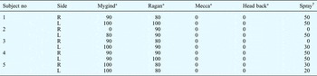
Eustachian Tube Orifice Dye Coverage For Each Head Position, For Drops And Spray

Data represent percentage of eustachian tube orifice area covered with methylene blue dye, unless otherwise specified. *Drops. †Mygind and Ragan positions. R = right; L = left
Study design
We undertook a prospective, five-period, cross-over study.
Subjects
Five healthy adult volunteers participated in the study.
Exclusion criteria
We excluded any subjects with nasal obstruction which prevented access of the fibre-optic endoscope, and also any with rhinitis.
Study procedure
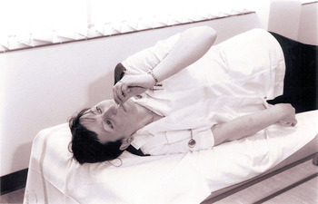
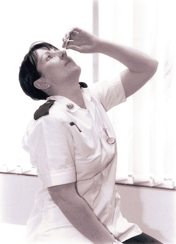
Five healthy volunteers were used to compare the following head positions: (1) Mygind (the subject lies supine with their head extended over the end of the bed) (Figure 1); (2) Ragan (the subject lies on their side without a pillow, thereby allowing the parietal eminence to rest on the bed, and the nose is tilted slightly upwards towards the opposite shoulder; drops are instilled into the lower nostril) (Figure 2); (3) Mecca (subject kneels with their head down and forward) (Figure 3); and (4) ‘head back’ (subject sits upright with the head extended) (Figure 4).
Mygind position.

Ragan position.

Mecca position.

‘Head back’ position.

Each subject initially adopted one of the above head positions, and maintained this for five minutes after the nasal cavities were sprayed with a topical anaesthetic agent. Six drops of dyed saline were administered into the nasal cavity, and photographic evidence of the degree of eustachian tube orifice staining was gathered, to enable comparison of the four head positions. Each head position was tested on a different day, to allow staining from the previous day's test to clear.
The most effective head positions for drops delivery to the eustachian tube orifice were then compared with drug delivery achieved with an intranasal spray (administered in the head back position).
Results were analysed by an independent observer.
Results
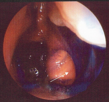
Our results (Table 1) indicated that both the Mygind and Ragan positions were greatly superior in delivering the administered drops to the lateral nasopharyngeal wall and eustachian tube orifice (Figures 5 and 6), compared with the Mecca and head back positions (Figures 7 and 8).
Naso-endoscopic view showing the left eustachian tube orifice (arrow) covered with methylene blue dye, following administration of drops into the nasal cavity with the head in the Mygind position.

Naso-endoscopic view showing the right eustachian tube orifice (arrow) covered with methylene blue dye, following administration of drops into the nasal cavity with the head in the Ragan position.

Naso-endoscopic view showing the right eustachian tube orifice (arrow) free of methylene blue dye, following administration of drops into the nasal cavity with the head in the Mecca position.

Naso-endoscopic view showing the left eustachian tube orifice (arrow) free of methylene blue dye, following administration of drops into the nasal cavity with the head in the ‘head back’ position.

Spray administration was found to be inferior (Figure 9) to drops administration via the Mygind and Ragan positions (Figures 5 and 6), as regards delivery to the eustachian tube orifice.
Naso-endoscopic view showing the right eustachian tube orifice (arrow) free of methylene blue, following administration of spray formulation into the nasal cavity with the head in the head back position.

Discussion
Middle-ear effusion may be defined as the presence of fluid material within the middle-ear space. This condition can be further characterised as either acute or chronic, and includes serous otitis media, secretory otitis media, haemorrhagic otitis media, and middle-ear accumulation of cerebrospinal fluid and perilymph.
A common factor in middle-ear effusions (except those due to trauma or surgical causes) is eustachian tube obstruction. Eustachian tube obstruction may be mechanical or functional.
Intrinsic mechanical obstruction may result from inflammation induced by infection or allergy, while extrinsic obstruction may result from adenoids or nasopharyngeal tumours. In an experimental study, the provocation of allergic rhinitis in patients with a history of allergy was associated with the development of eustachian tube dysfunction.Reference Friedman, Doyle, Casselbrant, Bluestone and Fireman9 A high prevalence of chronic otitis media with effusion has also been observed in patients with allergies.Reference Fireman1 Additional studies have documented eustachian tube dysfunction and middle-ear under-pressures in patients with allergic rhinitis during natural allergen exposure.Reference Skoner, Doyle, Boehm and Fireman10, Reference Skoner, Lee, Doyle, Boehm and Fireman11
The role of allergy in eustachian tube obstruction may involve one or more of the following mechanisms: (1) middle-ear mucosa as a target organ; (2) inflammatory swelling of the eustachian tube with resultant obstruction; (3) inflammation of the nose and nasopharynx; and (4) reflux, insufflation or aspiration of bacteria-laden allergic nasopharyngeal secretions into the middle-ear cavity.
The relationship between allergy and otitis media with effusion may involve inflammatory mediators, including cytokines and colony-stimulating growth factors released by mast cells and epithelial cells in the nose and nasopharynx. These mediators produce eustachian tube blockage through a number of mechanisms, which may include local injury, and vascular and/or neural mediated changes in the eustachian tube opening pressure and in middle-ear perfusion.
It is likely that the nasal allergic response predisposes to eustachian tube blockage, and that the subsequent middle-ear cavity under-pressure leads to middle-ear effusion. Bernstein suggested that between 30 and 40 per cent of children with recurrent otitis media with effusion and/or recurrent acute otitis media may have immunoglobulin E mediated disease as the cause of middle-ear inflammation.Reference Bernstein12 Other studies have shown that a relatively large number of children with recurrent middle-ear effusions have atopic disorders.Reference Mattucci and Barak13–Reference Kraemer, Marshall and Richardson15
The role of allergy in the development of otitis media with effusion is still controversial. This controversy will continue to exist until well controlled clinical studies show that, in selected populations, anti-allergy therapy is efficacious in the medical treatment of middle-ear effusion.
The treatment of middle-ear effusion and/or eustachian tube dysfunction may be medical (i.e. antihistamines, systemic or topical steroids, and antibiotics if concomitant otitis media exists) or surgical (i.e. myringotomy and grommets insertion).
Many studies have investigated the use of intranasal preparations in an attempt to improve eustachian tube function in a number of otological conditions.Reference Fireman1–Reference Danner7 Moran and associates reported that the use of oral antihistamine plus decongestant in conjunction with anti-infective drugs influences both the duration of nasal decongestion and the course of middle-ear effusion. Some of these authors' subjects experienced somnolence, dry mouth and blurred vision, thought to be related to the antihistamine.Reference Moran, Mutchie, Higbee and Paul16 These side effects could possibly have been avoided if topical drugs had been used, as systemic effects would be less likely. Achieving good local delivery, and therefore maximum effect on the eustachian tube orifice, is essential, and this is only possible if the drugs are administered intranasally with the head in an appropriate position.
• Intranasal medication delivery to the eustachian tube orifice has been adopted in the management of many otological conditions, but evidence for optimal head position is limited
• This study compared four different head positions (Mygind, Ragan, Mecca and ‘head back’) and also assessed spray versus drop formulation, via a prospective, five-period, cross-over study using methylene blue dyed saline in drops or spray, and naso-endoscopic assessment
• Maximal drops delivery to the eustachian tube orifice was obtained using the Mygind and Ragan positions
• Drops were more successful than spray in reaching the eustachian tube orifice
Tracy et al. studied the influence of intranasal beclomethasone as an adjunct to the treatment of chronic middle-ear effusion, using three randomly assigned treatment groups receiving either: (1) prophylactic antibiotics; (2) prophylactic antibiotics plus intranasal beclomethasone; or (3) prophylactic antibiotics plus intranasal placebo.Reference Tracy, Dermain, Hoffman and Goetz3 They found that resolution of chronic middle-ear effusion was far more frequent in the beclomethasone group (p ≤ 0.05) at both four and eight weeks. We believe that this improvement in the beclomethasone group could have been even greater if an appropriate method of maximising drug delivery had been used.
Under physiological conditions, mucociliary clearance transports mucus out of the paranasal sinuses onto the lateral nasal wall, bypassing the eustachian tube orifice. Therefore, drugs administered into the nasal cavity will not be automatically delivered to the eustachian tube orifice by nasal mucociliary transport.Reference Friedman, Doyle, Casselbrant, Bluestone and Fireman9 If clinicians advocate the use of topical preparations to improve eustachian tube function, then one would assume that this would be best achieved by delivery of medication directly to the eustachian tube orifice.
We hope this study will be valuable for research purposes when demonstrating the efficacy of topical nasal medication in eustachian tube dysfunction and related middle-ear pathology.
Conclusion
This study demonstrates that both the Mygind and Ragan positions enable superior delivery of intranasal medication to the eustachian tube orifice, compared with the Mecca and head back positions. In addition, we have demonstrated that drops preparations are superior to spray preparations for this clinical purpose.