Congenital heart disease (CHD) is the most common birth defect. Improvements in treatment have led to decreasing mortality, and the adult CHD population is growing. Reference Gilboa, Devine and Kucik1 Hence, investigations of late complications are needed. Liver complications are observed in CHD patients, and patients with so-called Fontan circulation are especially at risk. Reference Asrani, Asrani and Freese2 A number of studies have described Fontan-associated liver disease, which may manifest as liver fibrosis, cirrhosis, or hepatocellular carcinoma. Reference Gordon-Walker, Bove and Veldtman3 A full understanding of the pathophysiology of Fontan associated liver disease is still lacking, but elevated central venous pressure is present in all patients with a Fontan circulation and is considered pivotal. Reference Rychik, Veldtman and Rand4 Altered central venous pressure can also be seen in other patients with CHD, but investigations of liver disease as a possible consequence are sparse.
Patients with tetralogy of Fallot account for about 3.5 % of all CHD. Reference Apitz, Webb and Redington5 As tetralogy of Fallot primarily affects the right side of the heart, changes in central venous pressure can occur. Tetralogy of Fallot is a combination of four congenital heart defects: a ventricular septal defect, a pulmonary valve stenosis, an overriding aorta, and a thickened right ventricular wall. Corrective heart surgery in young age is essential for survival. Surgical repair of tetralogy of Fallot has been performed for about 50 years. Patients operated for tetralogy of Fallot as children are known to develop cardiac complications such as right ventricular dysfunction and arrhythmias. Especially at risk are patients with severe pulmonary valve stenosis, who often are operated with surgical techniques effectively removing the pulmonary valve, resulting in severe pulmonary regurgitation. Reference Cheung, Konstantinov and Redington6,Reference Chaturvedi, Kilner, White, Bishop, Szware and Redington7 This pulmonary regurgitation is usually tolerated well in childhood and often left untreated, but in young adulthood, signs of right ventricular failure may appear. Reference Rajpopat, Schmidt and Søndergaard8 Postponing surgery is preferred because repair of the pulmonary regurgitation involves a biological right ventricular to pulmonary artery conduit with limited duration, necessitating additional replacement surgeries later in life. How long surgery can safely be postponed is not clear and needs more research.
We know that patients with tetralogy of Fallot and pulmonary regurgitation have impaired right ventricular function. Reference Apitz, Latus and Binder9 Furthermore, elevated right ventricular end-diastolic pressure and high right atrial pressure are prevalent in tetralogy of Fallot cohorts where the majority of patients had severe pulmonary regurgitation. Reference Egbe, Bonnichsen, Reddy, Anderson and Borlaug10,Reference DiLorenzo, Hwang, Goldmuntz, Ky and Mercer-Rosa11 As both right ventricular end-diastolic pressure and right atrial pressure can be interpreted as measures of central venous pressureFootnote *, it is plausible that elevated central venous pressure is present in patients with tetralogy of Fallot and pulmonary regurgitation. Elevated central venous pressure causes passive venous congestion of the liver, potentially leading to hepatic dysfunction, Reference Asrani, Asrani and Freese2 hence these patients may be at risk of developing liver disease as a complication to their pulmonary regurgitation.
Studies describing extracardiac complications in tetralogy of Fallot patients are few. To our knowledge, the only report of liver disease as a late complication of tetralogy of Fallot is a recent retrospective study investigating liver fibrosis markers (type IV-collagen and hyaluronic acid) in 50 tetralogy of Fallot patients. Reference Yamamura, Sakamoto and Morihana12 In this study liver fibrosis markers were elevated compared to controls. Moderate and severe liver fibrosis was histopathologically confirmed in two patients, one of whom also had combined hepatocellular- and cholangiocarcinoma. Two previous case reports have also reported hepatocellular carcinoma in adult tetralogy of Fallot patients. Reference Mccabe, Farris, Hon, Ford and Book13,Reference Augustyn, Peng, Singal and Yopp14 However, we have not found any studies of tetralogy of Fallot patients where liver function, including radiological imaging and elastography, is tested, in order to uncover possible asymptomatic early stages of liver fibrosis. Reference Tsochatzis, Bosch and Burroughs15
We therefore wanted to thoroughly examine the liver function in adult patients with tetralogy of Fallot and a severe pulmonary regurgitation, to probe whether liver fibrosis may be an unrecognised complication.
Materials and methods
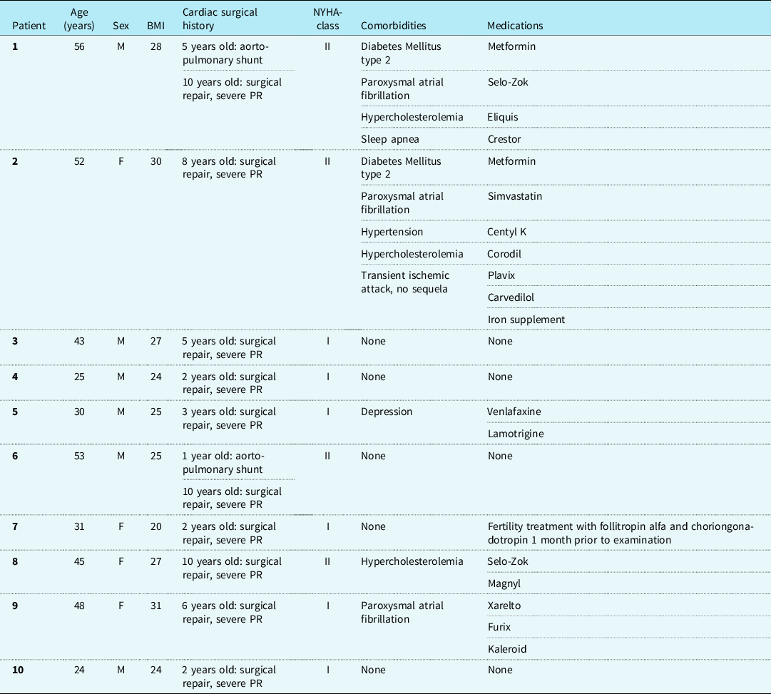
The study is a prospective, cross-sectional study of four women and six men with age ranging from 24 to 56 years (please refer to Table 1 for demographic data). They were all recruited from the outpatient clinic for Grown-Ups with Congenital Heart Disease at Copenhagen University Hospital, Rigshospitalet, Denmark. Only adult patients operated for tetralogy of Fallot with a resulting severe pulmonary regurgitation, no later interventions on the pulmonary valve, and no known liver disease were included in the study.
Demographic data

BMI = body mass index; F = female; M = male; NYHA-class = New York Heart Association Functional Classification; PR = pulmonary regurgitation.
All 10 patients were examined with the use of magnetic resonance imaging (MRI) Elastography of Liver, Hepatobiliary iminodiacetic acid scan, indocyanine green elimination by pulse spectrophotometry, elastography via FibroScan, abdominal ultrasound including liver elastography, blood samples including liver markers, echocardiography and an electrocardiogram. All investigations for liver condition were performed during the same day, with patients fasting a minimum of 6 hours. All patients had a cardiac MRI (cMRI) within 18 months of this study which was included in the data analysis.
The study complies with the 2013 Declaration of Helsinki made by the World Medical Association and was approved by The National Committee on Health Research Ethics (H-18009348). All participants provided written informed consent prior to enrolment.
Magnetic resonance imaging with elastography of liver
Liver stiffness measured by MRI elastography is validated as a diagnostic tool for liver fibrosis, Reference Singh, Venkatesh and Wang16 and has also been confirmed as a sensitive tool to evaluate for liver fibrosis in patients with CHD. Reference Sugimoto, Oka and Kajihama17 MRI with elastography of liver was performed on a 1.5-T MRI scanner (GE Optima™ 450W, General Electric Healthcare, Chicago Illinois, USA). Vibrations at 60 Hz were produced from an active driver and transmitted to a passive driver placed over the patient’s right hepatic lobe. Elastograms were generated by shear wave images with a modified phase contrast gradient echo sequence (slice thickness 8mm; gap 8mm; matrix 256 × 64; repetition time/echo time 50/21.9 ms; bandwidth ±31.25 kHz; flip angle 30; field of view 32–42cm). Processing and measurements of liver stiffness were made by one experienced MRI radiologist. Three MRI elastography slices were obtained per patient, with slices through the largest cross-section of the liver avoiding the dome and the inferior part of the liver. Avoiding liver edges, fissures, large vessels, wave interference and artifacts the largest possible region of interest was drawn based on the magnitude image as reference. Liver stiffness values were computed for each slice, and overall stiffness values for the whole liver were recorded as the median of all slices.
Hepatobiliary iminodiacetic acid scan
Hepatobiliary iminodiacetic acid scan with measurements of 99mTc-mebrofenin uptake rate is reported to be a valid method for assessment of liver function. Reference Erdogan, Heijnen and Bennink18 99mTc-mebrofenin was injected with a dose of given 150MBq. After injection of tracer, the patients were placed in the supine position under a two-headed gamma camera equipped with a high-resolution parallel-hole collimator. A dynamic scintigraphy was performed one minute per frame for 1 hour. Hepatocyte extraction fraction and hepatic clearance time was calculated from the dynamic scan.
Noninvasive indocyanine green clearance
Indocyanine green elimination is considered to correlate with hepatic function. Reference Vos, Wietasch, Absalom, Hendriks and Scheeren19 Indocyanine green elimination measurements were made by non-invasive pulse dye densitometry, which correlates with invasively determined values, which is considered the gold-standard. Indocyanine green (Verdye, Diagnostic Green, Aschheim-Dornach, Germany) was injected as a bolus of 0.25 mg/kg body weight Reference Sakka, Koeck and Meier-Hellmann20 in a peripheral vein catheter and flushed with saline. The minimally invasive pulse spectroscopy device LiMON (Pulsion, Maquet Holding B.V. & Co., Rastatt, Germany) was applied with the finger-clip attached to the patient’s index finger. The device conducted pulse spectroscopy for one minute prior to injection to obtain a baseline value and 7.5 min after injection. It provided a plasma disappearance rate (PDRLi,%/min) as well as (R15Li,%) as an extrapolation of the 7.5-minute spectroscopy.
FibroScan
The FibroScan device (Echosens®, Paris, France) was used to measure transient elastography. FibroScan is documented as an effective method for assessing liver fibrosis. Reference Castéra, Vergniol and Foucher21 Two operators performed the measurements. The patients were placed in supine position with the right arm elevated. The transducer was then placed over the right hepatic lobe. Reported values of liver stiffness in kilopascal (kPa) are based on 10 valid readings per patient, with an interquartile range less than 30% of the median liver stiffness.
Abdominal ultrasound with liver elastography
Ultrasound elastography is a documented tool to assess liver fibrosis. Reference Sigrist, Liau, El, Chammas and Willmann22 Abdominal ultrasound including 2D shear wave elastography was performed using LOGIQ E9 machines with a curved C1-6VN transducer (General Electric Healthcare, Chicago Illinois, USA). Standard B-mode examination of the liver and spleen including flow evaluation was performed on all patients. Ultrasound elastography was measured in the right hepatic lobe, 1–5 cm below the liver capsule with patients in supine position and elevated right arm. All examinations and calculation of elastography measurements were performed by one experienced radiologist. The median of 10 valid measurements was reported as the ultrasound elastography value.
Echocardiography and cardiac MRI
All patients are followed at the GUCH clinic at Rigshospitalet, with regular clinic visits including transthoracic echocardiography and cardiac MRI (cMRI). Data from the latest transthoracic echocardiography and cMRI made before the other examinations were reviewed retrospectively, with a maximum of 18 months between transthoracic echocardiography and liver function tests.
Liver biopsies
Liver tissue was formalin fixed and paraffin embedded. Sections 2–4 um thick were cut and a routine staining panel applied including: Haemotoxylin and Eosin (H&E), modified sirius, periodic acid-Schiff, Masson trichome, periodic acid-Schiff with diastase, iron, reticulin artisan and oxidized orcein. Liver tissue sections were immunohistochemically stained on 3 µm thick sections using the CK7 antibody from Dako/Agilent, GA619 (clone OV-TL12/30) following the manufacturer’s instructions. The staining took place on the Omnis from Agilent utilizing the EnVision Flex+ detection kit (GV800). The primary antibody was diluted using Antibody Diluent (Dako DM830) and were incubated for 20 minutes. The sections were counterstained with haematoxylin. Fibrosis scores are reported as Metavir-score. Reference Bedossa and Poynard23
Statistics
This is a descriptive study comprising 10 patients. It was designed as a pilot study, to indicate whether future larger studies should be performed. The group is too small for extensive statistics.
Results
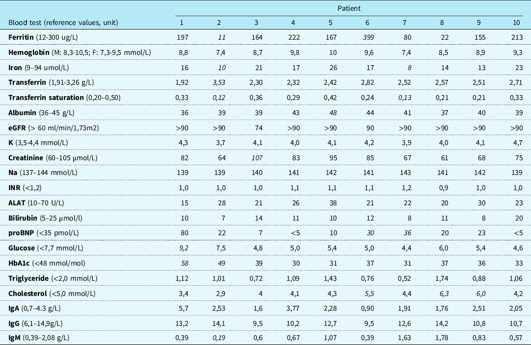
All patients completed all non-invasive tests, but liver biopsy was only performed for clinical indications. In one patient there was an artefact on the MR liver elastography making it unreadable. For another patient (with steatosis on abdominal ultrasound) the FibroScan could not be performed properly. All patient charts including prescription drug consumption were reviewed to control for other diseases or medicine potentially influencing liver function, including excessive alcohol use. Blood samples including liver markers were obtained from all patients, with all results within reference values (Table 2). All subjects tested negative for hepatitis B and hepatitis C (HBsAg, HCV-RNA). Patients were also examined and evaluated for possible liver stigmata, which were not present in any.
Blood test results

ALAT = alanine aminotransferase; eGFR = estimated glomerular filtration rate; HbA1c = glycated haemoglobin; IgA = immunoglobulin A; IgG = immunoglobulin G; IgM = immunoglobulin M; INR = international normalized ratio; Na = natrium; proBNP = pro-brain natriuretic peptide.
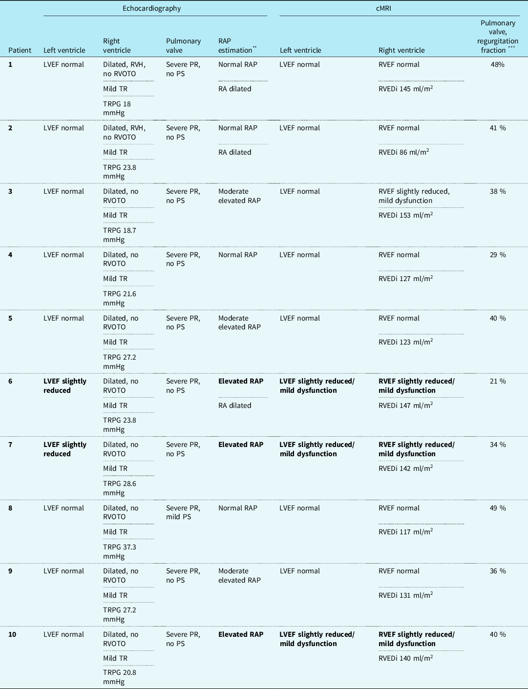
Cardiac history was obtained from patient charts (please refer to Table 3 for baseline cardiac data). Electrocardiogram was taken on the day of examination, and all patients presented with sinus rhythm. Three patients had a history of paroxysmal atrial fibrillation, other than this no clinical arrhythmia was registered.
Cardiac baseline information

cMRI = cardiac magnetic resonance imaging; LVEF = left ventricular ejection fraction; PR = pulmonary regurgitation; PS = pulmonary stenosis; RA = right atrium; RAP = right atrial pressure; RVEDi = right ventricular end diastolic volume index; RVEF = right ventricular ejection fraction; RVH = right ventricular hypertrophy; RVOTO = right ventricular outflow obstruction; TR = tricuspid regurgitation; TRPG = tricuspid regurgitation peak gradient.
** Normal RAP = inferior vena cava (IVC) small and inspiratory collapse > 50 %, suggesting RAP ≤ 5 mmHg; moderate elevated RAP = IVC > 21 mm, inspiratory collapse > 50%, suggesting RAP 5–10 mmHg; elevated RAP = IVC dilated, inspiratory collapse < 50%, suggesting RAP 10–20 mmHg. Reference Kutty, Peng and Danford27
*** Degree of pulmonary regurgitation is listed as mild if regurgitant fraction <20%; moderate if 20–40%; severe if >40%. Reference Bataller and Brenner28
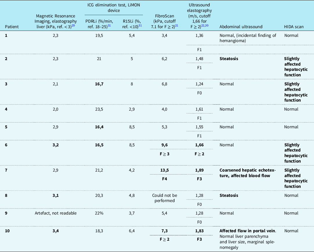
Of the 10 patients examined, four had results within normal values in all tests of liver condition (MRI elastography, hepatobiliary iminodiacetic acid scan, indocyanine green elimination by pulse spectrophotometry, abdominal ultrasound, Fibroscan). One male patient with otherwise normal results was referred for magnetic resonance cholangiopancreatography because of discrete delayed clearing of intrahepatic bile ducts on the Hepatobiliary iminodiacetic acid scan, with magnetic resonance cholangiopancreatography being normal. Two patients had mild signs of steatosis. One patient with known diabetes mellitus type 2, and one patient with known hypercholesterolemia. Three patients had results indicative of possible liver fibrosis (please refer to Table 4). These patients were referred to the Gastroenterology outpatient clinic at Copenhagen University Hospital for further evaluation from a hepatologist.
Test results from various liver function tests

F = fibrosis Metavir -score; HIDA = hepatobiliary iminodiacetic acid; ICG = indocyanine green; MRI = magnetic resonance imaging; ref. = reference value.
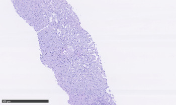
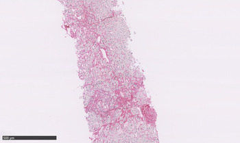
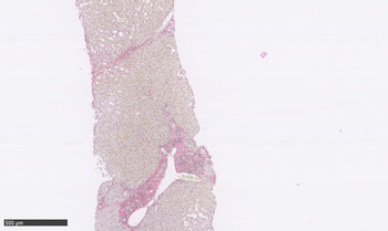
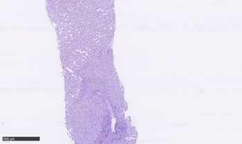
Patient 6, a 53-year-old male, had findings slightly outside the reference values on MRI liver elastography, indocyanine green elimination by pulse spectrophotometry, FibroScan, hepatobiliary iminodiacetic acid scan and ultrasound elastography, but otherwise a normal abdominal ultrasound. A new FibroScan measurement reporting liver stiffness of 8,9 kPa was later made in the outpatient clinic, with no other signs of liver disease. Patient 7, a 31-year-old female had signs of probable fibrosis on abdominal ultrasound and FibroScan, with hepatobiliary iminodiacetic acid scan indicating slightly affected hepatocytic function, but an MRI liver elastography just within the normal range, and normal indocyanine green elimination by pulse spectrophotometry. Repeated FibroScan measurement of 8,6 kPa and a liver biopsy was later made. Liver histology was described as irregular fibrosis minimum stage 2 and extensive centrilobular fibrosis and dilation indicating chronic venous stasis, Figures 1 and 2. Patient 10, a 24-year-old male had findings indicative of possible fibrosis on MRI liver elastography, FibroScan, ultrasound elastography and abdominal ultrasound, with normal findings on Hepatobiliary iminodiacetic acid scan and indocyanine green elimination by pulse spectrophotometry. Liver histology was described as fibrosis stage 1 and centrilobular fibrosis and dilatation indicating chronic venous stasis, Figures 3 and 4. Repeated FibroScan at the time of biopsy reported liver stiffness of 6,2 kPa.
Liver histology from patient 7. Haematoxylin-Eosin (HE) stain showing liver tissue with sinusoidal dilation.

Liver histology from patient 7. Modified sirius staining of connective tissue with extensive centrilobular perisinusoidal fibrosis.

Liver histology from patient 10. HE stain showing liver tissue with sinusoidal dilation.

Liver histology from patient 10. Modified sirius staining of connective tissue with portal fibrosis stage 1 and centrilobular perisinusoidal fibrosis.

Discussion
While Fontan associated liver disease is established as a common complication in Fontan patients, Reference Apitz, Latus and Binder9 liver fibrosis is currently not considered a clinically relevant complication in tetralogy of Fallot patients. Although hepatic fibrosis may be less severe and prevalent among tetralogy of Fallot patients compared to Fontan patients, two recent studies have addressed liver disease as a possible complication in patients with tetralogy of Fallot.
Zhang et al Reference Zhang, Li and Jani24 recently reported increased liver stiffness in 36 young adults with biventricular CHD, including 18 patients with tetralogy of Fallot. Liver stiffness was measured by shear wave elastography. The patients had a mean age of 27 years, and none of the included subjects had more than mild semilunar regurgitation. Still, the results yielded significantly higher liver stiffness in CHD patients compared to age-matched healthy controls. Based on vascular Doppler findings from the hepatic ultrasound, elevated central venous pressure is mentioned as a possible cause although no results from invasive haemodynamic measurements are available to confirm this. However, these are new and interesting findings, which support a hypothesis of biventricular CHD patients with elevated central venous pressure being at risk for developing liver disease.
Another recent study by Yamamura et al Reference Yamamura, Sakamoto and Morihana12 reported significantly elevated liver markers in both Fontan and tetralogy of Fallot patients, compared to controls with other CHD and no right side heart sequelae. Forty-five out of fifty tetralogy of Fallot patients had significant pulmonary regurgitation, making this cohort somewhat similar to the 10 patients included in the present study. Two out of twenty-two of the tetralogy of Fallot patients undergoing abdominal ultrasound had findings suggesting liver cirrhosis. These two, a 32-year-old male and a 50-year-old female, subsequently had a liver biopsy made which confirmed the presence of moderate (F2) and severe (F3) liver fibrosis, respectively. In comparison, the present study found paraclinical signs of possible fibrosis in as many as three out of ten patients, with histological confirmation of fibrosis stage F1 and F2 in the two patients who underwent liver biopsy. However, in Yamamura et al’s study only twenty-two out of fifty tetralogy of Fallot patients had abdominal ultrasound performed, and this was the only test evaluating liver function in addition to blood samples. This set-up could explain the lower fraction of patients presenting with fibrosis in their study compared to our patients, supported by the finding that they only identified patients with moderate and severe fibrosis. As liver fibrosis is considered asymptomatic and standard laboratory investigations are of limited value, a multimodal evaluation including both abdominal ultrasound and some form of elastography is considered necessary to diagnose early stages of fibrosis, as recommended when screening for liver complications among Fontan patients. Reference Gordon-Walker, Bove and Veldtman3 This approach is confirmed in the present study, with all subjects having normal biochemical liver parameters, and somewhat differing results in what test modality that best picked up on abnormal liver function. Still, the combination of MRI elastography, FibroScan, and abdominal ultrasound including elastography seems to be a good approach when looking for early signs of fibrosis in this patient group. However, it should be noted that in the two patients referred to liver biopsies, both FibroScan and Ultrasound elastography gave higher Fibrosis scores than the liver biopsies. This may be explained by an overestimation of liver stiffness by elastography measurements due to elevated central venous pressure in these patients.
Central venous pressure is known to influence liver stiffness measured by elastography. A previous study has shown elevated liver stiffness in Fontan-patients post-surgery, interpreted as an immediate response to hepatic congestion. Reference DiPaola, Schumacher, Goldberg, Friedland-Little, Parameswaran and Dillman25 Similarly, increased liver stiffness has been described in patients with decompensated heart failure, with a significant decrease after patients improved. Reference Millonig, Friedrich and Adolf26 Although this relation should be considered when interpreting liver stiffness in patients with known elevated central venous pressure, estimation of liver stiffness should still be considered relevant when looking for liver fibrosis. A study of liver function in 41 Fontan patients, including 10 with liver biopsy, found a statistically significant correlation between degrees of liver stiffness and histopathological fibrosis. Reference Kutty, Peng and Danford27
The literature reports progression from liver fibrosis to cirrhosis in the majority of patients after an interval of 15–20 years. Reference Bataller and Brenner28 In accordance, the degree of fibrosis increases with time in Fontan patients. Reference Rychik, Veldtman and Rand4 Cirrhosis is the main risk factor for development of hepatocellular carcinoma, and the annual risk of hepatocellular carcinoma in cirrhotic Fontan associated liver disease patients is estimated to 1.5–5%. Reference Gordon-Walker, Bove and Veldtman3 However, hepatocellular carcinoma is to our knowledge only reported in three previous tetralogy of Fallot cases. Nevertheless, the two case studies describing hepatocellular carcinoma in tetralogy of Fallot patients Reference Mccabe, Farris, Hon, Ford and Book13,Reference Augustyn, Peng, Singal and Yopp14 and Yamamura et al’s study which described a combined hepatocellular and cholangiocarcinoma could suggest that fibrosis secondary to hepatic congestion is associated with increased risk of liver malignancy. Importantly, however, no causality between liver congestion and liver malignancy has yet been demonstrated in tetralogy of Fallot patients. With the potentially malign progression of fibrosis over time in mind, it should however be noted that the two patients with confirmed fibrosis in this study were some of the youngest in the cohort, 30 and 25 years respectively. Therefore, although only stage F2/F1 fibrosis was demonstrated in these patients, liver fibrosis is not a static condition, and the findings may be considered clinically relevant.
This study examined tetralogy of Fallot patients with moderate to severe pulmonary regurgitation, with the hypothesis that a severe pulmonary regurgitation can predispose for liver congestion by means of elevated right atrial pressure. In the absence of central venous stenosis the right atrial pressure is equal to central venous pressure, and the association between elevated central venous pressure and hepatic fibrosis is known from studies on functionally normal hearts with right sided heart failure. Reference Xanthopoulos, Starling, Kitai and Triposkiadis29 Elevated central venous pressure is also considered an important part in the aetiology in Fontan associated liver disease, as Fontan patients are known to have increased central venous pressure. Reference Rychik, Veldtman and Rand4 Unfortunately, there are no heart catheterisation data on the patients included in this study. However, right atrial pressure estimated by inferior caval vein diameter and inspiratory collapse on transthoracic echocardiography was reviewed retrospectively. Interestingly, the same three patients that were referred to a hepatologist because of possible liver fibrosis, all had signs of elevated right atrial pressure on previous transthoracic echocardiography. On the contrary, estimated high right atrial pressure was not reported in any of the other seven subjects. In accordance, both patients with confirmed liver fibrosis in Yamamura et al’s study had an elevated right atrial pressure of 12 mmHg and 16 mmHg respectively, and in the multivariate analysis right atrial pressure had a significant association with elevated type-IV collagen. Furthermore, an elevated right atrial pressure of 35 mm Hg was also reported in both of the previously mentioned case studies on tetralogy of Fallot patients with hepatocellular carcinoma. Reference Mccabe, Farris, Hon, Ford and Book13,Reference Augustyn, Peng, Singal and Yopp14 Also of interest is a study reporting a significant association between liver stiffness measured by FibroScan and elevated central venous pressure in children and adults with CHD. Reference Jalal, Iriart and De Ledinghen30 However, this paper included patients with different types of CHD, and none of the subjects were further evaluated with liver biopsy to investigate the presence of actual fibrosis in those with elevated measurements on FibroScan. Thus, larger samples are needed to elucidate whether elevated right atrial pressure is associated with liver fibrosis in tetralogy of Fallot patients.
The presence of a severe pulmonary regurgitation, which in turn may be correlated with elevated central venous pressure, could also be considered an attributing factor in the development of hepatic congestion and fibrosis in tetralogy of Fallot patients. Five out of ten patients in this study had severe pulmonary regurgitation as measured on cMRI, but in the three patients with affected liver function only one had definitive severe pulmonary regurgitation, defined as pulmonary regurgitation fraction ≥40% in cMRI. All patients were however described to have a severe pulmonary regurgitation on transthoracic echocardiography, and it may be the presence of a significant pulmonary regurgitation, whether moderate or severe, that has the potential to cause non-favourable hemodynamics, possibly leading to hepatic congestion. Pulmonary regurgitation fraction measured by cardiac MRI also had a significant association with elevated type IV collagen in the mentioned study from Yamamura et al. One could speculate if this sub-population of tetralogy of Fallot patients is at special risk of developing liver fibrosis, given the quite high proportion of patients with signs of fibrosis in our study. Nevertheless, more studies including patients without pulmonary regurgitation are necessary to probe whether prolonged pulmonary regurgitation is a possible risk factor for later liver complications. However, a potential correlation between pulmonary regurgitation and hepatic dysfunction would certainly be of clinical relevance, as replacement of the pulmonary valve is a possible treatment option for these patients. The discussion of when to replace a dysfunctional pulmonary valve in tetralogy of Fallot patients is ongoing, Reference Rajpopat, Schmidt and Søndergaard8 and a potential risk of developing liver fibrosis would be an argument for earlier replacement of the pulmonary valve.
Lastly, it should be noted that the three patients with abnormal findings on liver tests all had mild biventricular dysfunction on cMRI and/or echocardiography, contrary to the other seven patients, who all had normal left ventricular function. As decreased cardiac output is considered part of the process in the development of Fontan associated liver disease, Reference Emamaullee, Zaidi and Schiano31 this is an interesting finding. It could indicate that decreased ventricular function may make some patients with tetralogy of Fallot vulnerable for hepatic injury.
It would be interesting to investigate the prevalence and natural course of liver complications in a larger cohort with patients with tetralogy of Fallot. Information on whether liver complications are prevalent in these patients is essential when planning future follow-up plans. Screening for liver complications in all patients, as recommended in Fontan patients Reference Emamaullee, Zaidi and Schiano31 is certainly not reasonable with our current knowledge, as we still do not know if mild forms of liver fibrosis are of clinical importance in patients with tetralogy of Fallot. Liver fibrosis is a condition with no specific treatment options, and it can be considered unethical to systematically investigate for an untreatable complication with unknown importance. However, more research is needed to establish whether liver complications should be added to the list of possible complications to look for in patients with tetralogy of Fallot.
Limitations
This study has several limitations. The results come from a small sample of 10 patients, and therefore no statistics calculations have been performed. With such a small sample and no control group, no conclusions can be made about whether the findings of mild liver fibrosis in selected patient is related to their CHD. Seven of the 10 patients had a BMI ≥ 25 which could be a bias when assessing liver function; however, the two patients with findings of mild liver fibrosis on biopsy both had normal body mass index. Patients with tetralogy of Fallot and pulmonary regurgitation can have elevated central venous pressure, which can overestimate liver stiffness on elastography measurements, why liver biopsies were performed to confirm the findings when relevant.
Conclusion
This study presents findings of fibrosis in two out of ten patients with tetralogy of Fallot and pulmonary regurgitation. Although only mild degrees of fibrosis were found, the potential progression of fibrosis to cirrhosis and possibly hepatocellular carcinoma, indicate that these findings should not be overlooked. As the ageing tetralogy of Fallot population grows, the finding of even mild liver fibrosis in young tetralogy of Fallot patients calls for further investigations and larger samples.
Acknowledgements
Assistance in performing the examinations provided by Anna Viktoria Setterberg, Mariam Hassan and Britt Maria Tolstrup, the technologists at Clinical Physiology, Nuclear Medicine and PET, Copenhagen University Hospital, Rigshospitalet was greatly appreciated. The authors are also grateful for the Department of Anesthesiology, Copenhagen University Hospital, Rigshospitalet to provide the LiMON device.
Financial support
The authors received no financial support for the research or authorship of this article.
Conflicts of interest
None.
Ethical standards
The authors assert that all procedures contributing to this work comply with the ethical standards of the relevant national guidelines on human experimentation (The Committee Act) and with the Helsinki Declaration of 1975, as revised in 2008, and has been approved by the institutional committees (The National Committee on Health Research Ethics).