Formal thought disorder (FTD), speech that is difficult for the listener to follow, sometimes to the point of complete incomprehensibility, occurs in 25%–75% of patients with schizophrenia (Roche, Creed, MacMahon, Brennan, & Clarke, Reference Roche, Creed, MacMahon, Brennan and Clarke2015). Multiple clinical abnormalities are considered to contribute to the phenomenon: in her widely used Thought, Language and Communication (TLC) scale, Andreasen (Reference Andreasen1979) distinguished 18 different elements. This author also argued for the existence of ‘positive’ (i.e. characterized by the presence of abnormality) and ‘negative’ (characterized by diminution of a normal function) forms of FTD, and factor analytic studies have tended to support this distinction, finding that two factors account for a large amount of the variance in TLC scores among schizophrenic patients (Roche et al., Reference Roche, Creed, MacMahon, Brennan and Clarke2015). One of these, termed ‘disorganization’ (Peralta, Cuesta, & de Leon, Reference Peralta, Cuesta and de Leon1992) or ‘fluent disorganization’ (Andreasen & Grove, Reference Andreasen and Grove1986), typically includes items such as tangentiality, derailment, incoherence, illogicality, circumstantiality, and loss of goal. The other, labelled ‘alogia’ (Andreasen, Reference Andreasen1982) or ‘emptiness’ (Andreasen & Grove, Reference Andreasen and Grove1986) has been found to load on poverty of speech and poverty of content of speech and less consistently on perseveration.
Among several different theoretical approaches to FTD (for reviews see Kircher, Brohl, Meier, & Engelen, Reference Kircher, Brohl, Meier and Engelen2018; McKenna & Oh, Reference McKenna and Oh2005), it has been proposed that at least some of the phenomenon reflects underlying executive or frontal lobe dysfunction (Chaika, Reference Chaika1990; Liddle, Reference Liddle1987; McGrath, Reference McGrath1991). According to this proposal, impaired planning and monitoring lead to the speech of patients with FTD being poorly formulated and prone to errors during execution, which then go unnoticed by the patient and uncorrected. Supporting this view, significant correlations have been found between scores on the disorganization syndrome in schizophrenia (the main symptom of which is FTD) and poor performance on a range of different executive tests (Dibben, Rice, Laws, & McKenna, Reference Dibben, Rice, Laws and McKenna2009). More recently, Bora et al. (Bora, Yalincetin, Akdede, & Alptekin, Reference Bora, Yalincetin, Akdede and Alptekin2019) meta-analyzed the neuropsychological correlates of positive FTD and alogia in 50 studies of schizophrenia and found that both were significantly associated with poor performance on a range of executive tests. However, in both these meta-analyses, correlations were also found with impairment in non-executive tests, as well as with global measures of cognition.
Another potential way of examining the frontal/executive hypothesis of FTD is by means of functional imaging. Wensing et al. (Reference Wensing, Cieslik, Muller, Hoffstaedter, Eickhoff and Nickl-Jockschat2017) meta-analyzed 18 whole-brain task-related PET and fMRI studies, 17 on schizophrenic patients and one employing healthy subjects who had been given ketamine. They found pooled evidence for activation changes associated with FTD in the temporal lobe (the left superior temporal gyrus was associated with both hyper- and hypo-activation in the individual studies, and the left posterior medial temporal gyrus was associated with hyperactivation), but there were no associations with activation changes in frontal regions. However, it should be noted that the tasks used in the studies included by Wensing et al. (Reference Wensing, Cieslik, Muller, Hoffstaedter, Eickhoff and Nickl-Jockschat2017) were wide-ranging (passive listening, semantic priming, making semantic judgements, word generation, free word association, the 1-back version of the n-back task, and the go/no-go task), and only three of them – word generation, the 1-back task and the go/no-go task – would normally be considered to make demands on executive function.
In this study, therefore, we further examined the brain functional correlates of FTD, using a relatively demanding executive task, the 2-back version of n-back task. We also employed a larger sample of patients than in previous studies, who showed a wide range of FTD scores. Finally, we were interested in the extent to which brain activations associated with FTD were related to the two main factor-analytically derived subsyndromes of the symptom, fluent disorganization and alogia.
Methods
Participants
The patient group consisted of adults meeting DSM-IV criteria for schizophrenia recruited from hospitals in the Barcelona area (Benito Menni CASM, Hospital Mare de Déu de la Mercè, Hospital Sagrat Cor de Martorell). They were excluded if they (a) were younger than 18 or older than 65, (b) were left-handed, (c) had a history of brain trauma or neurological disease, or (d) had shown alcohol/substance abuse within 12 months prior to participation. With respect to the last criterion, we also excluded those who reported habitual use of cannabis. Social use of alcohol was permitted, as was non-habitual use of cannabis. All the patients had either subchronic or chronic illness (duration 1–38 years). The patients were all taking antipsychotic medication (typical n = 5; atypical n = 44; both n = 23). The mean daily dose (in chlorpromazine equivalents) was 587.27 mg (s.d. = 420.95 mg).
IQ (premorbid IQ in the patients) was estimated using the Word Accentuation Test (Test de Acentuación de Palabras, TAP) (Del Ser, Gonzalez-Montalvo, Martinez-Espinosa, Delgado-Villapalos, & Bermejo, Reference Del Ser, Gonzalez-Montalvo, Martinez-Espinosa, Delgado-Villapalos and Bermejo1997; Gomar et al., Reference Gomar, Ortiz-Gil, McKenna, Salvador, Sans-Sansa, Sarro and Pomarol-Clotet2011), a test that requires pronunciation of Spanish words whose accents have been removed. The patients were also required to have a current IQ in the normal range (70+), as measured using four subtests of the Wechsler Adult Intelligence Scale III (WAIS-III): Vocabulary, Similarities, Block Design and Matrix Reasoning.
Although the main focus of the study was on correlations between FTD and activations in the patient group, we also examined a comparison group of healthy controls who were selected to be age- and sex-matched with the patients and met the same exclusion criteria. We used findings from this contrast to determine how far regions of activation related to FTD in the patients could be considered to be located within the network of regions normally activated (i.e. by the controls) during task performance, and also by the patients themselves.
All participants gave written informed consent prior to participation in accordance with the Declaration of Helsinki. All the study procedures were approved by the local Clinical Research Ethics Committee (Comité de Ética de Investigación Clínica de las Hermanas Hospitalarias).
Clinical assessment
Symptoms were assessed with the Positive and Negative Syndrome Scale (PANSS) (Kay, Fiszbein, & Opler, Reference Kay, Fiszbein and Opler1987). To rate FTD, the patients were administered a videotaped semi-structured interview. This lasted 15–25 min and consisted of three parts: in the first, patients were asked to freely talk about their childhood (being prompted if necessary); in the second, they were asked to interpret five Rorschach inkblots; and in the third, they had to tell one or more fairy tales (techniques used by Kircher et al., Reference Kircher, Liddle, Brammer, Williams, Murray and McGuire2001; Oh, McCarthy, & McKenna, Reference Oh, McCarthy and McKenna2002; Rochester & Martin, Reference Rochester and Martin1979). TLC ratings were then made by four clinicians who had been trained in its use (EP-C, BS-S, SS and LL-A); they worked in pairs and resolved differences in case of disagreement. We used the 1986 version of the TLC (Andreasen, Reference Andreasen1986) where items are rated from 0 (no more than occasional instances of less severe forms of abnormality such as circumstantiality, loss of goal, stilted speech) to 4 (extreme).
As well as employing the TLC global score item to assess overall FTD severity, we scored fluent disorganization (summed scores on derailment, loss of goal, tangentiality, illogicality, circumstantiality and incoherence) and alogia (summed scores on poverty of speech, poverty of content of speech and perseveration); inclusion of these items was based on the study of Peralta et al. (Reference Peralta, Cuesta and de Leon1992).
N-back task
Participants performed a sequential-letter version of the n-back task (Gevins & Cutillo, Reference Gevins and Cutillo1993) during fMRI scanning. The task consisted of two levels of memory load (1-back and 2-back) presented in a blocked design manner: in the 1-back condition, participants had to respond with a key press with their right index when the letter shown on the screen was the same as the one that was presented immediately before, whereas in the 2-back condition they had to respond when the letter was the same as that presented two letters previously. In each task block, 24 letters were sequentially shown every 2 s (1 s on, 1 s off), for a total block duration of 48 s. Each block contained five repetitions of targets requiring a response from the subject (1-back and 2-back depending on the block) located randomly within the block. In order to identify which task had to be performed, letters were shown in green in the 1-back blocks and in red in the 2-back blocks. Four 1-back and four 2-back blocks were presented in an interleaved way, and between them, a baseline stimulus (an asterisk flashing with the same frequency as the letters) was presented for 16 s, which provided a resting baseline period. All individuals went through a training session before entering the scanner.
Subjects' n-back performance was measured with the signal detection theory sensitivity index, d prime (d’) (Green & Swets, Reference Green and Swets1966), which takes into account the number of hits and false alarms during the task. Higher values of d’ indicate a better ability to discriminate between targets and distracters. Subjects with negative d’ values in either or both the 1-back and 2-back conditions were presumed to not be performing the task and were excluded a priori from the study.
Imaging data acquisition and analysis
Images were acquired in a 1.5 T GE Signa scanner (General Electric Medical Systems, Milwaukee, Wis) using a gradient echo-planar imaging (EPI) sequence depicting the blood oxygenation level-dependent (BOLD) contrast. The sequence consisted of 266 volumes, each containing 16 axial planes acquired with the following parameters: TR = 2000 ms, TE = 40 ms, flip angle = 70°, section thickness = 7 mm, in-plane resolution = 3 × 3 mm2, inter-slice gap = 0.7 mm. The first 10 volumes were discarded to avoid T1 saturation effects.
Imaging analyses were performed with the FEAT module included in the FSL software (Smith et al., Reference Smith, Jenkinson, Woolrich, Beckmann, Behrens, Johansen-Berg and Matthews2004). For the first-level analyses, images were motion-corrected using the MCFLIRT algorithm and co-registered to a common stereotactic space (Montreal Neurological Institute template). The data were high-pass filtered (130s cutoff) to remove low-frequency signal drifts. Before group analyses, normalized images were spatially filtered with a Gaussian filter (FWHM = 5 mm). Individuals with an estimated maximum absolute movement >3.0 mm or an average absolute movement >0.3 mm were excluded from the study to minimize unwanted movement-related effects.
Statistical analyses were performed by means of general linear models (GLMs) with two regressors of interest (1-back and 2-back conditions) and with movement parameters as additional regressors of non-interest (six in total, three rotations and three translations). Resting periods were not modelled and acted as implicit baseline. GLMs were fitted to generate individual activation maps for the 1-back v. baseline and 2-back v. baseline contrasts. Group comparisons were carried out with two sample t tests on the 2-back >baseline contrast with mixed-effects models (Beckmann, Jenkinson, & Smith, Reference Beckmann, Jenkinson and Smith2003).
To study the association between FTD severity and brain activity during the n-back task, regression analyses were performed in the patient group in the 2-back v. baseline contrast with the global score of the TLC and the two factor-analytic subscales, fluent disorganization and alogia. Sex, age and estimated premorbid IQ (TAP score) were included as covariates since these factors would be expected to independently influence fMRI findings. We chose the 2-back v. baseline contrast as the main analysis on the basis that the 1-back version of the task is easy and is unlikely to engage executive function/working memory to the same degree as the 2-back task.
The default threshold of z = 2.3 in FSL was used to define the initial set of clusters. All statistical tests were carried out with a significance level of p < 0.05, FWE-corrected for multiple comparisons at the cluster level by means of Gaussian random field methods, as standard in FSL.
Results
From an initial pool of 103 patients, 70 were finally included in the study. Two patients were excluded because of comorbidity: one had an illness that could have affected brain function and in the other, a structural scan revealed an incidental finding. Four patients were excluded because they were left-handed. Thirteen were excluded because of excessive head movement during the scanning, five because of poor behavioural performance on the n-back task (i.e. negative d’ scores) and eight because they had a current IQ lower than 70. These 70 patients were matched with 70 healthy controls. Demographic and clinical data for the final sample of patients and controls are shown in Table 1.
Demographic and clinical data

Values are means (s.d.), except for TLC scores, which were not normally distributed. Values for this variable are given as medians and inter-quartile ranges (IQR), with means and standard deviations also given in square brackets.
A breakdown of the global TLC scores revealed that 42 of the 70 patients scored 0 (absent/minimal) 9 scored 1 (mild), 11 scored 2 (moderate), 6 scored 3 (severe) and 2 scored 4 (extreme).
Behavioural performance
The mean d’ in the healthy controls was 4.32 ± 0.76 in the 1-back and 3.35 ± 0.89 in the 2-back conditions, respectively. The corresponding values for the patients were 3.41 ± 1.09 in the 1-back and 2.24 ± 0.87 in 2-back condition. A repeated measures ANOVA revealed a significant main effect of condition (F = 191.91, p < 0.001) indicating that performance was better in the 1-back than in the 2-back condition, and a significant main effect of group (F = 56.44, p < 0.001), indicating that the controls performed better than the patients. The group × condition interaction was not significant (F = 1.33, p = 0.25).
Global FTD score was correlated with 2-back performance at trend level (rs = −0.22, p = 0.06). The correlation between Global FTD and 1-back performance was slightly lower and not significant (rs = −0.19, p = 0.11). N-back performance was significantly negatively correlated with alogia both in the 1-back (rs = −0.40, p < 0.001) and 2-back versions of the task (rs = −0.33, p = 0.005), but fluent disorganization was not (1-back: rs = −0.02, p = 0.84; 2-back: rs = −0.02, p = 0.87).
fMRI: group comparison
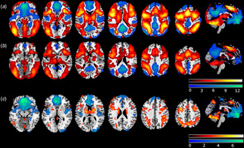
Mean activation maps for the 2-back v. baseline contrast are shown in Fig. 1A and B. The pattern of activations was broadly similar in the controls and the patients: both showed a large cluster of activation which encompassed bilateral dorsal fronto-parietal regions, the inferior frontal cortex and anterior insula, the anterior cingulate cortex, the thalamus, the putamen, the visual cortex and the cerebellum.
(A) Activation map for healthy subjects in the 2-back condition. (B) Activation map for schizophrenia patients in the 2-back condition. Warm colours indicate activation, cold colours indicate de-activation relative to baseline. (C) Group comparison. Warm colours indicate areas hypo-activated by the patients, cold colours indicate failure to de-activate in the patients . Colour bars depict Z-values. Images are shown in neurological convention (right is right).

The controls also showed areas of de-activation, seen in the medial prefrontal cortex and posterior cingulate/precuneus, amygdala, hippocampus and posterior temporal cortex, all bilaterally (Fig. 1A). In the patients, de-activations were notably less extensive in the medial prefrontal cortex, and they did not show de-activations in the amygdala, hippocampus or posterior temporal cortex (Fig. 1B).
The between-group comparison (Fig. 1C) revealed that the schizophrenic patients showed significantly reduced activation in the dorsolateral prefrontal cortex (DLPFC) and parietal cortex, and in the thalamus, putamen and cerebellum. They also showed significant failure of de-activation in the medial frontal cortex, insula and temporal cortex and also the mid-cingulate and superior parietal cortex. Details on the MNI coordinates, cluster sizes and p values can be found in online Supplementary Table 1.
Correlations with global TLC scores in the patient group
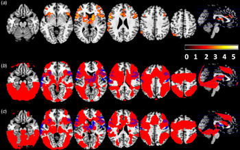
The findings are shown in Fig. 2. Controlling for the effect of sex, age and TAP-estimated premorbid IQ, there was a cluster of significant negative correlation (i.e. greater FTD, less activation) in the right inferior frontal cortex and DLPFC, extending to the frontal operculum (peak MNI coordinates x = 60, y = 20, z = 16; cluster size = 1956 voxels; Z = 4.06; p < 0.001). A second cluster was seen in the left postcentral gyrus extending into the left temporal cortex, left DLPFC and inferior frontal cortex (peak MNI coordinates x = −68, y = −6, z = 22; cluster size = 1442 voxels; Z = 4.86; p < 0.001). There were no clusters of significant positive association with TLC scores.
(A) Areas of significant negative correlation between global FTD scores and brain activity during the 2-back task. Colour bar depicts Z-values. (B) Overlap between brain regions engaged by the control group during the 2-back condition (red) and regions showing a negative correlation with global FTD scores (blue). Overlap is shown in purple. (C) Overlap between brain regions engaged by the patient group during the 2-back condition (red) and regions showing a negative correlation with global FTD scores (blue). Overlap is shown in purple. Images are shown in neurological convention (right is right).

It can be seen from the middle and bottom panels of Fig. 2 that the clusters of significant correlation with global FTD scores in the patient group mostly fell within the regions engaged by both the controls and the patients during performance of the task. The only exceptions were small areas in the temporal cortex in the case of the controls, and small areas in the temporal cortex and basal ganglia/thalamus in the case of the patients.
To examine for potential effects of drug treatment, the above correlational analyses were repeated adding antipsychotic dose (in chlorpromazine equivalents) as a covariate. After doing this, the regions of negative correlation with global TLC scores remained largely similar, with a cluster being seen in the right inferior frontal cortex and DLPFC, that now extended into the right caudate, putamen and thalamus and the left postcentral gyrus extending into the DLPFC and inferior frontal cortex, and now also including the left caudate and putamen. Once again, there were no regions of positive association.
Given that disorganization and FTD have been found to be associated with global cognitive impairment (Bora et al., Reference Bora, Yalincetin, Akdede and Alptekin2019; Dibben et al., Reference Dibben, Rice, Laws and McKenna2009), we also repeated the principal analysis adding current IQ (i.e. WAIS score) as a further covariate. The results remained largely unchanged in the right lateral prefrontal cortex, where a cluster of negative association with FTD scores continued to be seen. However, the cluster in the left lateral prefrontal cortex was no longer evident.
Correlations with fluent disorganization and alogia
Unlike global FTD, fluent disorganization scores were not associated with clusters of negative correlation. Two clusters of positive correlation (i.e. greater FTD, greater activation) were seen in this analysis, in the superior occipital cortex, extending into the white matter on the left (peak MNI coordinates x = −32, y = −70, z = 6; cluster size = 1357 voxels; Z = 4.44; p < 0.001) and right (peak MNI coordinates x = 40, y = −42, z = 6; cluster size = 1405 voxels; Z = 4.69; p < 0.001).
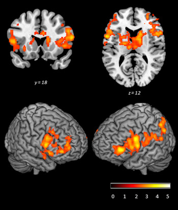
In contrast, the alogia subscale continued to show two clusters of negative correlation, one in the bilateral DLPFC, inferior frontal cortex and basal ganglia (left hemisphere: peak MNI coordinates x = −56, y = 10, z = 26; cluster size = 1956 voxels; Z = 4.33; p < 0.001; right hemisphere: peak MNI coordinates x = 38, y = 36, z = 10; cluster size = 3419 voxels; Z = 4.10; p < 0.001). This frontal region was largely coincident with the regions showing negative correlation with global TLC scores.
Inclusion of antipsychotic dosage in CPZ equivalents as an additional covariate did not significantly alter these findings. Inclusion of WAIS IQ as a covariate similar made little difference.
Areas of significant negative correlation between the alogia subscale and brain activity during the 2-back v. baseline task. Images are shown in neurological convention (right is right). Colour bar depicts Z values.

Correlations with individual alogia items
In order to further explore the associations with the three individual TLC items contributing to the alogia subscale (poverty of speech, poverty of content of speech, and perseveration) we defined the two clusters obtained in the above analysis as regions of interest (ROIs) and extracted the mean parameter estimates for activation in each patient. Correlations with each item, once again controlling for sex, age and TAP-estimated premorbid IQ, are shown in Table 2. All three items proved to be significant predictors of the activation levels of the two ROIs.
Regression analyses in the two ROIs showing association with the alogia subscale. Beta values represent the standardized regression coefficient for each item, covarying for sex, age and TAP

DLPFC: dorsolateral prefrontal cortex.
Discussion
In this study presence of FTD in schizophrenia was correlated with reduced activation in the DLPFC and the inferior lateral frontal cortex bilaterally, as well as in the left temporal cortex and other regions. Further analysis indicated that this correlation reflected an association with the alogia factor of FTD, but not with the fluent disorganization factor. These findings accordingly provide brain functional imaging support for the frontal/dysexecutive theory of FTD, but at the same time suggest that this theory will only ever explain a limited part of the phenomenon.
Our findings differ from those of Wensing et al. (Reference Wensing, Cieslik, Muller, Hoffstaedter, Eickhoff and Nickl-Jockschat2017), whose meta-analysis did not find pooled evidence for an association between FTD and task-related activations in frontal cortical regions. However, as noted in the Introduction, these authors pooled data from studies using many different tasks, only three of which could be considered to be sensitive to executive function. One of these three studies (Erkwoh et al., Reference Erkwoh, Sabri, Schreckenberger, Setani, Assfalg, Sturz and Plessmann2002) did not find associations with FTD within the territory of the prefrontal cortex in 20 patients but did find these elsewhere (the right precentral gyrus, the right ventral anterior thalamic nucleus, the left insula and the right occipital lobe). The other two studies found, like us, associations with reduced activation in frontal regions. Thus, Ragland et al. (Reference Ragland, Moelter, Bhati, Valdez, Kohler, Siegel and Gur2008) examined 13 patients while they produced words from a single semantic category (fruits, furniture, vegetables) or alternated between two semantic categories (e.g. furniture and fruits). Presence of FTD was associated with reduced activation in the right anterior cingulate cortex and cuneus for fluency compared to a baseline contrast of repeating a single word, and with reduced activation in the right superior frontal gyrus compared to a baseline of repeating overlearned lists of words such as the days of the week. Tagamets et al. (Reference Tagamets, Cortes, Griego and Elvevag2014) examined 11 patients during 1-back performance. Associations between a linguistically derived measure which they argued reflected coherence of speech and task-related activations were seen in the superior and middle temporal cortex bilaterally and parts of the lateral and medial frontal cortex, as well as in the posterior visual cortex.
The main finding in Wensing et al.'s (Reference Wensing, Cieslik, Muller, Hoffstaedter, Eickhoff and Nickl-Jockschat2017) meta-analysis was an association between FTD and left temporal cortex activation, something that we also found. However, whereas in their meta-analysis there were associations with both increased and reduced activations in this brain region, in our study the association was in the same direction as with the frontal changes, i.e. less activation with increasing FTD. This association, it should further be noted, fell outside the regions activated by the patients and the controls during n-back performance in our study, and also outside the working memory network identified in a meta-analysis by Owen et al. (Reference Owen, McMillan, Laird and Bullmore2005) in healthy subjects. For this reason its interpretation could be regarded as uncertain, but if genuine, would argue against FTD having a purely frontal/dysexecutive basis.
Further exploration of our findings indicated that reduced lateral prefrontal activation was associated only with the alogia component of FTD. This goes against findings from the neuropsychological literature, particularly the meta-analysis of Bora et al. (Reference Bora, Yalincetin, Akdede and Alptekin2019) cited in the Introduction, which have found small but significant negative correlations between a range of different executive test scores and both positive FTD and alogia scores. As far as we know, only one previous study has examined the brain functional correlates of alogia. Kircher et al. (Reference Kircher, Liddle, Brammer, Murray and McGuire2003) examined correlations between poverty of speech scores and activations in six schizophrenic patients while they gave a verbal description of Rorschach inkblots. Associations with reduced activation were found, but their localization – in the left hippocampal gyrus and fusiform gyrus – was quite different from the lateral frontal, insula, anterior cingulate and parietal cortex regions in our study. Correlations they found within the lateral frontal lobe cortex were with increased activity. Little further can be said about the discrepancy between our and Kircher et al.'s (Reference Kircher, Liddle, Brammer, Murray and McGuire2003) findings, however, given the fact that their study did not examine executive (or any other cognitive) task-related activations as such, but simply the correlates of producing thought-disordered speech.
Why executive impairment might only underlie certain aspects of FTD, such as poverty of speech, poverty of content of speech and perseveration is uncertain in the present state of knowledge. However, it is interesting to note that McGrath (Reference McGrath1991), in most detailed theoretical articulation of the frontal/dysexecutive proposal of FTD to date, focused almost exclusively on explanations of such features. Thus, he argued that one obvious consequence of a dysexecutive syndrome affecting speech would be a reduced ability to form an intention to speak, in other words poverty of speech. Forming an intention to speak also requires generation of a topic focus to guide the communication, and failure to do this could lead to the patient conveying little information, so also giving rise to poverty of content of speech. Failure to monitor expressed speech could exacerbate this latter problem by virtue of a failure to ensure that the listener is comprehending what is being communicated. Finally, McGrath (Reference McGrath1991) invoked perseveration to account for repetitiveness of speech and rigidity of theme, and also linked it to clang associations – he speculated that these could be understood as perseveration of the phonological features of words at the expense of their meaning. Importantly, there was no explanation in his account of key components of fluent disorganization such as neologisms, derailment and the breakdown of syntax and semantics within sentences in Andreasen's definition of incoherence.
It is also relevant that the syndrome of alogia in schizophrenia resembles the speech abnormality described in the literature on neurological patients with the frontal lobe syndrome. Such patients are well-recognized as showing both reduced speech output (speech adynamism) and perseveration (e.g. Baddeley & Wilson, Reference Baddeley and Wilson1988; Blumer & Benson, Reference Blumer, Benson, Benson and Blumer1975). Additionally, in a study that rated 11 patients with the frontal variant of fronto-temporal dementia using the TLC, Reference Ziauddeen, Dibben, Kipps, Hodges and McKennaZiauddeen et al. (2011) found that three showed poverty of content of speech, which was rated as mild in two cases and moderately severe in the third. In contrast, references to fluent disorganization-like abnormalities in frontal lobe patients are difficult to find: Novoa & Ardila (Reference Novoa and Ardila1987) and Alexander et al. (Reference Alexander, Benson and Stuss1989) mentioned in passing digressions, vagueness and rambling, and ‘free association of ideas’, and one of Blumer & Benson's (Reference Blumer, Benson, Benson and Blumer1975) patients - replied to questions in a way which they interpreted as suggesting an inability to maintain specific meanings. One of a series of seven patients with frontal dementia reported by Neary et al. (Reference Neary, Snowden, Northen and Goulding1988) was considered to show pressure of speech which was frequently off the point, and in another speech was described as being rapid with puns. In Reference Ziauddeen, Dibben, Kipps, Hodges and McKennaZiauddeen et al.'s (2011) series of 11 patients with - fronto-temporal dementia, pressure of speech (mild) was rated only in one patient and circumstantiality (mild) in another.
Some limitations of our study need to be acknowledged. The sample size was relatively large and the patients showed a full range of FTD, but there were few patients with the most severe grades of the symptom. We used a working memory task to examine fMRI activations, but there are of course other aspects of executive function beyond working memory, such as planning and error correction, which may be more relevant to FTD than working memory. We found two clusters of positive association between FTD and fluent disorganization in the superior occipital cortex; the significance of this finding has to be regarded as unclear. Finally, all the patients in the study were taking antipsychotic medication and we did not examine adherence to this.
Supplementary material
The supplementary material for this article can be found at https://doi.org/10.1017/S0033291720001063.
Acknowledgements
This work was supported by several grants from the Instituto de Salud Carlos III [Co-funded by European Regional Development Fund/European Social Fund) ‘Investing in your future’] [Miguel Servet Research Contract to R. Salvador (CPII13/00018) and to E. Pomarol-Clotet (CPII16/00018); Research Project to Peter J McKenna (PI14/01691)], a grant from the Ministerio de Economía, Industria y Competitividad [Juan de la Cierva Research Contract to P. Fuentes-Claramonte(FJCI-2015-25278)] and the Secretaria d'Universitats i Recerca del Departament d'Empresa i Coneixement de la Generalitat de Catalunya (2017SGR1271).
Conflict of interest
All authors declare that they have no conflicts of interest.