Introduction
Positron emission tomography (PET) with 18F-fluorodeoxyglucose has been increasingly used in the diagnostic investigation of patients with neoplasms of the head and neck. Combined PET and computed tomography (CT) scanning has the added advantage of providing a layer of anatomical data which facilitates surgical planning. More recently, physicians have been using combined PET and CT scanning not only in diagnosis but also in the surveillance of head and neck cancers and de-differentiated thyroid cancers.
Case report
A 48-year-old woman with a history of papillary thyroid cancer was referred to our clinic with recent onset of hoarseness and a suspicious laryngeal lesion.
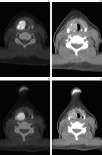
Her endocrinologist had ordered a combined PET and CT scan to investigate a sustained elevation of her thyroglobulin level and an enlarged thyroid nodule, in light of a negative radioiodine scan. The integrated PET and CT scan had revealed an 18F-fluorodeoxyglucose avid lesion at the level of the right true vocal fold, suspicious for laryngeal cancer (Figures 1a and 1c). No other 18F-fluorodeoxyglucose avid lesions had been seen. The CT portion of the scan had shown a mass at the level of the right vocal fold (Figures 1b and 1d).
(a), (c) Axial integrated positron emission tomography (PET) and computed tomography (CT) scanning images at the level of the larynx, showing a strongly 18F-fluorodeoxyglucose avid, right-sided mass. (b), (d) Axial CT scan (without PET overlay) showing the lesion.

Upon further questioning, the patient reported that she had undergone a thyroidectomy for papillary cancer 21 years ago, which resulted in unilateral vocal fold paralysis. Shortly after her initial surgery, she had undergone a medialisation procedure of her right vocal fold, using Teflon®. She had done well since that time.
Examination of the patient's neck revealed no significant neck masses or swelling.
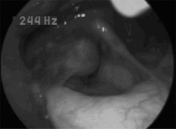
Flexible laryngoscopy showed impaired right vocal fold motion, with a smooth, well circumscribed, 2 cm mass on the right aryepiglottic fold and false vocal fold (Figure 2).
Lesion seen on videostroboscopy.

Given the findings and the suspicious nature of the laryngeal lesion, the patient was taken to the operating theatre for direct laryngoscopy and biopsy of the lesion. The fragments of tissue exhibited a rubbery consistency, with a mixed dull and shiny component.
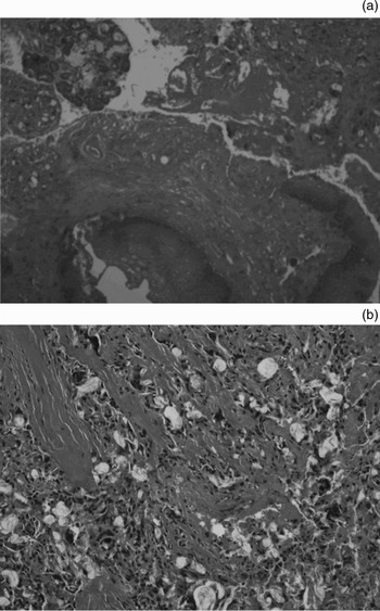
Histopathological examination demonstrated normal squamous epithelium overlying a marked foreign body giant cell reaction to exogenous material, consistent with a Teflon granuloma (Figures 3a and 3b).
(a) Photomicrograph showing squamous epithelium overlying the translucent Teflon material and a foreign body giant cell reaction (H&E;×10). (b) Photomicrograph showing translucent Teflon foreign body and numerous multinucleated giant cells (H&E; ×10).

Discussion
Positron emission tomography provides physiological and biochemical data regarding the patient's disease. Eighteen F fluorodeoxyglucose is distributed throughout the body on the basis of intracellular intake via glucose transporters or transport proteins. The degree of 18F-fluorodeoxyglucose uptake has been shown to be strongly related to neoplastic growth, due to the overexpression of glucose transport proteins and certain key enzymes.Reference Rosenbaum, Lind, Antoch and Bockisch1 A traditional PET scan lacks the anatomical detail needed for operative planning, but an integrated PET and CT scan allows the surgeon to localise the disease more precisely. Recently, 18F-fluorodeoxyglucose PET–CT scanning has become a useful adjunct in surveillance for thyroid cancer, detecting disease that can no longer concentrate radioiodine.Reference Schluter, Bohuslavizki, Beyer, Plotkin, Buchert and Clausen2–Reference Nahas, Goldenberg, Fakhry, Ewertz, Zeiger and Ladenson4 A recent study by Shammas et al. examined the utility of 18F-fluorodeoxyglucose PET–CT scanning for detecting and localising recurrent or metastatic differentiated thyroid cancer in a cohort of 61 patients (59 of whom had negative radioiodine scans); they found an overall sensitivity and specificity in predicting histological disease of 68.4 and 82.4 per cent, respectively.Reference Shammas, Degirmenci, Mountz, McCook, Branstetter and Bencherif5
While 18F-fluorodeoxyglucose PET–CT scanning can be extremely useful for imaging head and neck cancer, the radiographic findings for some disease processes can be confusing even with fused imaging techniques. There are several sources of false positive results. Infectious, autoimmune or granulomatous disease may show increased uptake owing to the high glucose turnover seen in activated granulocytes, lymphocytes and macrophages.Reference Rosenbaum, Lind, Antoch and Bockisch1 In a recent review, Rosenbaum summarised the many reports of false positive findings of 18F-fluorodeoxyglucose PET, which included cases of laryngeal uptake associated with vocalisation during the uptake period (generally this uptake is symmetrical), autoimmune thyroiditis, supraclavicular brown fat, physiological and post-chemotherapy hyperplasia uptake, post-operative inflammation, and some granulomatous disorders such as sarcoidosis, tuberculosis and fungal infections.Reference Rosenbaum, Lind, Antoch and Bockisch1, Reference Shammas, Degirmenci, Mountz, McCook, Branstetter and Bencherif5 Reports have also described PET-positive osteoradionecrosis in patients with a past history of nasopharyngeal carcinoma.Reference Liu, Chang, Ng, Chan and Yen6
One of the recognised non-neoplastic causes of 18F-fluorodeoxyglucose uptake is a granulomatous reaction to Teflon (i.e. teflonoma). Teflon (polytetrafluoroethylene) was used in the past to facilitate medialisation of the paralysed vocal fold. Teflon granulomas of the larynx are a known complication of vocal fold medialisation, occurring in approximately 2–3 per cent of patients.Reference Loehrl and Smith7 The development of Teflon granulomas, even long after a technically successful injection, was one of the problems that led to the virtual abandonment of this procedure for vocal fold medialisation in the late 1980s. Teflon granulomas have previously been reported in a number of the head and neck sites. Beginning as early as 1980, Teflon granulomas have been described as mimicking thyroid nodules and neoplasms,Reference Sanfilippo, Shelburne and Ingram8, Reference Wassef, Achouche, Guichard and Tran Ba Huy9 posterior fossa masses (following microvascular decompression with Teflon),Reference Megerian, Busaba, McKenna and Ojemann10 and laryngeal and neck lesions.Reference Wenig, Heffner, Oertel and Johnson11, Reference Moukarbel, Youssef, Shab and Hamdan12
Microscopically, Teflon appears as a translucent, amorphous aggregate. As described in many major surgical pathology textbooks, when macrophages encounter a foreign object too large to be phagocytosed (such as Teflon), they fuse to form larger foreign body giant cells composed of up to several dozen individual macrophages. This fusion is a hallmark of the foreign body giant cell reaction. Macrophages and foreign body giant cells tend to remain at the surface of an implant for the duration of its residence. Encapsulation can also occur, involving a generally avascular collagen shell being deposited around a foreign body, effectively isolating the foreign body from the host tissues.
Radiographically, Teflon granulomas are characterised by hyperattenuated, poorly defined masses on unenhanced CT scans, intermediate signal intensity on T2-weighted magnetic resonance images (less signal intensity than expected for a tumour) and avid 18F-fluorodeoxyglucose PET uptake. Eighteen F fluorodeoxyglucose avidity is not directly related to the Teflon, but is believed to be a by-product of the inflammatory reaction in response to the foreign body.
Unfortunately, these lesions can easily mimic neoplasms. Perhaps the best differentiator between benign disease and neoplasm in this situation is a careful patient history. One reason that Teflon was abandoned was its propensity to induce granulomas. Fortunately, newer injectable materials used for vocal fold medialisation are more biocompatible and less likely to cause long-standing inflammation.
Teflon granulomas have been reported in several head and neck sites. Positron emission tomography positive Teflon granulomas have previously been reported in the nasopharynx (secondary to Teflon injections for repair of velopharyngeal insufficiency),Reference Harrigal, Branstetter, Snyderman and Maroon13 and two reports have described teflonomas located in or adjacent to the vocal folds.Reference Hewitt, Singh and Wareing14, Reference Yeretsian, Blodgett, Branstetter, Roberts and Meltzer15 In one case report, the diagnosis was confirmed histologically after biopsy, and in the other a Teflon granuloma was presumed on the basis of the patient's history and the relative stability of the lesion over time, seen on PET–CT imaging.
• Combined positron emission tomography (PET) and computed tomography (CT) scanning is being increasingly used in diagnosing recurrent or persistent thyroid cancer
• A variety of benign or inflammatory lesions may produce false positive results on combined PET and CT scanning
• Teflon, a substance once used in vocal fold medialisation, produces an 18F-fluorodeoxyglucose avid lesion on combined PET and CT scanning, which may be falsely interpreted as recurrent thyroid disease
Our patient had a history of papillary thyroid cancer, an elevated thyroglobulin level and a negative radioiodine scan. Her 18F-fluorodeoxyglucose PET–CT scan showed an 18F-fluorodeoxyglucose avid laryngeal lesion suspicious for malignant disease. Subsequent biopsy was negative for malignancy, revealing a Teflon granuloma. While the lesion was suspicious for Teflon granuloma prior to surgical intervention, and there had been prior reports of 18F-fluorodeoxyglucose avid Teflon granulomas, we felt that a biopsy was warranted due to our patient's positive PET–CT scan and persistently elevated thyroglobulin level.
Conclusion
Eighteen F fluorodeoxyglucose PET–CT scanning is becoming an extremely important diagnostic tool in head and neck surgery and thyroid cancer. Radiologists and head and neck surgeons should be aware of the potential false positive laryngeal finding in those patients with a history of laryngeal Teflon injection.