INTRODUCTION
Toxoplasma gondii and Neospora caninum are closely related Apicomplexan parasites which are morphologically similar but possess some structural, molecular and antigenic differences (Ellis et al. 1998; Speer et al. 1999; Howe and Sibley, 1999). They have similar life-cycles with different definitive hosts, the felids (Hutchinson, 1965) and canids (McAlister et al. 1998; Gondim et al. 2004) respectively, and have similar intermediate hosts including a wide range of mammals (Dubey, 1999; Tenter, Heckeroth and Weiss, 2000). The ingestion of infective oocysts from the environment and tissue cysts from raw meat is considered to be a major route of infection of T. gondii (Tenter et al. 2000) and vertical transmission from mother to offspring a major route of infection of N. caninum (Davison, Otter and Trees, 1999). Both parasites cause significant disease and economic loss in the farming industry with T. gondii primarily causing abortion and foetal abnormality in sheep, and N. caninum causing abortion and foetal abnormality in cattle. T. gondii is also an important pathogen of humans while, in contrast, there is currently little evidence of human infection caused by N. caninum (Graham et al. 1999).
The balance of similarities and differences between these parasites raises the question as to whether frequent co-infection of individual hosts occurs and the degree to which these parasites may interact by acting antagonistically or synergistically during infection and disease. For example, does infection with one parasite prevent infection with the other or is the outcome of infection influenced by co-infection? Previous serological investigations have shown that antibodies to both parasites were present in several species including coyotes (Lindsay et al. 1996) and non-domestic felids from southern Africa (Cheadle, Spencer and Blagburn, 1999) but few studies have investigated this question in other species. Previous in vitro studies (Sunderman and Estridge, 1999) have demonstrated that Toxoplasma and Neospora can co-exist in tissue culture, and both species could invade the same host cell and undergo endodyogeny. Immunological studies (Innes et al. 2001) showed that, although there was evidence of induction of cross-reactive immune recognition following immunization with T. gondii in sheep, this was not sufficient to prevent foetal loss following subsequent N. caninum oocyst challenge.
The main objectives of this study were to develop a sensitive nested PCR technique for the detection of N. caninum DNA in host tissue, to use this to determine the prevalence of N. caninum in 3 naturally infected mammalian populations in Britain (rats, mice and sheep), and to determine the levels of N. caninum co-infection with T. gondii in these species.
MATERIALS AND METHODS
Collection of samples and DNA extraction
House mice (Mus domesticus) used in this study were captured within domestic dwellings in the Cheetham Hill area of Manchester, UK as part of a pest control programme (Marshall et al. 2004), brown rats (Rattus norvegicus) were captured in urban areas of Manchester. Tissue was obtained from lambs (Ovis aries) which were either aborted, dead at birth or died shortly after birth from 2 farms and contained individuals from 3 breeds, Suffolk cross, Charolais and Lleyns as described by Duncanson et al. (2001), Williams et al. (2005) and Morley et al. (2005). Approximately 1 g of brain tissue was dissected from mice, rats and lambs and, in the case of lambs, tissue was from the midbrain area along with samples of umbilical cord, tongue and heart. Cord tissue was not available for all lambs due to farming constraints during sampling and this was especially the case for the N. caninum-infected aborted lambs of which very few were obtained. All tissue samples were aseptically collected, using sterile instruments with great care being taken to exclude contamination either across tissues or from the surrounding environment, and stored frozen until DNA could be extracted under clean laboratory conditions as described by Williams et al. (2005).
DNA was extracted using a proteinase K lysis followed by phenol chloroform, ethanol precipitation method as described previously (Duncanson et al. 2001). DNA was resuspended in 100 μl of T.E. buffer, pH 8·0 and stored at 4 °C.
PCR detection of Toxoplasma gondii
Detection of T. gondii was carried out using nested PCR amplification of the Surface Antigen Gene 1 (Savva et al. 1990) as modified by Morley et al. (2005). The PCR product from one positive example of each species was extracted from the gel using the GENECLEAN 2 kit (Q-BIOgene) and sequenced in both directions (Lark Technologies Inc.) to confirm the detection of Toxoplasma (Accession nos. DQ077665 (sheep), DQ077664 (rat) and DQ077666 (mouse)).
PCR detection of Neospora caninum
The Nc5 region of N. caninum has been cloned and shown to be highly specific to N. caninum by DNA hybridization or PCR and is clearly distinguishable from T. gondii (Kaufmann et al. 1996; Yamage, Flechner and Gottstein, 1996). This gene was selected as the target for PCR amplification and detection using primers Np21 PLUS (5′ GGG TGT GCG TCC AAT CCT GTA AC 3′) and Np6 PLUS (5′ CTC GCC AGT CAA CCT ACG TCT TCT 3′) (Liddell, Jenkins and Dubey, 1999) both alone and as a nested technique combined with primers Np6 (5′ CAG TCA ACC TAC GTC TTC T 3′) and Np7 (5′ GGG TGA ACC GAG GGA GTT G 3′) (Yamage et al. 1996; Baszler et al. 1999) in the 2nd round. Both primer sets had previously been shown to be N. caninum specific, we further ensured the specificity of the technique by confirming there was no amplification using our nested technique, from 4 different isolates (RH, Martin, 17695 and COR), representing the 3 lineages of T. gondii. The 1st round reaction was performed as described by Liddell et al. (1999) with minor modifications as described below, 50 μl reactions contained 5 μl of 10× PCR buffer (HT biotechnology) (100 mM Tris-HCl, pH 9·0, 15 mM MgCl2, 500 mM KCl, 1% Triton X-100, 0·1% (w/v) stabilizer), 0·5 mM MgCl2, 12·5 nM each dNTP, 10 pM each primer, 2·5 units Taq polymerase (Bioline) and 1 μl of test DNA or water. After an initial denaturation step of 5 min at 95 °C, 40 cycles of PCR were performed with cycle times of 40 sec at 94 °C, 40 sec at 63 °C, 1 min 10 sec at 72 °C, followed by a final step of 10 min at 72 °C. The second round PCR was carried out as described by Baszler et al. (1999), reaction conditions were the same as the 1st round except 25 pM of primers Np7 and Np6 were used. Two microlitres of 1st round product was used as the template and cycling conditions were as the 1st round except that an annealing temperature of 56 °C was used. These PCR methods were compared to the semi-nested technique of Baszler et al. (1999) to calculate the performance of the test. All PCR reactions were performed using a Stratagene ROBOCYCLER™. PCR products were run on a 2% Agarose TBE gel containing ethidium bromide and visualized on an Alpha imager™ 1220. All PCR reactions were performed a minimum of 3 times to ensure comprehensive sampling of the test DNA. Due to the sensitive nature of the nested PCR technique negative controls were included in all PCR reactions and contamination could easily be tracked since many samples were naturally negative and therefore most positive reactions were interspaced between negative reactions. Positive PCR products from each host species were extracted from the gel using the GENECLEAN 2 kit (Q-BIOgene) cloned using the TOPO TA cloning® kit (Invitrogen) and sequenced (Lark technologies Inc.) to confirm the presence of DNA corresponding to the N. caninum Nc5 region. All negative PCR samples underwent a Tubulin PCR (Terry et al. 2001), targeting host DNA, to check the sample was suitable for PCR.
RESULTS
Evaluation of a nested PCR detection system for Neospora caninum
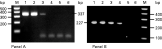
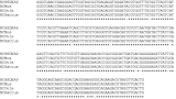
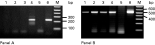
Previous work on T. gondii detection (Duncanson et al. 2001) suggested that the use of a nested PCR reaction would be required for the accurate detection of apicomplexan parasites directly from mammalian tissues. Although single-round PCR techniques (e.g. Liddell et al. 1999) and a semi-nested technique (Baszler et al. 1999) are available for the detection of N. caninum, we decided to develop a more sensitive detection method which can detect small quantities of Neospora DNA, in the presence of huge excesses of mammalian DNA. A series of dilution experiments were carried out using the NP21PLUS, NP6PLUS, NP7 and NP6 primers to determine the sensitivity of this nested PCR approach and compared it with the semi nested technique of Baszler et al. (1999). A series of dilutions of N. caninum DNA in genomic rat DNA was used as the template in nested PCR reactions using primers NP21PLUS and NP6PLUS in the first round followed by NP7 and NP6 in the second round. The results presented in Fig. 1 show that in the first round Neospora DNA could be detected at a dilution of 1 in 25 (lane 3) and following the nested PCR, detection was increased 5-fold to a 1 in 125 dilution (lane 4). We found the semi-nested technique of Baszler et al. (1999) was 4-fold less sensitive than our nested technique when used in the presence of host DNA (data not shown). An initial comparison of the single-round and nested PCR approaches on actual tissue samples from sheep revealed that the single-round approach underestimated the number of positive samples by 36% when compared to nested PCR. Even after many repeated tests of the same samples we were still unable to detect 14% of positive tissue samples using the single round alone, when compared with the nested technique. Furthermore when testing DNA extracted from infected tissue samples, bands from the single round PCR were always faint and usually difficult to distinguish clearly. Tissues positive for N. caninum usually produced a clear strong band following the second round, allowing easier interpretation of gels. Sequencing of the N. caninum PCR product amplified from an example of each host species (Accession nos. DQ077661 (sheep), DQ077662 (rat) and DQ077663 (mouse)) confirmed the specificity of the PCR with greater than 97% homology with the published Nc5 sequence (Accession no. X84238) (Yamage et al. 1996) (Fig. 2).

Fig. 1. Comparison of the detection of Neospora caninum DNA, using single and nested PCR techniques. Rat DNA was spiked with dilutions of N. caninum DNA and PCR amplification was carried out using single round PCR using primers Np21PLUS and Np6PLUS (Panel A) and nested PCR 1st round primers Np21PLUS and Np6PLUS, 2nd round primers Np6 and Np7 (Panel B). Lane 1 Undiluted N. caninum DNA, Lane 2 1/5 dilution of N. caninum DNA, Lane 3 1/25 dilution of N. caninum DNA, Lane 4 1/125 dilution of N. caninum DNA, Lane 5 1/625 dilution of N. caninum DNA Lane 6 negative control, Lane M Hyper ladder1 marker (Bioline).

Fig. 2. Sequence alignment of PCR fragments amplified from naturally infected mammalian brains using Neospora caninum-specific primers compared with part of the Nc5 region of N. caninum (Yamage et al. 1996) NCSSDNAP (Accession no. X84238), NCMus, PCR product amplified from a naturally infected mouse (Accession no. DQ077663), NCOvis, PCR product amplified from a naturally infected sheep (Accession no DQ077661), NCRattus, PCR product amplified from a naturally infected rat (Accession no. DQ077662). [star ] Denotes exact match between all sequences, [bull ] denotes a mismatch with at least 1 sequence.
Neospora caninum and Toxoplasma gondii co-infection
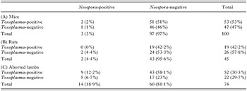
To compare rates of co-infection of T. gondii and N. caninum, nested PCR was carried out on DNA extracted from brain tissue from the mice, rat and lamb populations. Three (3·0%) of the 100 mice tested positive by PCR for N. caninum and 53 (53·0%) were positive for T. gondii. Two (4·4%) of the 43 rats were positive for N. caninum and 19 (42·2%) were T. gondii positive. Of the 74 aborted lambs 14 (18·9%) tested N. caninum positive and 52 (70·3%) T. gondii positive (Table 1). As shown in Table 1, both co-infection and the absence of co-infection were present in the mice with 2 mice (2·0%) testing positive for T. gondii and N. caninum, and 1 (1·0%) testing positive for N. caninum alone. Neither of the 2 N. caninum-positive rats tested positive for Toxoplasmosis. The numbers of N. caninum-positive samples in these 2 species was too small to enable further statistical analysis.

Of the 74 lambs 9 (12·2%) were positive for N. caninum and T. gondii, 5 (6·7%) for N. caninum alone, 43 (58·1%) for T. gondii alone and 17 (23%) were negative for both parasites. Statistical analysis using the χ2 contingency table test for association and Yates' correction for continuity demonstrated there was neither positive or negative association between N. caninum and T. gondii infection in this population of lambs (P=0·85). Within our sample of 3 breeds, Suffolk cross, Charolais and Lleyns, we found individuals from all 3 breeds infected with N. caninum showing that there was no association with breed.
Tissue distribution of Neospora caninum in lambs
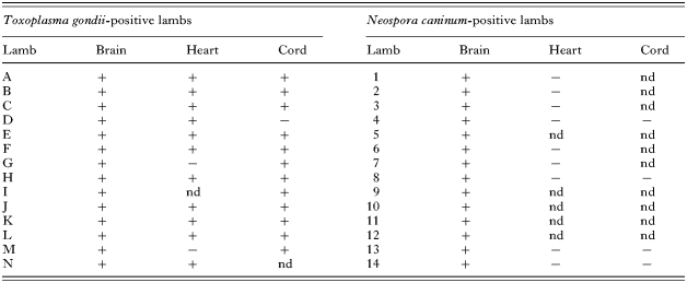
As differences have been reported in the tissue distribution of T. gondii and N. caninum (Duncanson et al. 2001; Pereira-Bueno et al. 2002) we investigated this aspect using the aborted lamb tissue samples. Samples for brain, heart and cord, where available, were collected from N. caninum-positive lambs and analysed for N. caninum presence, a similar number of T. gondii-positive lambs tissues were compared. Although T. gondii could be detected reliably in a number of tissues, N. caninum was only detectable in the DNA extracted from brain tissue (Table 2). Fig. 3 shows PCR results of N. caninum and T. gondii specific PCRs from 1 lamb that was positive for both N. caninum and T. gondii. The results presented clearly show that N. caninum was only detectable in DNA extracted from brain tissue but T. gondii was detectable in DNA extracted from brain, heart, cord and tongue tissues.
Table 2. Comparison of sites of successful detection of Toxoplasma gondii and Neospora caninum in different tissues of Toxoplasma and Neospora positive lambs respectively (“+” Denotes successful SAG1 or Nc5 PCR, “−” denotes unsuccessful SAG1 or Nc5 PCR, “nd” denotes no sample available.)


Fig. 3. Detection of Toxoplasma gondii and Neospora caninum DNA in different lamb tissues. Lamb DNA samples were amplified by PCR to detect N. caninum DNA (Panel A) and T. gondii DNA (Panel B). DNA was extracted from tongue (Lane 1), cord (Lane 2), heart (Lane 3), brain (Lane 4), Lane 5 negative control, Lane 6 positive control, Lane M Hyper ladder1 marker (Bioline).
DISCUSSION
Since N. caninum was identified as a separate species a number of methods of identification have been developed, including serological, immunohistochemical labelling and molecular methods including PCR. PCR is recognized as a specific sensitive technique used to identify species-specific parasite DNA sequences (Wastling, Nicoll and Buxton, 1993). A number of PCR methods have been used to assess Neospora infection e.g. Payne and Ellis (1996), Lally, Jenkins and Dubey (1996), Ho et al. (1997) and Gottstein et al. (1998). We further developed a nested PCR technique based on the highly repeated Nc5 region specific to N. caninum (Kaufmann et al. 1996) using primers NP21PLUS, NP6PLUS (Liddell et al. 1999) combined with NP7, NP6 (Yamage et al. 1996; Baszler et al. 1999). This increased the sensitivity of detection 5-fold and also allowed the use of greater quantities of host DNA as template without masking the final result. In common with previous authors (Ho et al. 1997; Almeria et al. 2002) we found that the same samples did not always test positive and a minimum of 3 PCR reactions were carried out on separate occasions. Examination of field samples demonstrated that, despite repeated testing, the single-round PCR underestimated the number of Neospora-infected individuals by 14·3%, when compared to the nested technique. In contrast Baszler et al. (1999), in a study of N. caninum prevalence in foetal tissues from spontaneous bovine abortions, found that although semi-nested PCR increased the sensitivity of detection, it was unnecessary for the detection of infection. Two possible explanations for this discrepancy exist. Firstly, different host species (i.e. bovine vs ovine) may have a different parasite density and may therefore require different test conditions. Secondly, Baszler et al. (1999) compared only single-round and semi-nested PCR techniques. We have shown that semi-nested PCR is 4-fold less sensitive than our technique, clearly demonstrating that a nested PCR technique is required for diagnosis in sheep. Previous studies using primers Np21PLUS and NP6PLUS have shown a detection limit of 1 tachyzoite in 0·5 g brain tissue (Almeria et al. 2002) by adding the second round of PCR using primers NP7 and NP6, we increased the sensitivity by 5-fold to the equivalence of 0·2 tachyzoites in 0·5 g brain tissue.
In our samples we found very low levels of N. caninum infection in both mice (3/100, 3%) and rats (2/45, 4·4%). It is interesting to note that a recent study of rats from cattle farms with N. caninum associated abortion found serological evidence of exposure to N. caninum in 9/55 rats, with N. caninum DNA detected by a single-round PCR in 2 of these serologically positive individuals (Huang et al. 2004). In both host species levels of T. gondii infection were significantly higher (53/100, 53% and 19/45, 42·2% respectively). This suggests that there is no particular linkage in the epidemiological factors that determine prevalence levels of T. gondii and N. caninum in mice and rats. The rats and mice in our study were collected from an urban environment, investigation of similar samples from a rural area might provide insight into the importance of different modes of transmission, for example, it might be expected in an urban area with a greater density of dogs that infection by the ingestion of oocysts may increase levels of infection when combined with vertical transmission whereas in rural areas with fewer dogs vertical transmission may be the more important transmission route. Further work is needed to address this question.
In our studies of aborted lambs we found high levels of infection with Neospora (14/74, 18·9%). Naturally occurring Neospora infection of ovines detected either by serological survey or PCR have been reported as rare (Hemphill and Gottstein, 2000). However, Hassig et al. (2003) detected N. caninum using a single-round PCR method in the brain tissue of 4 out of 20 (20%) aborted lambs and our data are in accordance with these figures. Further work may be required to investigate this variability in reported prevalence.
A number of serological investigations into co-infection of Neospora and Toxoplasma have been reported in the literature including co-infection rates of 4/53 (7·7%) of coyotes (Lindsay et al. 1996), 4/68 (5·9%) of non-domestic felids from southern Africa (Cheadle et al. 1999), 0/66 camels (Hilali et al. 1998), 0/221 red foxes (Jakubek et al. 2001), 1/549 red foxes (Hamilton et al. 2005), 12/144 (10·5%) sheep (Hassig et al. 2003) and 3·5% of 597 sheep (Figliuolo et al. 2004). In our experiments we found co-infection with DNA from both parasites in 2% of mice but not in any of the 43 rats tested, however the prevalence of Neospora was low and, therefore, statistical analysis was inappropriate. Of the aborted lambs 9/74 (12·2%) were co-infected, confirming previous serological results (Hassig et al. 2003; Figliuolo et al. 2004) that co-infection can occur in sheep. However, our results were in contrast to a PCR based study in Switzerland (Hassig et al. 2003) where there was no observed co-infection with toxoplasmosis in the 4/20 (20%) aborted lambs infected with Neospora. This is probably due to the small numbers of animals that exhibited either N. caninum or T. gondii (n=7). On the other hand our larger data set of infected animals (n=57) allowed us to address the question of co-infection and we demonstrated there was no statistical association between infection with Neospora and Toxoplasma (P=0·85). Our results support the in vitro, immunological and serological evidence that there is no exclusivity of infection and that co-infection would appear to be a random event.
We investigated tissue distribution in aborted lambs to answer a number of questions including whether or not Neospora could be detected in cord tissue. Lamb cord is foetally derived and if Neospora could be reliably detected in it we could use this as a means of screening live births for Neospora infection. Neospora has been amplified from many experimentally and naturally infected bovine tissues again with the most successful amplification from brain tissue (Gottstein et al. 1998; Baszler et al. 1999). Ho et al. (1997) suggested that experimentally infected bovines appear to have a wider tissue distribution of Neospora sp. DNA than naturally infected animals, with natural infection being detected in brain and spinal cord and rarely in other tissue. Our studies support this hypothesis in the case of aborted lambs.
Our investigations demonstrated that naturally occurring infection of N. caninum occurs in all three species of mammals investigated but at a much lower prevalence than T. gondii. We also found evidence that co-infection with T. gondii was occurring but that there was no specific association. The incidence of N. caninum-infected mice and rats has implications for the translocation of Neosporosis across wider geographical areas due to the spread of infection through rodent and other pest populations. Furthermore, the apparent broad host range of Neospora raises the question as to if or when it might appear as a human parasite. Further research is needed to investigate these questions.
We would like to acknowledge The University of Salford, Perry Foundation and The Wellcome Trust for providing funding for the work conducted in this study. Thanks go to A. M. Tenter and J. P. Dubey for the control Neospora caninum DNA. We gratefully acknowledge the Cheetham and Broughton SRB, Peter Marshall and Allan Maiden and family for their involvement in this research.