Introduction
The normal Eustachian tube is closed in its resting state, and only opens for short periods because of the action of tensor veli palatini and levator veli palatiniReference Poe1 during processes such as yawning and swallowing. An abnormal patency of the Eustachian tube is known as a patulous Eustachian tube.Reference Poe2 This results in the direct and abnormal communication of sound and air pressure between the nasopharynx and middle ear.
The main symptoms are autophony of voice and breathing when sound passes through the patent Eustachian tube from the nasopharynx to the middle ear. These symptoms can reduce quality of life to such an extent that some patients report not wanting to speak so as to avoid them. Other symptoms include aural fullness, and, less frequently, hearing loss and vertigo.
Patients will often describe factors that worsen or improve their symptoms. Exacerbating factors include rapid weight loss, dehydration, alcohol or caffeine, prolonged talking, and exercise. Symptoms can be relieved by lying supine or holding the head in a dependent (head down) position, or by nasal irrigation. It is believed that venous engorgement accounts for the resolution of symptoms when the head is dependent.Reference Mackeith and Bottrill3
The key examination finding is medial and lateral movement of the tympanic membrane with respiration. This is most easily seen using magnification with a microscope or endoscope, and the examination should be performed in the upright position. Pure tone audiometry findings are usually normal.
The prevailing theory regarding the aetiology of patulous Eustachian tube is a reduction of volume in Ostmann's fat pad, which is located in the lateral wall of the cartilaginous Eustachian tube and acts as a closing valve. This theory is supported by the high prevalence of patulous Eustachian tube in patients who have experienced rapid and significant weight loss following bariatric surgery.Reference Muñoz, Aedo and Der4 Other risk factors include scarring from previous surgery (e.g. adenoidectomy), or following radiotherapy and neuromuscular disorders that result in muscle loss.Reference Poe2 An important differential diagnosis for patulous Eustachian tube is superior semi-circular canal dehiscence, which also causes autophony of voice and aural fullness. A high-resolution computed tomography (CT) scan of the temporal bones may, therefore, be indicated to exclude semi-circular canal dehiscence and to establish any dehiscence of the carotid canal if Eustachian tube surgery is planned.Reference Mackeith and Bottrill3
The symptoms of patulous Eustachian tube often have a severe negative impact on the patient's quality of life. In addition, it is often diagnosed late (an average of 6.7 years between the initial presentation of symptoms and treatment in one seriesReference Mackeith and Bottrill3).
Treatment options involve explanation of symptoms and reassurance. Conservative management to address exacerbating factors, such as avoiding dehydration, reducing diuretics like alcohol and caffeine, or regaining lost weight, are also important. For patients with persistent and significant symptoms, the most established surgical treatment option is reinforcement of the Eustachian tube via an endoscopic transnasal approach. This can involve: Eustachian tube cautery;Reference O'Connor and Shea5, Reference Robinson and Hazell6 Eustachian tube ligation;Reference Rotenberg, Busato and Agrawal7 or injection augmentation of the Eustachian tube with a range of implant materials including but not restricted to cartilage,Reference Oh, Lee, Goh and Kong8 calcium hydroxyapatiteReference Vaezeafshar, Turner, Li and Hwang9 and polydimethylsiloxane elastomer (e.g. Vox).Reference Mackeith and Bottrill3, Reference Schröder, Lehmann, Sudhoff and Ebmeyer10
In 2014, we published a case report of a novel strategy for managing patulous Eustachian tube. This involved a CT-guided, transcutaneous approach to reinforce the Eustachian tube, using a silicone (polydimethylsiloxane) elastomer suspension implant (Vox Implants; Uroplasty, Minnetonka, Minnesota, USA).Reference Rodrigues, Waddell and Cook11 We described a 36-year-old woman with disabling autophony secondary to patulous Eustachian tube (semi-circular canal dehiscence was excluded with CT imaging). Endoscopic transnasal Eustachian tube augmentation using Vox had failed to provide symptomatic relief after two attempts. However, the subsequent placement of the Vox implant in the lateral aspect of the Eustachian tube using a CT-guided approach was effective, and remained so at the six-month follow up. Since then, we have performed the procedure in a series of 8 patients, with a total of 12 ears. Here we report on the outcomes of this novel approach.
Materials and methods
The authors assert that all procedures contributing to this work complied with the Helsinki Declaration of 1975, as revised in 2008. This research received no specific grant from any funding agency, commercial or not-for-profit sectors.
The patients all presented with autophony of voice and respiration, with a significant impact on quality of life. Our patients’ comments regarding the effect of patulous Eustachian tube on their quality of life included the following: ‘At the time no other treatments had worked. At times I even felt suicidal, unable to consider how I could learn to live with my symptoms’, and ‘The condition is debilitating and affects everyday life, particularly when trying to have a conversation’.
Some had attempted prior treatment with endoscopic augmentation of the Eustachian tube via the nasopharynx, but in all cases this had been unsuccessful. Therefore, the external CT-guided approach was offered. All patients gave full, informed and written consent having been warned of the risks of: Eustachian tube occlusion, and subsequent middle-ear effusion, facial nerve injury, and numbness or pain secondary to trigeminal nerve injury.
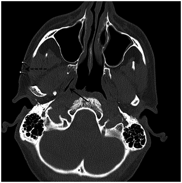
All procedures were carried out under the joint care of the senior radiological and otolaryngological authors, as local anaesthetic day-case procedures. The relevant Eustachian tube was identified with a Somatom Sensation 16 multi-detector row CT scanner (Siemens Medical Solutions, Erlangen, Germany), without the use of contrast medium. A site on the cheek was selected that allowed access to the lateral aspect of the Eustachian tube by passing a needle over the mandibular notch and just posterior to the lateral pterygoid plate, and this site was indicated with a radio-opaque skin marker (Figure 1).
Axial computed tomography scan showing right-sided patulous Eustachian tube (arrow) and radio-opaque skin marker at injection site (dotted arrow).

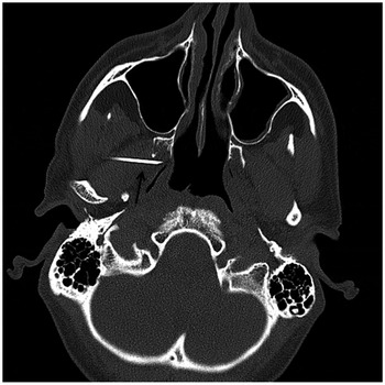
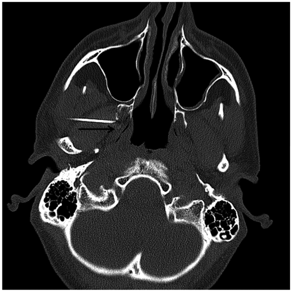
Under aseptic technique, 5 ml of 1 per cent lignocaine was progressively injected into the skin and deep tissues around the relevant Eustachian tube using a 21 gauge needle. Then, under CT-guidance, a 70 mm, 18 gauge needle (Medex; Smiths Medical, Ashford, UK) was advanced until its tip was seen immediately lateral to the Eustachian tube (Figure 2). Up to 2 ml of silicone (polydimethylsiloxane) elastomer suspension implant (Vox Implants) was then injected (Figure 3). The volume of implant injected was titrated to be sufficient to narrow the lumen of the Eustachian tube, but not to completely occlude it (Figure 3). This minimised the chances of a middle-ear effusion following the procedure, whilst still providing symptomatic relief. The images in Figures 1–3 were all taken at the same spinal level to allow for direct comparison.
Axial computed tomography scan showing needle tip positioned at the lateral wall of the Eustachian tube prior to Vox injection (arrow).

Axial computed tomography scan following Vox implant injection (arrow). Note the narrowed Eustachian lumen as compared to Figure 1.

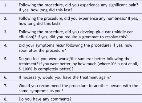
We subsequently performed telephone or e-mail follow up of all patients using the structured questionnaire shown in Table 1. Our main outcome measure was whether the autophony symptoms had resolved following the CT-guided implant injection and to what degree. In addition, we established whether any adverse outcomes or complications had occurred, and whether the procedure was considered acceptable by this group of patients.
Structured questionnaire used for patient follow up after CT-guided Vox injection for patulous Eustachian tube

CT = computed tomography
Results
A total of 8 patients underwent this treatment: 5 females and 3 males, with an age range of 41 to 73 years, and a mean age of 52.4 years. Four patients had bilateral treatment and 4 had unilateral treatment, resulting in a total of 12 ears (7 left ears and 5 right ears). Three ears had the treatment performed twice because of symptom recurrence. The previous treatments included endoscopic transnasal Eustachian tube injections, grommets and tympanic membrane fillers. The follow-up interval from the final treatment until the questionnaire interview ranged from 6 to 62 months, with a mean follow-up interval of 29 months.
The treatment was initially immediately effective for all 12 ears. At the time of the questionnaire follow up, 100 per cent symptom resolution was maintained in 7 of the 12 ears (58 per cent). Of the remaining five ears, at the time of questionnaire follow up, one ear still had 90 per cent resolution of symptoms, one ear had 50 per cent and two ears had maintained 25 per cent improvement of symptoms. Only one ear showed no symptom resolution at all by the time of the questionnaire follow up. This gives an overall (complete and partial) symptom resolution rate of 91 per cent for this CT-guided transcutaneous approach for patulous Eustachian tube treatment.
With regard to complications, only one ear (9 per cent) developed a middle-ear effusion following treatment, and a grommet was required to resolve the effusion. Other reported complications included a single report of significant ear pain lasting 7 days following treatment. Numbness occurred post-treatment in 4 ears in the distribution of the maxillary division of the trigeminal nerve. Three patients had numbness of the face on the side of the treated ear and one had numbness of the tongue. Every case of numbness resolved within six months.
All patients reported that, if necessary, they would have the treatment again, and they would recommend it to another person with the same symptoms as them. The comments made by those patients for whom the treatment was effective highlighted the impact of patulous Eustachian tube on their quality of life and, therefore, the high value they place on effective treatment. Patients’ comments regarding the effect of successful treatment on their quality of life included: ‘There was for me no doubt in my mind but to try it. I have been fixed and have no side effects. My hearing is normal, I have no pain, my Eustachian tubes appear to function normally’, ‘I would recommend it to anyone. I previously didn't want to talk to anyone – I had really given up hope – it revolutionised my life’, and ‘I'm a million times better, it's been really good. I hope it never comes back, it's given me my life back’.
Discussion
Patulous Eustachian tube is a difficult condition to treat. It is associated with debilitating symptoms that have a significant impact on the patient's quality of life (as confirmed by our patients). Most of the currently available interventions require operating theatre time and often necessitate a general anaesthetic.Reference Mackeith and Bottrill3, Reference Rotenberg, Busato and Agrawal7, Reference Vaezeafshar, Turner, Li and Hwang9, Reference Schröder, Lehmann, Sudhoff and Ebmeyer10 We had previously described a novel intervention, which we have shown to be effective in treating patulous Eustachian tube in this series of patients.Reference Rodrigues, Waddell and Cook11
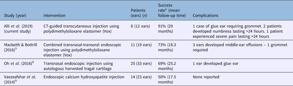
The procedure can be performed under local anaesthetic in the radiology suite, without the use of valuable operating theatre time. Our series shows that in 58 per cent of cases (7 of 12 ears), symptoms were controlled completely following treatment; a further 33 per cent of cases (4 of 12 ears) had partial resolution of symptoms after a mean follow up of 29 months. Thus, the overall symptom resolution rate was 91 per cent, with 11 of 12 patients having complete or partial resolution of symptoms after a mean follow up of 29 months. This success rate is comparable with other recently published series reporting transnasal injection augmentation techniques with a range of implant materials (Table 2).
Success rates and complications in this series and recent studies of transnasal Eustachian tube injection augmentation for patulous Eustachian tube

*Complete and partial resolution of autophony. CT = computed tomography
There were no long-term complications arising from this technique in our series. There was significant pain in 1 of 12 ears, facial or tongue numbness of several months duration in 2 of 12 ears, and glue ear in 1 ear. This complication profile is comparable to endoscopic transnasal approaches (Table 2).
Injection of implant material (Teflon™) into the internal carotid artery has been reported previously.Reference O'Connor and Shea5 However, to our knowledge, this serious complication has not been described in this era of the endoscopic approach, which affords excellent visualisation of the Eustachian tube. We feel that the use of CT image guidance for accurate positioning of the needle tip for implant placement, as described in this series, should protect against the complication, just as the use of endoscopic guidance has done.
Regarding the patients in whom only partial symptom resolution was achieved, performance of the procedure with the patient supine may have caused enlargement of the Eustachian tube walls because of venous engorgement. This could result in CT images showing a lateral Eustachian tube wall that appears thicker than it would do in the upright position, which is when symptoms are most prevalent. This could impact negatively on the site of the Vox implant injection, resulting in suboptimal placement of the implant material. This might explain the less successful outcomes and failures.
• Patulous Eustachian tube is a benign but notoriously difficult condition to treat successfully
• Symptoms include autophony of voice and breathing, and aural fullness
• For persistent and significant symptoms, the established surgical treatment is Eustachian tube reinforcement via an endoscopic transnasal approach
• Computed tomography guided Vox injection is a viable and effective alternative treatment for patulous Eustachian tube
• This technique has success rates and complication profile comparable to currently available treatment options
• Furthermore, the treatment is highly acceptable to patients with this condition
All patients in this series, including the single patient for whom there was no long-term symptom resolution of any degree, felt that this was a procedure they would undertake again, and they would recommend it to another person with patulous Eustachian tube. This 100 per cent acceptability rate is an important factor in our assertion that CT-guided transcutaneous implant injection is a viable alternative modality for treating patulous Eustachian tube.
Competing interests
None declared