I. INTRODUCTION
Energy storage devices such as lithium-ion batteries (LiBs) are important for portable devices, electrical vehicles, and power tools.Reference Armand and Tarascon1,Reference Whittingham2 The increasing demand has led researchers to focus on improving energy density, durability, cycling ability, charging time, and low-cost LiBs.Reference Smith, Burns, Zhao, Xiong and Dahn3–Reference Smith, Dahn, Burns and Dahn8 So far, various kinds of positive and negative electrodes have been developed, and readily available cost-efficient silicon materials have come into the spotlight.Reference Park, Cho, Lee, Yoo, Song and Cho7,Reference Smith, Dahn, Burns and Dahn8 Silicon is electrically conductive and can swell modestly to accommodate the lithium ions, exhibiting superior properties as a LiB anode.Reference Kasavajjula, Wang and Appleby9 However, the large volume expansion (>300%) during the lithiation process leads to the pulverization of silicon materials along with considerable microstructural degradation, resulting in the disruption of electron-conducting paths, and loss of reversible capacity.Reference Zhang10–Reference Gu, Li, Li, Hu, Zhang, Xu, Thevuthasan, Baer, Zhang, Liu and Wang13
Mesoporous silicon sponge (MSS) with its preformed meso/macropore structures and the grafted carbon on the surface has been demonstrated as a robust anode material to accommodate the large volume expansion that accompanies the lithiation process in our previous studyReference Li, Gu, Hu, Kennard, Yan, Chen, Wang, Sailor, Zhang and Liu14 by in situ transmission electron microscopy (TEM) and continuum media mechanical calculations. MSS has a highly porous structure with thin crystalline Si walls surrounding pores on the order of 50 nm in size and the carbon-grafted MSS was prepared using chemical vapor deposition of acetylene on MSS at a temperature of >600 °C to improve the electrical conductivity and mechanical/electrical integrity of the particles during cycling. The grafted MSS anodes exhibited excellent electrochemical performance with high capacity, good capacity retention, and less irreversible capacity loss. For example, it can deliver a capacity of up to ∼750 mA h/g based on the total electrode weight with 480% capacity retention over 1000 cycles.
More recently, extensive effort has been devoted to stabilize the surface of silicon to improve its suitability for various energy, electronics, sensor, and biological applications.
The surface-functionalized MSS film has been investigated in our recent studyReference Kim, Joo, Pan, Boarino, Jun, Ahn, Arkles and Sailor15 where organic trihydridosilanes were grafted to the hydrogen-terminated porous Si nanostructures via the thermally induced silane dehydrocoupling reaction with no catalyst at mild temperature. These modified surfaces are stable to corrosive aqueous solutions and common organic solvents with retention of the electronic quality of the original Si–H surface. These surface-functionalized MSS films are especially useful in bio-imaging and drug delivery.Reference Li, Gu, Hu, Kennard, Yan, Chen, Wang, Sailor, Zhang and Liu14
Pore structures and connectivity of bare MSS materials and grafted MSS materials prepared via the thermally induced silane dehydrocoupling reactionReference Li, Gu, Hu, Kennard, Yan, Chen, Wang, Sailor, Zhang and Liu14 are investigated using hyperpolarized (HP) 129Xe nuclear magnetic resonance (129Xe NMR) in this study. The detailed information on the pore structure and connectivity will help better understand and predict the materials properties.
The pore geometry is complex in most porous materials, even in ordered mesoporous silicon, making it difficult to characterize the interconnections and channels.Reference Moudrakovski, Terskikh, Ratcliffe, Ripmeester, Wang, Shin and Exarhos16 Mixed analysis methods have been applied using small-angle X-ray scattering, nano X-ray tomography, neutron scattering, high-resolution TEM, and nitrogen adsorption–desorption isotherm analysis,Reference McDowell, Ryu, Lee, Wang, Nix and Cui12,Reference Gu, Li, Li, Hu, Zhang, Xu, Thevuthasan, Baer, Zhang, Liu and Wang13 but these methods provide no direct information of how channels and cages are connected.
Over the years, 129Xe NMR has developed into a powerful and robust method for studying porous solids.Reference Ripmeester17 The large chemical shift range of 129Xe is strongly dependent on both local environmental and chemical factors such as the composition of the matrix, nature, and concentration of adsorbed molecules, and the shape and size of resident void spaces.Reference Ripmeester17–Reference Driehuys, Cates, Miron, Sauer, Walter and Happer22 In addition, the combination of HP 129Xe by optical pumping and continuous flow (CF) measurement conditions dramatically enhances the sensitivity up to a factor of 104 by minimized contribution of Xe–Xe interaction.Reference Ruset, Ketel and Hersman23,Reference Knagge, Smith, Smith, Buriak and Raftery24 Since HP 129Xe gas percolates through the interconnected pores of MMS, HP 129Xe NMR has the unique advantage of directly probing not only these buried interfaces but also the interconnectivity between the pores.Reference Terskikh, Mudrakovskii and Mastikhin25–Reference Wang, Wang, Liu, Exarhos, Pawsey and Moudrakovski28
HP 129Xe NMR has been used to the study functionalized ordered mesoporous silica (SiO2) in our previous work.Reference Kim, Joo, Pan, Boarino, Jun, Ahn, Arkles and Sailor15 It is interesting to compare our results for grafted mesoporous Si sponge materials prepared via the thermally induced silane dehydrocoupling reaction with those for the functionalized mesoporous SiO2 prepared using hydrolytic condensation. HP 129Xe NMR was also applied to probe the porosity of the nanoparticle Si electrode in our previous workReference Mao, Song, Hopson, Karan, Guduru and Wang29 where the pores are made by the aggregation of the Si nanoparticle. The information on the porosity and connectivity of the mesoporous Si sponge with and without surface modification along with the result from the previous study of Si nanoparticle electrode materialsReference Mao, Song, Hopson, Karan, Guduru and Wang29 will help better understand the silicon-based porous material’s properties for making new materials in future.
II. EXPERIMENTAL
A. MSS-H material preparation
MSS-H was prepared by constant current anodization of heavily boron-doped P-type (<1 mΩ cm resistivity) polished (100 face) single crystal silicon wafers in ethanolic hydrofluoric acid electrolytes. CAUTION: HF is highly corrosive to the eyes and skin and proper precautions should be followed when handling. The preparation followed a published “perforated etch” procedure.Reference Sailor30,Reference Qin, Joo, Gu and Sailor31 Prior to preparation of the porous layers, the wafer surfaces were cleaned using a sacrificial etch consisting of electrochemical anodization (180 s, 87.1 mA/cm2) in an electrolyte consisting of 3:2 (v:v) 48% aqueous HF:absolute ethanol, followed by ethanol rinse, then dissolution of the porous film with aqueous KOH (1 M). The wafer was rinsed with water, then ethanol. The perforation etching waveform consisted of a square wave (current density of 87.1 mA/cm2 applied for 120 s, current density then increased to 174.2 mA/cm2 for 0.636 s, and lower values of current density of 0 A/cm2 for 30 s), repeated for 70 cycles. The multilayered porous Si film was removed from the substrate by application of a current pulse of 4.36 mA/cm2 for 500 s in an electrolyte consisting of 1:29 (v:v) of 48% aqueous HF:absolute ethanol. The freestanding porous Si film (∼300 mg) was then fractured by ultrasonication (50T, VWR International, Radnor, Pennsylvania) for 90 min in ethanol (7 mL), and the resulting colloidal dispersion of porous silicon micron particles was allowed to settle at room temperature overnight. The upper 30% portion of the colloidal dispersion was discarded, and the remaining portion was filtered through filter paper (Whatman, Cat. No. 1002-070, Clifton, New Jersey). The filter paper was dried, and the brown colored particles were collected from the paper and used without further purification.
B. MSS-C18 material preparation
Grafted MSS material (MSS-C18) was prepared by following a published “dehydrogenative coupling” procedure.Reference Kim, Joo, Pan, Boarino, Jun, Ahn, Arkles and Sailor15 The prepared MSS material (MSS-H, ∼300 mg) was transferred to a 20 mL vial, and 2 mL of octadecylsilane (H3Si-C18) reagent was added. The vial was capped and subjected to oil bath at 100 °C for 12 h with vigorous stirring. After 12 h, the resulting particles were washed several times with n-hexane and ethanol. Between rinsing steps, the nanosize particles were separated from the rinsing liquid by centrifugation (Eppendorf Centrifuge Model 5424R) at 15,000 rpm for 15 min. Washed particles were degassed under vacuum overnight and stored in vacuum.
C. HP 129Xe NMR
1. 129Xe NMR
HP 129Xe NMR experiments were carried out on a Bruker Advance DMX 300, Billerica, Massachusetts spectrometer operating at 82.98 MHz (magnetic field 7.05 T) using a variable temperature static probe with a CF of HP xenon. For HP 129Xe NMR experiments, a single-pulse (SP) Bloch-decay method was used, and MSS powders (MSS-H, MSS-C18) were loaded into 7 mm Zirconia rotors. The powders were packed into the NMR rotors using a hammer and care was taken to apply similar force to ensure that the material inside the rotor was packed tightly and consistently. The rotor cap contained a small drilled hole that allowed HP 129Xe gas to pass into the materials by means of small plastic tubing. The HP 129Xe NMR experiments were performed without spinning. SP spectra were collected with a 1 µs 129Xe pulse and a repetition delay of 0.5 s.
D. Instrumentation
Etching waveforms were generated in a computer program written in Labview (National Instruments, Inc., Austin, Texas), and the electric current was driven by using a Keithley 2651A Sourcemeter, Solon, Ohio power supply interfaced to LabView program. A 50T ultrasonic bath (VWR International) was used for ultrasonic fracture of the pSi films. Particles were collected by centrifugation (Eppendorf Centrifuge Model 5424R, Hamburg, Germany). Attenuated total reflectance Fourier transform infrared (ATR-FTIR) spectra were recorded using a Thermo Scientific Nicolet 6700 FTIR instrument fitted with a Smart iTR diamond ATR fixture, Waltham, Massachusetts. Scanning electron microscopy (SEM) images were obtained with an FEI XL30 UHR (Ultra High Resolution; FEI, Hillsboro, Oregon) SEM operating in the secondary electron mode. TEM images were obtained with a JEOL-1200 EX II 120 kV instrument, Tokyo, Japan. Nitrogen adsorption–desorption isotherms of the MSS materials were recorded at 77 K using a Micromeritics ASAP 2020 instrument, Norcross, Georgia. Prior to the nitrogen adsorption experiment, the MSS materials were degassed under vacuum overnight. The analysis result was determined using the BET (Brunnauer–Emmett–Teller) method.
A custom-built 129Xe polarizer was used in the HP 129Xe NMR experiments. The design of this high efficiency polarizer was based on a report by Hersman and co-workers.Reference Ruset, Ketel and Hersman23 Unlike most of the HP Xe polarizers currently in use, this design maintains a low-pressure environment to enable efficient transfer of spin from the excited Rb electron manifold to the Xe nucleus. Efficient pumping from the diode laser (100 W at 795 nm) was provided by means of a long path-length cell (1 m), high Rb vapor density, and by counter flowing the spin exchanged optically pumped gas in a direction opposite to that of the laser propagation direction. This arrangement achieved a remarkably high polarization rate (>35%), even at high flow velocity. A xenon–helium–nitrogen mixture with a volume composition of 1:66:33 was used in all CF HP experiments. The flow rate was maintained at approximately 300 s/min (gas flow normalized to standard conditions). In the CF HP experiments, the HP xenon flow was delivered directly from the polarizer to the coil region of the NMR probe through 1.5 mm ID plastic tubing. Variable temperature NMR experiments in the 173–373 K range were performed using a liquid nitrogen cooling assembly and PID temperature controller. All measured 129Xe NMR chemical shift data were referenced to the free xenon gas chemical shift at 0 ppm.
III. RESULTS AND DISCUSSION
A. Characterization of materials
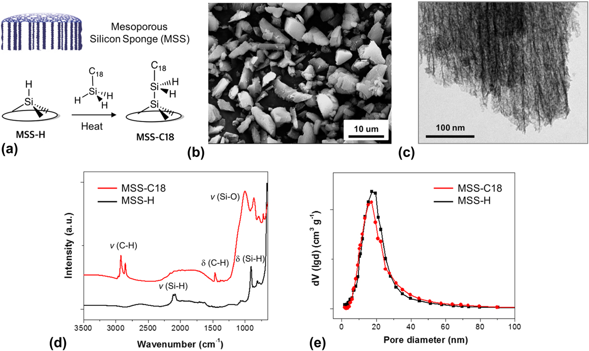
A schematic graph is displayed in Fig. 1(a) to illustrate a simplified structure of the MSS material and its grafting of surface via thermally induced dehydrogenative coupling with octadecylsilane reagent (H3Si-C18). SEM images of the MSS sample after the grafting reaction (MSS-C18) and the TEM images for the edge region of MSS-C18 are given in Figs. 1(b) and 1(c), respectively. The SEM and TEM images show nonuniform micron-sized particles (<10 μm in length) and nanometer sized channels in grafting MSS-C18, demonstrating that the open pore structure was preserved after grafting of MSS-C18. Comparative Fourier-transform infrared (ATR-FTIR) spectra of the as-prepared MSS (MSS-H) and grafted product (MSS-C18) [Fig. 1(d)] proved the existence of grating of C18 in MSS-C18 based on the observation of C–H stretching and bending bands associated with the grafted carbon source on the MSS surface. Furthermore, nitrogen adsorption–desorption isothermal analysis of MSS materials shows pore size distribution peaks at 18 nm center (mesoporous) with no significant changes after grafting reaction [Fig. 1(e)].
(a) Simplified structure of the MSS material and its grafting of surface via thermally induced dehydrogenative coupling with octadecylsilane reagent (H3Si-C18). (b) SEM images of the MSS sample after the grafting reaction (MSS-C18). (c) TEM images for the edge region of MSS-C18 in (b). (d) ATR-FTIR spectra of the as-prepared MSS (MSS-H) and carbon-grafted product (MSS-C18). Assignments of Si–H, Si–O, and C–H stretching and C–H bending (scissor), and Si–H deformation modes are indicated. Symbols: v = stretching and δ = bending. (e) Pore size distribution of MSS materials from nitrogen adsorption–desorption experiments.

B. CF HP 129Xe NMR at 300 K
HP 129Xe NMR spectra of MSS materials (MSS-H, MSS-C18) using the CF HP method were taken under identical conditions to examine any changes in porosity and connectivity of MSS materials before and after grafting. By using the HP 129Xe CF method, a very low partial pressure of Xe is present in the gas mixture (around 10 mbar, or 1% of total), precluding any contributions of Xe–Xe interactions in the analysis.Reference Ruset, Ketel and Hersman23,Reference Knagge, Smith, Smith, Buriak and Raftery24 Thus, the resulting 129Xe NMR spectra only represent inherent characteristics of the resident framework and the surface properties of the porous material. Moreover, the very low concentration of Xe gas reduces the rate of exchange between the different adsorption regions, affording the detection of inhomogeneity in the pore space of the materials.
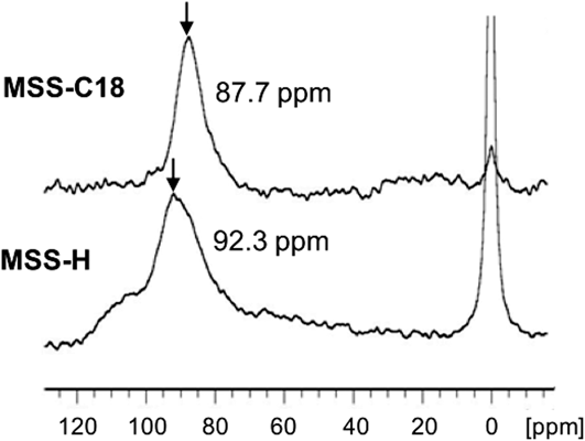
Figure 2 displays CF HP 129Xe NMR spectra of MSS materials (MSS-H, MSS-C18) taken at 300 K. The narrow peak at 0 ppm is associated with the free xenon gas and the broader down-field peaks with nonzero chemical shifts are associated with adsorbed xenon gas within the pores of MSS materials. One dominant peak at 92.3 ppm with a broad linewidth, and one shoulder peak at 101.0 ppm were observed for the MSS-H before grafting. After the surface grafting (MSS-C18), the peak position is shifted up-field to 87.7 ppm, and the linewidth of the peak becomes narrower. The presence of two resonance peaks for the MSS-H sample suggests that there are at least two different pore environments for adsorbed Xe in MSS-H materials. In comparison, a single resonance peak observed for Xe adsorbed onto the grafted MSS-C18 sample suggests that the grafting of C18 into the MSS-H material changes the porosity and connectivity of the pores. In general, when pore sizes are smaller as a result of grafting long-chain molecules inside the mesoporous channels, we should have observed a lower field shift (or higher chemical shift) for the MSS-C18 than for the MSS-H samples. However, an upper field shift (or lower chemical shift) was observed for the MSS-C18 after grafting C18. A similar up-field shift was also observed previously for C10 grafted mesoporous silica (MCM-41)Reference Moudrakovski, Terskikh, Ratcliffe, Ripmeester, Wang, Shin and Exarhos16 and was explained in terms of a screening of the surface micropores, or the “roughness” of the internal surface of the mesoporous SiO2 channels. The grafting of the C10 in mesoporous channels blocks most of these strong adsorption micropore centers on the surface of C10-coated mesoporous channels, resulting in an up-field shift. The existence of such “micropores” in the walls of bare mesoporous silicon has also been reported in a previous study using a combination of SET, TEM, N2 adsorption, and 129Xe NMR techniques.Reference Mao, Kim, Joo, Sailor, Hopson and Wang32 Therefore, the up-field shift for MSS-C18 is caused by the blocking of these stronger micropore centers due to the formation of C18 grafted layers. The relative large chemical shift change (about 5 ppm) suggests that the grafting significantly modified the pore surfaces. To further understand the changes in porosity and connectivity of the pores upon grafting, temperature-dependent CF HP 129Xe NMR analysis of MSS materials was also performed.
CF HP 129Xe NMR spectra of MSS-H (bottom) and MSS-C18 (top) taken at 300 K. The numbers indicate the maximum peak position (black arrow).

C. Temperature-dependent HP 129Xe NMR
In temperature-dependent 129Xe NMR spectra, the xenon peak position and linewidth are related to the dynamics of adsorbed xenon and its ability to penetrate into the pore space. Typically, the xenon peak is shifted down-field by the decreasing temperature because of enhanced xenon adsorptions (reduced thermal dynamic factor) onto the pore surface. When the surface of the main pores contains many micropores, Xe is mainly adsorbed in micropores at lower temperatures. As the temperature increases, Xe enters the larger mesopores and undergoes fast-exchange with the different pore environments. In this case, Xe is distributed mostly in the mesopore channels at higher temperatures and the chemical shifts of the Xe peak is from the average contributions from both meso- and micropores, showing a characteristic average peak and shift.
The reduction in Xe gas exchange at low temperatures allows Xe to access nonuniform pore environments. In the absence of chemical shift anisotropy, generally low-temperature linewidths are related to pore nonuniformity, while at higher temperatures of ≥300 K, where fast-exchange of Xe atoms is present, the linewidths are representative of both pore nonuniformity and interconnectivity among the different pores and channels because Xe can undergo fast-exchange among different interconnected pores or channels. A higher degree of connectivity between nonuniform pores results in a narrower linewidth with an increasing temperature for adsorbed Xe as a result of the fast-exchange that averages out the effects of pore nonuniformity. On the basis of a previous study, where large linewidths were associated with chemical shift anisotropy,Reference Sears, Wasylishen and Ueda33 the much narrower linewidths observed in this study suggest negligible effects of chemical shift anisotropy present in MSS materials. Consequently, the comparative variable-temperature 129Xe NMR linewidth analysis conducted here provides an excellent gauge of pore nonuniformity and connectivity.
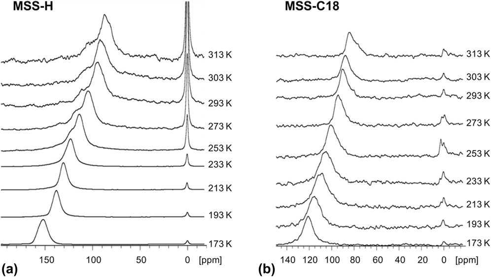
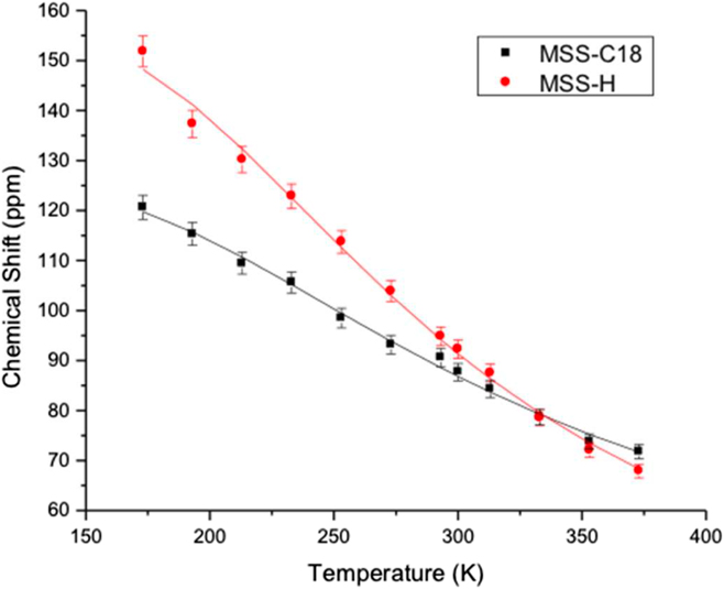
Temperature-dependent HP 129Xe NMR spectra for MSS-H and MSS-C18 are given in Fig. 3. The up-field shifts of resonance peaks and linewidth broadening with increasing temperature are noticeable for MSS-H material along with appearance of a new shoulder peak at the down-field side at a temperature of 253 K and above. MSS-C18 exhibited a similar up-field shift of peaks with increasing temperature, however without significant line broadening and with no additional peaks observed. This different temperature dependence of the 129Xe NMR spectra for MSS-H and MSS-C18 suggests a change in porosity and connectivity of the pores upon the grafting of C18 inside the mesoporous channels. Temperature-dependent chemical shift plots in Fig. 4 also show very different curves for MSS-H and MSS-C18, further indicating the significant change in porosity and connectivity of MSS materials as a result of C18 grafting.
CF HP 129Xe NMR spectra of (a) MSS-H and (b) MSS-C18 taken at 173–313 K (bottom to top).

129Xe NMR chemical peak shift plots as a function of temperature (173–373 K) for the MSS materials. Data were obtained from the spectra of Fig. 3, and each point represents maximum intensity position of chemical shift.

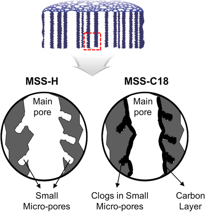
To help better understand these different temperature dependencies of 129Xe NMR spectra, a schematic model of porous structure for each MSS is provided in Fig. 5, illustrating the presence of small micropores embedded in larger mesoporous channels and the formation of a thin grafted layer on the main mesopore channels that covers up the underneath micropores. This model for the bare MSS-H material is consistent with the results from the previous study of MSS materials made using a similar method.Reference Mao, Kim, Joo, Sailor, Hopson and Wang32 The proposed structure for the MSS-C18 material will be confirmed at the end of this paper based on our experimental results and analysis.
Illustration of proposed pore morphology in the MSS structures, depicting the presence of small micropores embedded in larger mesoporous channels. The grafting of carbon blocks the underneath micropores due to the formation of thin carbon layers on the main pore surface.

A narrow single resonance peak at low temperature (<253 K) for MSS-H indicates the relative uniform micropores that are present in MSS-H as shown in Fig. 5. As the temperature increases, Xe enters the larger mesopores and undergoes fast-exchange with different pore environments. The linewidths at higher temperature are related to the uniformity of larger mesopore channels and the connectivity among different pores. The broader linewidths and shoulder peaks found in MSS-H at higher temperatures indicate that there are at least two different mesopore environments in MSS-H materials. This shoulder peak is present at temperatures at and above 253 K, indicating the existence of two types of pores that are not well connected because the fast-exchange of Xe atoms at higher temperature did not average out the effect of these two different pore environments. These two types of pores are most likely from two different domains of pores. By contrast, a single 129Xe NMR peak is observed at all temperatures for MSS-C18. The presence of two peaks in MSS-H and a single narrower peak in MSS-C18 indicates that pores are better connected in the MSS-C18. The higher connectivity in MSS-C18 is mostly a result of removal of the micropores due to the grafting of C18 onto the surface of the mesopores, resulting in faster exchange of adsorbed Xe among different pore environments. The hydrocarbon chains in the MSS-C18 material not only block the xenon from access to the micropores but also alter the surface composition and morphology of the silicon mesoporous channels. Therefore, it is not surprising to observe a significant difference in chemical shifts at low temperature between MSS-H and MSS-C18 materials. For example, the MSS-H and MSS-C18 showed 129Xe NMR peaks at 152 ppm and 121 ppm at 173 K, respectively. The fast reorientation of the surface-bound C18 hydrocarbon chains at higher temperatures effectively prevents xenon from reaching the surface, more effectively reducing its interaction with the microspores on the meso-silicon surface. At lower temperatures, the motion of the chains is significantly reduced, and the surface of bare MSS materials is not as effectively covered. Xe may be able to penetrate a small amount of micropores at low temperature. Therefore, at low temperature, Xe in the MSS-C18 material samples different pore environments including some partially blocked micropores, resulting a broader linewidth than those at high temperature.
Due to the increased adsorption at lower temperatures, a larger signal for Xe adsorbed on the pore surfaces is detected as compared to the signal for Xe in the void space (0 ppm). The signals associated with the smaller pores will be enhanced due to the higher surface area to free volume ratio of the micropores. Therefore, it is understandable that we observe the enhanced adsorbed Xe signal with respect to the free Xe gas signal in the void space at low temperature. However, the degree of the enhancement in the adsorbed signal with respect to the free Xe gas is significantly different for MSS-H and MSS-C18 samples. It was found that the spectral intensity ratio (obtained by integration of the peak areas) of adsorbed Xe to free Xe gas for the MSS-H materials at 173 K was 27 times greater than that at 300 K, whereas the intensity ratio of adsorbed Xe to free Xe gas for the MSS-C18 materials at 173 K was only 3 times larger than that at 300 K. The results show that the MSS-H adsorbed much more Xe than MSS-C18 at low temperature (<250 K), due to the penetration of Xe to the higher number of the micropores present in MSS-H as compared with the MSS-C18 samples. In MSS-C18, the grafted layer of hydrocarbon chains blocks most entrances of the micropores (as proposed in Fig. 5), inhibiting Xe diffusion into the strong adsorption micropore centers. These blocked pores will also lead to a decrease in the observable signals associated with Xe inside the mesoporous channels at temperatures above 253 K.
D. Parameters derived from the temperature-dependent HP 129Xe NMR spectra
The temperature-dependent 192Xe spectra can also be used to obtain physical parameters related to the xenon adsorption properties toward MSS materials. Variations of the 129Xe resonance chemical shifts at different temperatures can be fitted to extract parameters related to the adsorption properties using a model based upon Henry’s Law.Reference Ratcliffe19,Reference Knagge, Smith, Smith, Buriak and Raftery24,Reference Terskikh, Mudrakovskii and Mastikhin25 In the fast exchange approximation with weak adsorption, the temperature dependence of the observed 129Xe chemical shifts for arbitrary pore sizes can be expressed as
 $$\delta = {\delta _{\rm{s}}}{\left( {1 + {B \over {RT}}{{\rm{e}}^{{{ - {\rm{\Delta }}{H_{{\rm{ads}}}}} \over {RT}}}}} \right)^{ - 1}}B = \left( {{{{V_{\rm{g}}}} \over {S{K_0}}}} \right)\quad ,$$
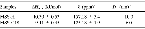
where V g is the free volume inside the porous material, T is the temperature, S is the specific surface area, K 0 is the pre-exponential term of Henry’s constant, R is the universal gas constant, ∆H ads is the heat of adsorption, and δs is the component of the observed 129Xe chemical shift characteristic of the interaction between xenon and the surface. Although Eq. (1) contains 3 variables, only two parameters, ∆H ads and V g/S, have a pronounced variation and effect on the fit. The situation can be further simplified by well-defined possible range of δs from the low temperature measurements, and our broad temperature screening experimental results support sufficiently constrained δs values to reduce the error range for the other two-parameter fitting. From the experimental data (see Figs. 3 and 4) and fitting of Eq. (1), the basic parameters such as ∆H ads and δs for each MSS materials were derived (Table I).

MSS-H and MSS-C18 show 10.3 kJ/mol and 9.4 kJ/mol for the ∆H ads values, respectively, which is consistent with physical adsorption of Xe on solid surfaces. No significant difference of these two values for MSS materials suggests that the grafting on the surface has very less effect of the adsorbing nature of xenon on the solid surface, whereas the values of δs for MSS-H (157.18 ppm) and MSS-C18 (125.18 ppm) are comparatively large, and it suggests that the sufficient coverage of grafted layers on the surface because δs values are strongly dependent on the surface compositions. The pore diameter value, D, was also derived from the NMR analysis results and equation. MSS-H and MSS-C18 show 10.0 nm and 6.0 nm of pore diameter, respectively, and these are much smaller in size than those from nitrogen adsorption–desorption analysis [∼18 nm, Fig. 1(e)]. The value D indicates average pore size of MSS materials, so it represents high sensitivity of HP 129Xe NMR analysis for porous materials having main mesopore channel framework along with micropores. The existence of micropores on the walls of mesoporous silica has also been reported in our previous study.Reference Moudrakovski, Terskikh, Ratcliffe, Ripmeester, Wang, Shin and Exarhos16 Our results show that 129Xe NMR is more sensitive to the micropores than the N2 adsorption measurement, resulting in smaller D values. 129Xe NMR probes pores ranging from 0.1 to 30 nm, whereas the N2 absorption mainly represents the distribution of pores from 2 to 50 nm. Unlike the condensation and multilayer adsorption in N2 adsorption measurements, HP 129Xe NMR taken at room temperature allows only submonolayer adsorption of the Xe atom on the surface, making it extremely sensitive to pore environment. Also, significantly decreased D values of MSS-C18 (6 nm) compared with MSS-H (10 nm) suggest the generation of grafting layer (∼2 nm) on the channel surface of MSS as shown in Fig. 5. Based on our results and analysis of temperature-dependent spectra along the TEM, SEM, FTIR, and N2 adsorption techniques, the proposed model given in Fig. 5 accurately illustrates the porosity and pore structures of MSS materials before and after C18 grafting.
IV. CONCLUSION
Pore structure and interconnectivity of grafted MSS materials were analyzed using temperature-dependent CF HP 129Xe NMR spectroscopy. A variation of uniformity and interconnectivity was observed when the MSS materials were grafted via a thermally induced dehydrogenative coupling method. The 129Xe NMR chemical shift data indicate the existence of micropores inside the larger mesopore channels in MSS, and the effects of grafting on the pore surfaces. Partitioning of Xe between the micropores and the mesopores in the MSS materials is temperature-dependent, with Xe principally occupying the micropores at lower temperatures. At higher temperatures, Xe exchanges through several different types of pore environments. However, the grafting of C18 on the surface of the micropores in MSS materials reduces the nonuniformity of porous channels. The temperature-dependent study provides insights on the differences in structure and interconnectivity of the pores between bare MSS and grafted MSS material. Such information can be useful for obtaining a better understanding of the relationship between porous structures and the design and performance of electrode materials.
ACKNOWLEDGMENTS
This work was supported by the EPSCoR Implementation grant DESC0007074, Office of Basic Energy Sciences, and U.S. Department of Energy (US DOE). L-Q. Wang thanks the advance grant HRD-0548311 for the purchase of new equipment used in this study. M.J. Sailor thanks the fund supporting by the National Science Foundation under Grant Nos. DMR-1210417 and CBET-1603177, and by the Defense Advanced Research Projects Agency (DARPA) under Cooperative Agreement HR0011-13-2-0017. D. Kim thanks the UCSD FISP program and the financial support by Basic Science Research Progam through the Korea NRF funded by the ministry of Education (NRF-2018R1D1A1B07043383). The content of the information within this document does not necessarily reflect the position or the policy of the Government.