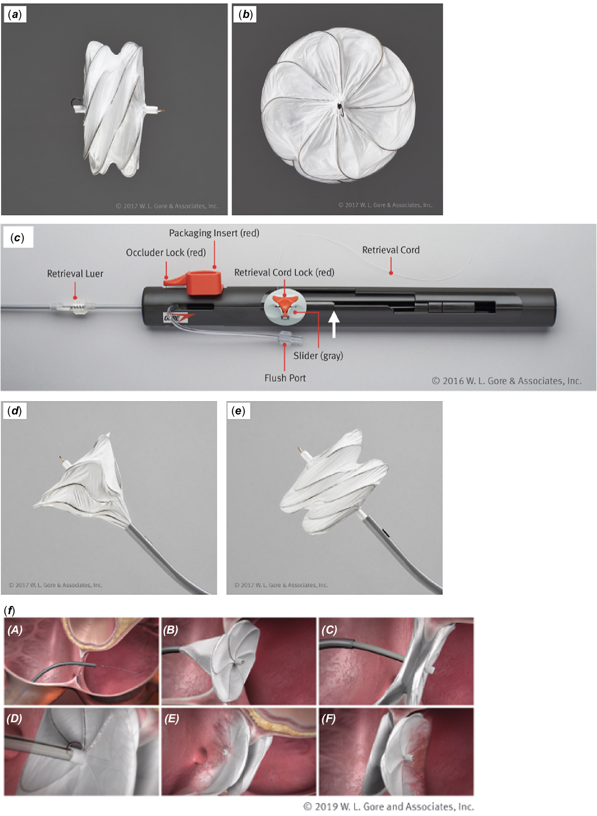
Atrial septal defects (ASD’s) are common with a reported incidence of 56 per 100,000 live births. Reference Hoffman and Kaplan1 Ostium secundum atrial septal defects constitute about 75% of these lesions. Reference Kirklin and Barratt-Boyes2 For ostium secundum atrial septal defects that need closure, surgical or transcatheter device closure are the two available options. Today transcatheter closure is the first-line treatment approach at most centres in the United States of America for appropriately selected patients with ostium secundum atrial septal defects. The first transcatheter closure of atrial septal defect in dogs was reported by King and Mills in 1974. Reference King and Mills3 Since then, there have been major modifications in the available devices as well as the transcatheter delivery systems. Multiple devices used in the past like the CardioSEAL (NMT Medical, Inc., Boston, MA), Reference Kaulitz, Peuster, Jux, Paul and Hausdorf4 clamshell occluder (C.R. Bard Inc., Murray Hill, New Jersey), Reference O'Laughlin5 Sideris Buttoned device (Custom Medical Devices, Inc.), Reference Rao, Berger and Rey6 Das-Angel Wings occlusion device (Microvena Corporation, White Bear Lake, MN), Reference O'Laughlin5 and the BioSTAR device (NMT Medical Inc., Boston, MA) Reference Hoehn, Hesse, Ince and Peuster7 have been discontinued. Currently, there are three devices available for closure of atrial septal defects that have been approved by the US Food and Drug Administration (FDA). The first is the AmplatzerTM (Abbott Laboratories, IL) atrial septal occluder device. Reference Turner, Owada, Sang, Khan and Lim8 Another device used until recently was the Gore Helex septal occlude. Reference Latson, Zahn and Wilson9 This device was replaced by a newer modified version referred to as the Gore® Cardioform septal occlude Reference Gillespie, Javois, Moore, Forbes and Paolillo10 (W. L. Gore & Associates, AZ) approved initially for closure of ASD’s <17 mm in 2015. The indication was later expanded to include patent foramen ovale (PFO) closures in 2017. The newest addition to the family of ASD closure devices in the United States of America is the Gore® Cardioform ASD occluder Reference Sommer, Love and Paolillo11 (W. L. Gore & Associates, AZ), approved by FDA in May 2019 (Fig 1). Similar to the Amplatzer atrial septal occluder, this device is self-centering (Fig 1 ) with a central waist that fills the atrial septal defect. Reference Sommer, Love and Paolillo11 The waist of the Gore Cardioform ASD occluder is believed to be “anatomically adaptable” so it conforms to the shape of the defect, rather than altering the defect to fit a circular waist. Reference Sommer, Love and Paolillo11 One of the perceived benefits of the Gore family of devices is that they are softer than the Amplatzer atrial septal occluder and potentially avoids the risk of device erosions with only two cases of cardiac erosion reported with a Gore Septal Occluder device after PFO closure Reference Kumar, Orford and Tobis12 as opposed to 0.3% reported with the Amplatzer atrial septal occluder. Reference Turner, Owada, Sang, Khan and Lim8 The ASSURED clinical trial Reference Sommer, Love and Paolillo11 demonstrated the safety and efficacy with the Gore Cardioform Atrial Septal Occluder with no device erosions reported in the 6 months follow-up for the study. Of note, there was 36% incidence of wire frame fracture. This was not associated with any clinical consequence, new shunts, or device instability. Reference Sommer, Love and Paolillo11 Long-term outcomes of these wire frame fractures need to be studied in the future.
Gore Cardioform atrial septal occluder device side view ( a ) and “en face” view ( b ). The various parts of the delivery handle ( c ) are labelled for ease of understanding. Special note should be made for the slight step made to depict the complete deployment of the left atrial disc and the waist (marked with a white arrow). At this point, the left atrial disc and the central waist are completely advanced as shown in ( d ). This is the step when the device should be pulled towards the atrial septum prior to exposing the right atrial disc. ( e ) shows the device being completely advanced resulting in the formation of the two discs with a central waist. ( f ) depicts the deployment steps of the Gore Cardioform ASD Occluder across the atrial septal defect. Images were provided courtesy of W. L. Gore & Associates.
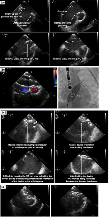
( a ) Complete evaluation of the atrial septal defect using transesophageal echocardiogram depicting the six rims (right upper pulmonary vein, atrioventricular valve, posterior, retroaortic, superior vena cava (SVC), and inferior vena cava (IVC) rims). Retroaortic rim is deficient in most patients and is not a contraindication for device placement. Authors recommend using a Mullins sheath (see Figure 6 and text) in patients with absent retroaortic rim. ( b ) Balloon sizing using “stop flow technique” on transesophageal echocardiogram (19.3 mm) and fluoroscopy (18.9 mm). Please note the calibration markers on the balloon used to calibrate the fluoroscopic measurements. ( c ) depicts the relatively perpendicular orientation of the device prior to locking the device (left panel) and the difficulty in accessing the rims (especially the IVC rim). This however was much easier after the device was locked (right panel) ( d ) depicts the transesophageal echocardiogram post-device deployment prior to release with favourable re-orientation of the device.

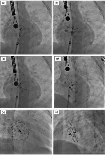
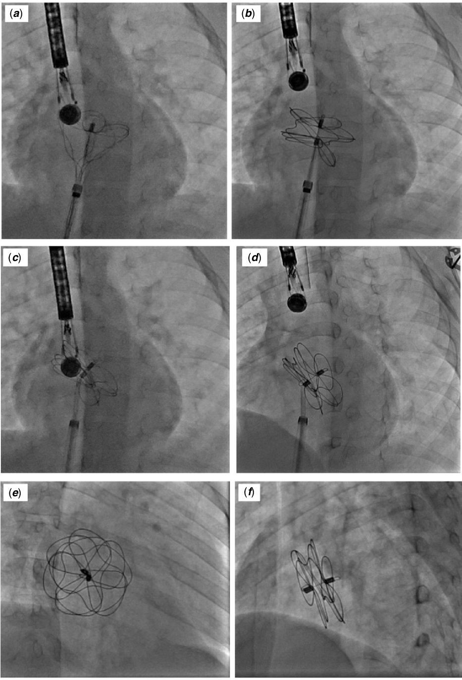
Fluoroscopic visualisation of the Gore Cardioform atrial septal occluder. ( a ) shows the tulip-shaped configuration of the left atrial disc and at this time the device should be pulled against the atrial septum. ( b ) Post-deployment of the left and right atrial discs but prior to locking. Please note the locking loop still inside the delivery catheter. ( c ) Change in the orientation of the device after locking the device in place. Note the locking loop is now engaged (better seen in ( d ); white arrow). ( d ) Device orientation just after the release. ( e ) Right anterior oblique/caudal projection and ( f ) left anterior oblique/cranial projection showing the final device position in the atrial septum. Note the splaying between the left and right atrial disc which suggests that the device has captured the atrial septum (this should also be confirmed using echocardiographic visualisation).
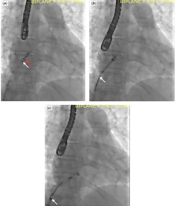
Retrieval of the device post-locking the device prior to releasing the retrieval cord. ( a ) In this case, we used a long Mullins sheath (tip marked by white arrow) for device deployment and used it to assist in device retrieval as well. The tip of the grey delivery catheter is marked by red arrow. The right atrial disc of the device is pulled using the retrieval cord (white arrow) inside the delivery catheter whose tip is overlapping the tip of the Mullins sheath ( b ). We retrieved the left atrial disc of the device inside the Mullins sheath in this case ( c ) with the tip of the delivery catheter now pulled further inside the Mullins sheath (white arrow).

As early adopters to the new device use in our patients, we realised that there is a learning curve to the use of this device and the delivery system. We have utilised this device as a part of the Gore continued access trial and as early adopters of the device post-FDA approval in 2019. We recently published our early outcomes of the use of this device on 98 consecutive patients from June 2017 to December 2019. Reference Eilers, Gowda and Gowda13 In the entire cohort, successful closure of the atrial septal defect was achieved in 92 patients. Five patients were referred for surgical closure and one patient underwent ASD closure with an Amplatzer septal occluder device. Reference Eilers, Gowda and Gowda13 In this cohort, there was one device embolisation, two patients had atrial fibrillation during procedure needing cardioversion, four patients had transient atrial tachycardia. Use of a MullinsTM sheath facilitated closure of large secundum ASDs with challenging anatomy. ASD size, device size, and maximum sheath size were significantly larger in those patients in whom a Mullins sheath was used. Reference Eilers, Gowda and Gowda13 We hereby describe learning points from our early experience with the aim of helping implanters across the world trying to use this device in their practice. These do not substitute the Instructions for Use (IFU) provided with the device.
Device description
The Gore Cardioform Atrial Septal Occluder device consists of an implantable occluder and a delivery system (Fig 1). The occluder is comprised of a platinum filled nickel-titanium alloy (nitinol) wire frame covered with expanded polytetrafluoroethylene. The expanded polytetrafluoroethylene is hydrophilic facilitating visualisation under echocardiography. It is also thrombo-resistant and has considerably lower risk of cardiac tissue erosion. The occluder comes in five different configurations with respect to the outer disc diameter (27, 32, 37, 44, and 48 mm) for the closure of defects ranging in size from 8 to 35 mm. The smaller sizes have six petals on each disc and the 44-mm and 48-mm device have eight petals. As mentioned above, this device is self-centering, that means that it has a central waist which fills the defect. This waist is highly flexible and compliant and achieves closure of atrial septal defects by occupying the defect.
The delivery system is similar to the Gore Cardioform septal occluder (used for PFO and small ASDs) comprised of an 80-cm long delivery catheter with outer diameter from 10 French to 14 French coupled to a handle. The delivery catheter carries a control catheter, coated with a Hydrophilic Lubricious Coating, inside it. The handle consists of a slider, a retrieval cord, and locks for the occluder and the retrieval cord. The slider aids deployment, positioning, locking, capture, and retrieval of the occluder (Fig 1). In general, in small to moderate sized defects, the prior generation Gore Septal Occluder can still be used, provided the rims are adequate and the defect is 17 mm or less.
Procedure details
The procedure is commonly done in children under general anaesthesia with transesophageal echocardiography. Some centres with experience in sedated transesophageal echocardiography can perform the procedure with conscious sedation or use intracardiac echocardiogram for evaluation of the defect. Unfractionated heparin is used to achieve an activated coagulation time >250 seconds. Biplanar fluoroscopic guidance (when available) is recommended throughout the procedure.
We routinely perform balloon sizing of the atrial septal defect in all our patients using the “stop-flow technique.” Reference Bartakian, El-Said, Printz and Moore14 The sizing balloon [Amplatzer sizing balloon® (AGA medical corporation, Plymouth, MN) or PTS® and PTS-X® sizing balloons (NuMed, Inc., Hopkinton, NY)] is measured on both fluoroscopy and echocardiogram and are usually within 1 mm of each other. We use the largest measured diameter for choosing the Gore Cardioform atrial septal occluder device.
Device selection
The manufacturer has given guidelines about the appropriate size of the device for various sizes of atrial septal defect (Table 1). However, there are a number of factors that go into the decision-making process for device selection. In the presence of deficient rims (particularly 2 or more), upsizing the device may be advantageous provided the septal length and cardiac anatomy allows this.
Guidelines from the manufacturer of the Gore Cardioform atrial septal occluder regarding device sizing based on atrial septal defect size (as measured by stop flow technique). Please note that the authors tend to use the 37-mm, 44-mm, and 48-mm device for defects larger than 18-mm, 23-mm, and 28-mm, respectively (if the atrial septal length allows)

Table obtained from Gore Cardioform ASD Occluder website: https://www.goremedical.com/resource/AY0110-EN1.
Device preparation
Once the appropriate device is selected, we recommend performing an activated coagulation time at this time to ensure therapeutic levels prior to device advancement. The delivery catheter is flushed with heparinised saline. At this time, the device is advanced out of the delivery catheter, inspected and deaired. While continuously flushing through the flush port (Fig 1), we withdraw the right atrial disc into the delivery catheter. There is a small step-down (felt on the slider) made to provide a tactile feel when it is time to deploy the right atrial disc (Fig 1c) but in this may sometimes be hard to feel. Therefore, a mark may need to be made on the delivery handle using a sterile marker (Fig 1c) to indicate this point. The fluoroscopic and echocardiographic (transesophageal echocardiography/intracardiac echocardiogram) appearance of the device when the LA side of the device is fully deployed should be appreciated by the operator (Fig 1d and corresponding fluoroscopic image Fig 3a). Note, at this stage during device deployment, the device should be pulled towards the atrial septum before deploying the right atrial disc.
Transesophageal echocardiogram and device deployment
Deployment of the Gore Cardioform atrial septal occluder can pose particular challenges for the echocardiographer since the device has less metal and also sometimes has a tendency to “swell-up” with blood during deployment. These factors can make the visualisation of the septal rims challenging, especially while trying to ascertain that the rims are adequately captured by the device. Therefore, allowing echocardiographer adequate time to visualise the atrial septal defect and familiarise with the appearance of the rims is crucial (Fig 2). Adequate visualisation of these rims and documenting appropriate capture of the rims is essential prior to device release (Fig 2). The authors prefer to use biplane fluoroscopy for device deployment. Splay between the left and right atrial disc due to capture of atrial septum in between the discs should be confirmed prior to the release. Ideally, on fluoroscopy, in a left anterior oblique/cranial projection, separation/splay should be seen of the right atrial and left atrial sides of the device at the anterior/superior rim and posterior/inferior rims (Fig 3). This is particularly helpful if the echocardiogram cannot adequately demonstrate the device capturing the rims of the atrial septal defect (which can happen particularly with imaging of the inferior rim). It is important to note that it is not infrequent for the device to appear relatively perpendicular to the atrial septum prior to locking the device on transesophageal echocardiography, but a favourable shift is usually noted with locking (Fig 3b–d). As long as the rims are adequately captured, it is advised to lock the device, as only then will the final device orientation be appreciated. Once the device is demonstrated to be in correct position, it is locked in place using the lock mechanism on the delivery handle (Fig 1c). Care should be taken that the device delivery slider is all the way forward and then slightly downwards prior to attempting the locking mechanism. At this stage, the implanter should pay special attention to avoid rotating the delivery handle as the torque may be transmitted to the device and may result in device malposition. Once locked in position, repeat echocardiographic visualisation is performed by 2D and colour flow doppler. After confirmation, the device can be released by releasing the retrieval cord. Care should be taken to keep the retrieval cord locked till the final device position is confirmed after locking. The Gore Cardioform ASD Occluder device is bulkier than the Gore Septal Occluder device and though protrusion in the superior vena cava can be seen (particularly in patients with small superior vena cava rims and superior defects), we have not encountered any cases in which the device caused clinically significant impairment of superior vena cava flow in those cases. This however, the superior vena cava flow should be carefully evaluated, in addition to other relevant surrounding structures. The device can still be retrieved after the locking loop is engaged using the retrieval cord if the device position is deemed unsatisfactory. This is accomplished by unscrewing at the retrieval luer (Fig 1c) and slowly pulling the device inside the outer grey catheter. It important to try and keep the right atrial eyelet coaxial with the delivery catheter to facilitate retrieval (Fig 4).
Challenging anatomy: absent/deficient rims and large atrial septal defects
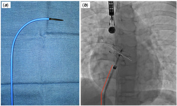
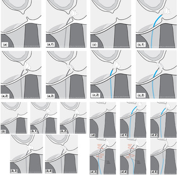
As much as 75–80% of ostium secundum atrial septal defects have a deficient retroaortic rim (defined as <5 mm rim). This deserves special attention as the relatively straight delivery cable and catheter can make the device slip through in the retroaortic area leading to device malposition. To avoid this (and prolapse of the device similarly in other areas due to other deficient rims) and help delivery of large devices, we prefer using a Mullins sheath® (Medtronic – Minneapolis, MN) to align the device parallel to the atrial septum (Fig 5) after deployment of left atrial disc and device waist (same French size as recommended when using short sheath). Clockwise rotation on the Mullins sheath and Gore Cardioform ASD Occluder delivery catheter may further help orient the device, Reference Eilers, Gowda and Gowda13 Figure 6. Use of a Hausdorf sheath can also help facilitate device alignment in cases where closure of the atrial septal defect was not successful despite the use of Mullins sheath. Reference Eilers, Gowda and Gowda13 Lately, we have also tried hand shaping the delivery cable (to exaggerate the curve at the tip) and it has worked well for device positioning and avoiding the use of Mullins sheath. This however should be done very carefully and may result in device malfunction.
( a ) Use of Mullins sheath® (Medtronic – Minneapolis, MN) in select patients (deficient/absent rims, large atrial septal defect). This helps provide a better angle (shown by red line in b ) and thereby help orient the device parallel to the atrial septum (dotted white line in b ) especially in patients with deficient retroaortic rim and large devices.

( a ): The GORE® CARDIOFORM ASD Occluder is placed in a defect with relatively small defect, and good rim tissue. (1) The left disc of the device is formed. (2) The left disc is pulled against the existing rim tissue. Note that the tension pulls the catheter toward the medial side of the IVC. The force of the occluder gaining apposition to the rims is entirely in the inferior direction. However, good apposition is gained here due to good rims. (3) The right disc is deployed, capturing both rims. Some tension is released on the delivery system allowing the delivery catheter to move laterally. ( b ): The GORE® CARDIOFORM ASD Occluder is placed in a defect with a larger defect, and smaller rim tissue. (1) The left disc of the device is formed. (2) The device is pulled inferiorly by the operator, until the superior portion of the disc comes into contact with the aortic rim. The catheter moves medially in the IVC as tension is applied. Note that the inflection of the wires that will form the occluder waist are still not making contact with the posterior-inferior rim. If the right disc were deployed in this position, petals forming the inferior portion of the right disc are likely to be entirely in the left atrium and not capturing the rim. (3) The operator applies more tension in order to bring the inferior portion of the disc into contact with the rim tissue; however, this causes the superior portion of the device to slip off the aortic rim into the right atrium. (4) If the right disc is deployed as depicted, petals from the left disc of the device will be entirely within the right atrium and will not appropriately capture the aortic rim. ( c ): The GORE® CARDIOFORM ASD Occluder is placed in the same anatomy as Figure 6 ( b ), but with the assistance of a long Mullin’s type sheath. Here, the curvature of the sheath augments the curvature of the delivery catheter of the occluder device. (1) The left disc of the device is formed. (2) The device is pulled inferiorly by the operator, until the superior portion of the disc comes into contact with the aortic rim. The catheter and sheath move medially in the IVC as tension is applied, largely straightening out the curve of the Mullins. In this depiction, the slight assist provided by the Mullin’s curvature allows the inferior portion of the occluder to come into appropriate contact with the rim tissue. (3) The right disc of the device is deployed, some tension is released from the system, and the occluder is appropriately capturing the septal rim tissue. ( d ): The GORE® CARDIOFORM ASD Occluder is placed in a yet more difficult anatomy a larger defect, and smaller rims. Here, another technique must be applied to gain septal apposition. (1) The left disc of the device is formed, with the Mullin’s sheath near the tip of the delivery catheter across the defect. (2) Both the Mullin’s sheath and handle of the device are pulled inferiorly until the superior portion of the device appears to contact the aortic rim. However, the inferior portion of the device is not in contact with the rim. Any further tension will pull the delivery catheter and sheath medially in the IVC, and the superior side of the device will pull into the right atrium. (3) The Mullin’s sheath is now pulled inferiorly until the tip is lower in the right atrial space or in the superior portion of the inferior vena cava. If the sheath position is stationary, then the tip of the sheath can now be considered a “fixed point.” The handle of the occluder delivery system may be rotated or withdrawn and will move the left cone of the device relative to this fixed point (note that the tip of the sheath may move somewhat as it is more flexible compared to the GORE® CARDIOFORM ASD Occluder delivery catheter). A posterior rotation of the occluder delivery system handle may aid in alignment depending on specific septal geometry. Pulling the handle inferiorly at the position shown the figure may still prolapse the petals of the occluder into the right atrial space; in this instance we may attempt the manoeuvre shown in ( d ). (4) Holding the position of the occluder handle, the Mullin’s sheath is rotated 180°. This moves the fixed point of the tip of the sheath to a more lateral position in the right atrial space. The curvature of the Mullin’s now pushes the delivery catheter laterally off the medial side of the inferior vena cava. Since the fixed point of the sheath tip is now pointed towards the lateral wall of the right atrium, the motion of the overall system when the handle is pulled inferiorly must be translated in direction. In effect, a virtual pulley is created around this curvature in the top-right of the s-shape. Now, when the operator pulls the handle of the device (holding the Mullin’s in the same position), the inferior motion is translated slightly around the “pulley” and is more perpendicular to the septum. The device can now be brought into contact with the rims. Again, posterior rotation of the handle (with or without further rotation of the Mullin’s sheath) may aid in achieving optimal apposition. (5) The right disc is deployed, successfully capturing the deficient rims.

In conclusion, as with any other new device, there is a learning curve in the use of the Gore Cardioform atrial septal occluder. Special consideration should be made while using the device sizing chart as in the instructions for use. The authors prefer the larger device at the upper end of the atrial septal defect size (if permitted by the atrial septal length). Marking the location on the delivery handle where the left atrial disc and the waist is fully deployed has helped the authors in device positioning. Lastly, using the curve of the Mullins sheath can make device deployment easier in challenging anatomy like absent retroaortic rim or large atrial septal defects. Careful attention to these points has helped authors and hopefully help the readers in the management of their patients.
Acknowledgements
Figure 1, 5 and Table 1 were provided courtesy of W. L. Gore & Associates.
Financial support
This research received no specific grant from any funding agency, commercial or not-for-profit sectors.