I. INTRODUCTION
Intentional incorporation of impurities into colloidal nanocrystals is essential to tune the electronic, optical, and magnetic properties of their corresponding bulk semiconductors.Reference Erwin, Zu, Haftel, Efros, Kennedy and Norris1–Reference Beaulac, Archer, Ochsenbein and Gamelin3 The unique properties of these nanocrystals, arising from the small volume and quantum confinement effects, promise several potential applications in biolabeling,Reference Bruchez, Moronne, Gin, Weiss and Alivisatos4 lasers,Reference Htoon, Hollingworth, Malko, Dickerson and Klimov5 light-emitting diodes,Reference Kim, Lee and Yang6 etc. Especially, doping nanocrystals with transition metal ions such as Mn2+ produces so called diluted magnetic semiconductor that could be used in spintronic devices because of the effect termed as the giant Zeeman splitting, which results from the exchange interaction between the transition metal ions and the electronic states of the host nanocrystals.Reference Yu, Liu, Kweon, Joo, Park, Ko, Lee, Shen, Tivakornsasithorn, Son, Park, Kim, Hwang, Dobrowolska, Furdyna and Hyeon2, Reference Efros, Rashba and Rosen7 As a result, the control of doping nanocrystals with transition metal ions is of fundamental interest in scientific research and in the application of spintronics.
Nucleation doping strategy is an effective doping method for synthesizing the core/shell structure, which has several advantages. First, the core nanocrystals are passivated with a wider gap material to improve the fluorescence efficiency and stability against photo-oxidation, this is because, in such a system, the surface-related trap states acting as nonradiative process were suppressed.Reference Beaulac, Archer and Gamelin8 In addition, either the lattice mismatch between the core and shell or the large splitting of Mn2+ excited state resulted from the thin thickness of the shell could induce the color tunability, which is the red shift or blue shift, respectively,Reference Zuo, Sun, Zhao, Jiang and Gao9, Reference Pradhan and Peng10 thus widening the window of Mn2+-doped semiconductor phosphors. It is known that such onion-like structure can be constituted by different compositions such as MnS/CdS,Reference Zuo, Sun, Zhao, Jiang and Gao9 MnS/ZnS,Reference Zheng, Yuan, Ikezawa, Jing, Liu, Zheng, Kong, Zhao and Masumoto11 and MnSe/ZnSe.Reference Zeng, Rutherford, Xie, Zou and Peng12 However, in these heterojunctions, the core and shell are in the same structure phase. To our best knowledge, there is no report on the synthesis of nanocrystals with core and shell in different structure.
Systematic researches have been carried out to study the effects of ligands on the formation of CdSe nanocrystals in different structures.Reference Al-Salim, Young, Tilley, McQuillan and Xia13 In all the cases to synthesize CdSe nanocrystals, it is well known that the condition of selenium precursors prepared with alkylphosphine would lead to wurtzite structure.Reference Murray, Noms and Bawendi14 However, such alkylphosphine is pyrophoric, expensive, and toxic, the experimental condition needs carefully controlled. Then greener protocols were developed by dispersing selenium in octadecene (ODE), which resulted in a blende structure of CdSe.Reference Jasieniak, Bullen, van Embden and Mulvaney15 Consequently, it is still challenging to synthesize CdSe nanocrystals in a wurtzite structure without the involvement of alkylphosphine. Herein in our work, by selecting the appropriate phosphine-free selenium precursor, through a nucleation doping strategy, we achieved such core/shell MnSe/CdSe nanocrystals with the CdSe shell in wurtzite structure while the MnSe core still in blende structure, but the pure CdSe nanocrystals obtained using the same anion precursor remained in blende structure. Thereby, we believe the strain induced by the lattice mismatch, as well as the ligands, play important roles in the formation of such structure. In this article, we will demonstrate that the MnSe/CdSe nanocrystals could be stabilized with MnSe in a zinc blende structure and CdSe blende or wurtzite.
II. EXPERIMENTAL SECTION
A. Chemicals
All chemicals, cadmium oxide (CdO, 99%), selenium powder (Se, 99.95%), 1-octadecene (90%), oleic acid (OA), and oleylamine (ODA), were used without further purification.
B. Synthesis of MnSe/CdSe core/shell nanocrystals
(i). Preparation of manganese stearate (MnSt2): The manganese precursor, MnSt2, was synthesized by the method reported in Ref. Reference Pradhan and Peng10.
(ii). Preparation of stock solutions: SeODE solution was prepared by adding 2-mmol Se and 20-mL ODE in a three-neck flask and then heating the mixture under nitrogen atmosphere at 200 °C for 2 h. The selenium precursor was prepared by adding 3-mmol Se and 9-mL ODA into a three-neck flask and heating the mixture to 200 °C until it was completely dissolved. The Cd precursor was prepared by mixing 2-mmol CdO, 3-mL OA, and 17-mL ODE in another three-neck flask and subsequently heating this solution to 150 °C and keeping for 3 h under nitrogen.
(iii). Typical synthesis of MnSe/CdSe core/shell nanocrystals through the nucleation doping strategy. ODE (10 mL) and 0.02-g MnSt2 were loaded into a 50-mL three-neck flask and degassed for 15 min at 110 °C by bubbling with nitrogen. Then the resulting colorless clear solution was heated to 300 °C. The SeODE (4 mL) was injected into the flask. Immediately after the injection, the color of the solution changed to faint yellow which showed the formation of MnSe nanocluster. The reaction mixture was allowed to cool down to 280 °C and annealed for 30 min. Subsequently, the cadmium precursor solution (4 mL) was injected, and the temperature was set at a designed numerical value for the growth of CdSe shell. Aliquots (about 2 mL) were taken at different time intervals for UV–VIS, photoluminescence (PL), transmission electron microscope (TEM), and x-ray diffraction measurements after purification.
(iv). The synthesis of MnSe/CdSe nanocrystals with shell in wurtzite phase was similar to that described above. After the mixture of ODE (10 mL) and 0.02-g MnSt2 was heated to 270 °C, 1.2-mL SeODA was added by injection and the temperature dropped to 260 °C. Five minutes after the injection, the Cd precursor was added and then aliquots were taken at different time intervals for characterization.
C. Measurements
The resulting materials were characterized by x-ray powder diffraction (XRD) on a Rigaku D/Max-2400 x-ray diffractometer (Rigaku, Tokyo, Japan) using Cu Kα radiation (40 kV, 60 mA). TEM and high-resolution transmission electron microscopy (HRTEM) measurements were obtained on a Tecnai-G2-F30 TEM (FEI, Hillsboro, OR). The UV–VIS spectra and PL spectra were obtained on a Shimadzu UV–VIS-nir 3600 (Shimadzu, Kyoto, Japan) and an FLS-920T fluorescence spectrophotometer with Xe 900 (450W xenon arc lamp, Edinburgh Instruments, Livingston, UK) as the light source, respectively.
III. RESULTS
A. Synthesis of nanocrystals with SeODE
The formation of the MnSe core is essential to both the nucleation doping strategy and the investigation of the shell growth mechanism, whereas the synthesis process of the core is significantly affected by the reaction condition, thus the resulted products will perform distinct emission properties. To get the MnSe/CdSe core/shell nanocrystals in blende structure, SeODE was prepared for the anion precursor. The chemical reactivity of the SeODE strongly depends on the heating temperature and timeReference Bullen, van Embden, Jasieniak, Cosgriff, Mulder, Rizzardo, Gu and Raston16; thus in our experiment, the heating time for the mixture was chosen to be 2 h at 200 °C to get the balance of time between the dissolution and the deactivation of the precursor. Furthermore, the different mole ratio of Mn to Se will result in different-sized MnSe nanocrystals eventually.
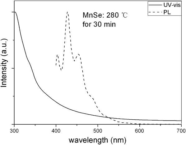
Figure 1 shows PL and UV–VIS spectra of MnSe cores annealed at 280 °C for 30 min. The absorption edge is over 400 nm, which is in accordance with the results reported previously.Reference Pradhan, Goorskey, Thessing and Peng17 Besides, a weak shoulder at about 345 nm is observed, the corresponding band gap is significantly larger than that for MnSe in rock salt structure, but comparable to zinc blende MnSe.Reference Qin, Lu, Wei, Qi, Peng, Yang and Qian18, Reference Sines, Misra, Schiffer and Schaak19 In the PL spectrum, there are three observable emission peaks at around 403, 427, and 454 nm. Since the characteristic emission peaks do not belong to octahedrally and tetrahedrally coordinated MnSe,Reference Heimbrodt, Goede, Tschentscher, Weinhold, Klimakow, Pohl, Jacobs and Hoffmann20 these emission peaks possibly originate from the defects in the nanocrystal cores, which is assumed to be caused by the metastability of MnSe in zinc blende structure. Unfortunately, the reaction mechanism is not clear as yet and further studies are needed to clarify the origin of the emission phenomenon.
Photoluminescence (PL) and UV–VIS spectra of MnSe annealed at 280 °C for 30min.

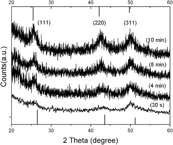
Time-dependent experiments were carried out to explore the evolution process of the nanocrystal. Figure 2 shows representative XRD patterns of the products obtained after various overcoating times, and the black line on the top and bottom axes represents the bulk diffraction peaks for blende CdSe and MnSe, respectively. The cores were annealed at 280 °C for 30 min, whereafter the overcoating temperature was set at 260 °C. The typical patterns shown in Fig. 2 correspond to aliquots taken at different overcoating times of 0.5, 4, 8, and 10 min. In the 10-min sample, three peaks located at 25.6°, 42.5°, and 50.1° are clearly displayed, corresponding to (111), (220), and (311) of zinc blende structure.Reference Kwak, Sung, Kim and Chae21 It can be seen that because of the low crystallization grade of the MnSe core as discussed above, the (111) and (220) diffraction peaks are not strong at first; however, as the time increased, the contour of the peaks become sharper and the position shifts to the low 2θ angle gradually. This trend implies the growing thickness of the shell.
X-ray powder diffraction patterns of MnSe/CdSe core/shell nanocrystals after growth time of 0.5, 4, 8, and 10 min, respectively.

In Fig. 3 are shown typical TEM and HRTEM images of nanocrystals taken out 4 min after injection with the cores annealed for 1 h. As seen from Fig. 3(a), these nanocrystals exhibit quasisphere morphology with a diameter about 8.0 nm. The corresponding electron diffraction pattern (inset) indicates these nanocrystals are in zinc blende structure, and these rings represent the (111), (220), and (311) planes, respectively, which is consistent with the XRD results. The HRTEM image [Fig. 3(c)] shows an interplanar distance of 0.347 nm, corresponding to the (111) plane spacing of zinc blende CdSe (0.351 nm), which indicates the d-dots with the appearance of CdSe. The smaller interplanar spacing is regarded as the result of transition from MnSe lattice to CdSe lattice, because the lattice constant of MnSe in zinc blende structure (about 0.5880 nm) is smaller than its analog of CdSe (about 0.6077 nm). Remarkably, a twin boundary is demonstrated in Fig. 3(b), which indicates that the shell is not a single-crystalline and is different from most of the particles previously reported.Reference Zheng, Yuan, Ikezawa, Jing, Liu, Zheng, Kong, Zhao and Masumoto11 In a two-step nucleation model, the amorphous polynuclei or polymolecules aggregate together in the first step, and in the second step, these amorphous structures start to adjust the positions of atoms and reorganized these clusters into the crystalline form.Reference Bryan and Gamelin22 Similarly, here we hypothesize the shell growth including two stages as below, in the initial stage of overcoating process, the cores adsorb monomers onto the surfaces to form amorphous layers and then in the second stage the layers start to crystallize. If two regions on the surface crystallize at the same time in the second step of surface nucleation, then they could not fuse into one single crystal, the twin boundary is formed.
Typical transmission electron microscope (TEM) and high-resolution transmission electron microscope (HRTEM) images of MnSe/CdSe nanocrystals acquired 4 min after injection. (a). TEM image and electron diffraction pattern of d-dots (inset). (b). HRTEM image of a twin boundary. (c). Illustration of interplanar distance of (111).

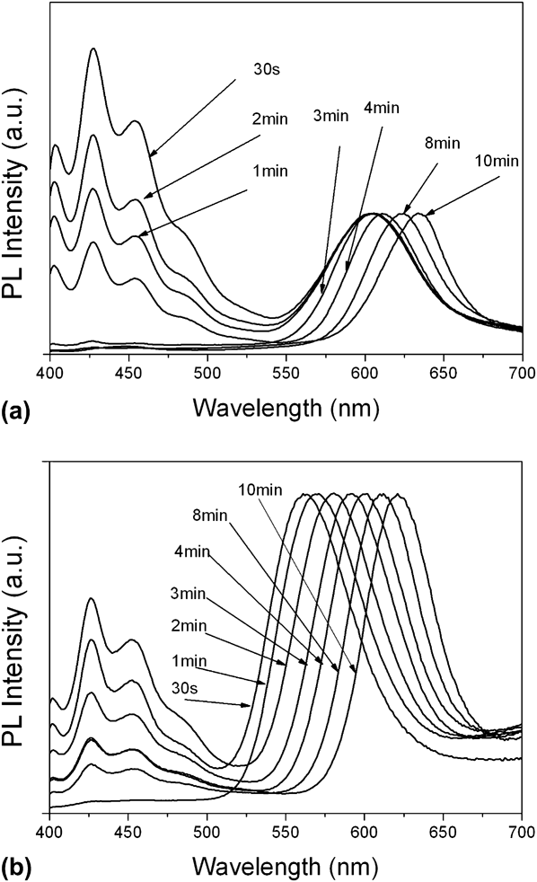
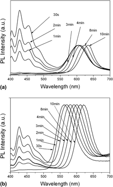
Manganese ion (Mn2+)-doped CdSe nanocrystals provide an ideal model for studying the doing process because of its allowance to tune the semiconductor band gap energy across the dopant excited state levels, which can be readily characterized by absorption and PL spectroscopy.Reference Beaulac, Archer and Gamelin8 When the CdSe shell is thin, which is in the strong confinement regime and the characteristic Mn2+ emission is observed. While the shells grow thicker, the band gap energy become smaller than the Mn2+-excited state energy and the excitonic emission will be dominant in nanocrystal luminescence. Figure 4 shows the temporal evolution of PL spectra of d-dots which were synthesized under different conditions. It can be seen that most of the spectra consist of two parts: the emissions located around 426 nm originate from the defects in cores as discussed above, and the normalized peaks involve the Mn2+ ion and exciton luminescences. If the cores were annealed only 30 min, as presented in Fig. 4(a), in the first few minutes after injection, the peak positions around ∼600 nm do not change much. However, if the annealing time lengthened to 1 h, the time-dependent Mn2+ ion emission is clearly observed immediately after injection as displayed in Fig. 4(b). It is shown that in both these figures, with the overcoating time more than 3 min, the emission peak position gradually red shifted. Based on the model described above, if the overcoating time increases, the CdSe shells grow thicker and the excitons locate in a weaker confinement regime, the exciton luminescence will thus become dominant in the spectra, manifesting the time-dependent red shift of emission peak positions. Therefore, according to which of emission is dominant, the evolutions of PL spectra could be divided into two stages in terms of Mn2+ and exciton luminescences. Furthermore, according to the peak position of 4 min in Fig. 4(b), the size of the nanocrystals was calculated using the empirical fitting functions provided by Yu,Reference Yu, Qu, Guo and Peng23 the result (about 4 nm) is much smaller than those observed in TEM (∼8 nm) which supports the assumption there is a MnSe core inside the nanocrystal with the appearance of CdSe.
PL spectra of d-dots (which is normalized) versus reaction time ranging from 0 to 10 min. (a). The cores annealed at 280 °C for 30 min, the shells grew under 260 °C. (b). The cores annealed at 280 °C for 1 h, the shells grew under 260 °C.

The emission of Mn2+ ion is significantly affected by the crystal field around the dopant. If the dopant experiences larger electric field difference along different directions, the Mn2+ ion has larger energy level splitting of the excited state, which manifests a blue-shift emission. Otherwise, the symmetric crystal field for a very dopant will result in a smaller splitting of energy levels. In Fig. 4(a), the emission peak positions which do not change much at the beginning suggest the crystal field for the dopant almost remains the same with the growth of CdSe. It is believed that the residual manganese precursor in the solution resulted in a growth doping process, and the dopants in the host which have smaller splitting of energy levels than those near the interface of MnSe and CdSe is the origin of the emission. Therefore, the emission property is similar to the traditional Mn-doped CdSe nanocrystals. However, in an ultrasituation, the complete consumption of manganese precursor exclude out the possibility of the doping in CdSe growth process. Thus the only environment for the dopant is near the interface of MnSe and CdSe where the crystal field for the dopant is different along different directions. As shown in Fig. 4(b), such situation resulted in blue-shifted Mn2+ emission comparing to Fig. 4(a). With annealing time increasing, the position of emission peak is red shifted, which suggests the d-dots have a core/shell structure since the crystal field for the dopant become symmetric in long range when the shell grow thicker. The different phenomena of PL are caused by the difference of local condition for the irradiative Mn2+ ions.
B. Synthesis of nanocrystals with SeODA
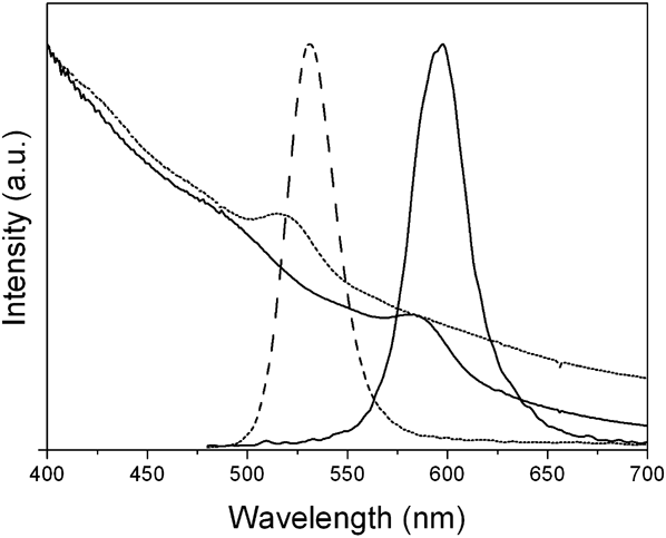
By altering the anion precursor, we got the MnSe/CdSe nanocrystals with shell in wurtzite phase. However, the protocol was not optimized. It can be seen that in Fig. 6(a) and 6(f) corresponding to 30-s and 10-min samples, respectively, the ensemble nanocrystals consist of two parts, the smaller ones and larger ones. It is assumed that the smaller ones indicate the formation of pure nanocrystals and the larger ones are the d-dots. The PL and absorption spectra corresponding to these two samples are shown in Fig. 5, where the dash lines belong to 30-s sample and the solid lines belong to 10-min sample. The narrow full width at half maximum of the spectra (∼ 25 nm) and the small stokes shift (∼15 nm) indicate that the emissions come from pure CdSe nanocrystals,Reference Pradhan and Peng10, Reference Jasieniak, Bullen, van Embden and Mulvaney15 which is consistent with our assumption. To further confirm that the emissions originate from smaller parts (pure CdSe nanocrystals), then the peak position from UV–VIS spectra was used to calculate the average size of the nanocrystals,Reference Yu, Qu, Guo and Peng23 and the 30-s sample is ∼2.5 nm while the 10-min sample is 3.9 nm, which are in accordance with the size of smaller parts in Figs. 6(a) and 6(f). No Mn2+ ion emission is observed because of low PL efficiency or some reason else. Figures 6(h) and 6(i) show the HRTEM image of a pure CdSe nanocrystal that is well crystallized and its fast Fourier transformation (FFT) corresponding to [111] zone axis is characteristic of blende structure according to interplanar spacing of (110).
UV–VIS and PL spectra of d-dots synthesized with SeODA which were acquired 30 s and 10 min after injection of Cd precursor, respectively.

TEM, HRTEM, and fast Fourier transformation (FFT) images of d-dots. (a, f) TEM images of 30-s and 10-min samples. (b, g) HRTEM images of 30-s and 10-min samples. (d)–(f) FFT images corresponding to different regions marked in (b). (h, i) HRTEM image and corresponded FFT of pure CdSe.

The composition of the ensemble nanocrystals were further determined by atomic absorption spectroscopy and energy dispersive x-ray fluorescence spectrometer, and the results showed that manganese element exists in the nanocrystals. Thus, it is argued that the high activity SeODA leads to the formation of large MnSe cores in a short time after injection. It is possible that the manganese carboxylate precursor was activated by the oleylamine and produced large MnSe. Meanwhile, the nucleation rate of intrinsic CdSe dots was also enhanced by the oleylamine. Therefore, two different-size particles coexisted in the solution. In Fig. 6(b), different lattice fringes were observed in a nanocrystal, which indicate different crystal structures. To confirm the phase of the core and shell in larger nanocrystals, different regions of nanocrystals shown in Fig. 6(b) were carefully examined by FFT. In Fig. 6(e), the FFT of region “e” corresponds to the [10 ] zone axis, characteristic of blende structure. The bright diffraction spots correspond to {311} crystal planes, and the interplanar spacing is 0.178 nm, which is consistent with the blende MnSe. The weak diffraction spots of {111} planes imply the stack faults happening to these planes. It is shown in Fig. 6(d) that the FFT of region “d” presents coexistence of wurtzite [002] and blende [10
 ] axis, which indicates the phase transformation from blende to wurtzite. The FFT of region “c” [Fig. 6(c)] signifies the wurtzite structure which is well crystallized, by presenting the {100} planes along c-axis with interplanar spacing of 0.372 nm. Figure 6(g) reveals that the overall nanocrystals are covered by wurtzite CdSe in the 10-min sample, which further confirms our conclusion that the growth of wurtzite CdSe shell on MnSe core.
IV. DISCUSSION
Different anion precursors were used to synthesize MnSe/CdSe core/shell nanocrystals. It is well documented that the SeODE results in blende structure when it is used to synthesize CdSe nanocrystals.Reference Jasieniak, Bullen, van Embden and Mulvaney15 Herein, the SeODA produced the blende CdSe nanocrystals as well, which is consistent with previous report.Reference Al-Salim, Young, Tilley, McQuillan and Xia13 However, distinct situation happened when such precursors were used to grow CdSe shell. The CdSe shell prepared with SeODA is in wurtzite structure while the shell prepared with SeODE remains in blende structure. Hence, the phenomenon of crystalline transition is believed to attribute to the effects of ligands and strain induced by lattice mismatch. It is reported that olelylamine could cause the crystalline transition of CdSe nanocrystals from blende to wurtzite.Reference Mahler, Lequeux and Dubertret24 The higher surface-to-volume ratio induced the more effective crystalline transition.Reference Mahler, Lequeux and Dubertret24 It is reasonable to assume that smaller nanocrystals may have larger surface tension, and in MnSe/CdSe case, the strain induced by lattice mismatch plays the same role as the surface tension in the crystalline transition process. Overall, the shell growth on MnSe cores is more like a second nucleation process, rather than the epitaxial growth because of the phenomena of inhomogeneous shell growth and crystalline transition between core and shell.
V. CONCLUSION
MnSe/CdSe core/shell nanocrystals were synthesized by nucleation doping with different anion precursors. Those MnSe/CdSe nanocrystals could be stabilized with MnSe in a zinc blende structure and CdSe blende or wurtzite under the influence of ligands. The wurtzite CdSe shells were achieved with the together effects of ligands and strain, and the route is much safer without using the alkylphosphine. Through analysis, it is proposed that the shell growth is more like a second nucleation process, rather than the epitaxial growth. This conclusion is believed to shed some light on the nucleation doping process.
ACKNOWLEDGMENTS
This work is supported by the National Science Foundation of China (Grant No. 61006001 and 60536010) and the Fundamental Research Funds for the Central Universities (Grant No. Lzujbky-2010-84).