Introduction
Transcanal endoscopic ear surgery has gained popularity in the field of otology, and has been increasingly adopted to perform tympanoplasty, stapedectomy and cholesteatoma surgery. The enhanced image quality and potential for a wider view of the surgical field has led to the adoption of transcanal endoscopic ear surgery, either as an adjunct to microscopic surgery or as a replacement. The safety profile of endoscopic ear surgery appears to be comparable to that of microscopic surgery, with low complication rates and equivalent success rates.Reference Kozin, Gulati, Kaplan, Lehmann, Remenschneider and Landegger1,Reference Fradeani, Milner and Iyer2 However, some studies have highlighted a potential operative risk: the heat generated by the light sources used in endoscopes.Reference Mitchell and Coulson3–Reference Tomazic, Hammer, Gerstenberger, Koele and Stammberger10
Multiple studies have highlighted the impact of temperature rises in ear surgery, including alteration of cochlear microphonics,Reference Kozin, Lehmann, Carter, Hight, Cohen and Nakajima4,Reference Kahana, Rosenblith and Galambos6 reduced otoacoustic emissions and auditory brainstem responses,Reference Aksoy, Dogan, Ozturan, Eren, Veyseller and Gedik7 and production of a caloric effect.Reference Bottrill, Perrault and Poe8 Furthermore, studies have found evidence of direct tissue damage when the tip of these endoscopes comes into contact with local tissue.Reference Tomazic, Hammer, Gerstenberger, Koele and Stammberger10 A study by Turner et al. also attributed reactivation of herpes simplex virus and facial nerve palsy to the raised temperatures in endoscope use.Reference Turner, Nayak, Kuhn and Roehm9
Various studies have assessed the risk of elevated intra-operative temperatures in endoscopic ear surgery. These were carried out in a variety of settings, including three-dimensional (3D) printed temporal bone models,Reference Ito, Kubota, Takagi, Watanabe, Futai and Furukawa5 in vivo animal models,Reference Bottrill, Perrault and Poe8 human cadaver modelsReference Kozin, Lehmann, Carter, Hight, Cohen and Nakajima4 and live patients.Reference Das, Mitra, Agarwal and Sengupta11 However, many of these studies only investigated light intensity of 50–100 per cent using a 0-degree telescope, even though a previous study had shown that transcanal endoscopic ear surgery can be safely performed at light intensities as low as 10 per cent.Reference McCallum, McColl and Iyer12 A comprehensive assessment of the different factors that affect endoscope temperature would help in fully addressing this question.
This is the first study of its kind to measure luminosity using a lux meter rather than solely relying on light source settings. This study also aimed to gain greater clarity on: low light settings starting at 10 per cent, the impact of closer proximity reached by angled endoscopes to middle-ear structures compared with 0-degree endoscopes, and the effect of new endoscopes compared with older endoscope models. These variables have previously not been considered together in a succinct experimental study.
Materials and methods
Study setting
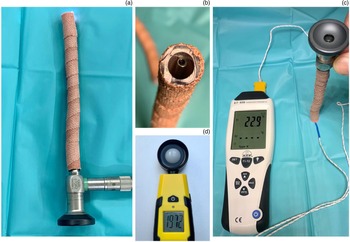
This study was undertaken at the University Hospital Monklands, with all measurements recorded by a single researcher. Transcanal endoscopic ear surgery was simulated in an operating theatre using 7 mm plastic suction tubing coated in insulating tape to simulate an external ear canal, with the length of the tubing either 5 mm longer than or equal to the tip of the endoscope. All measurements were recorded on the same surface (Figure 1). Operating theatre lighting and temperature were standardised throughout the study, and confirmed by baseline temperature and luminosity testing.
(a) The set up used to simulate transcanal endoscopic ear surgery using a 7 mm plastic suction tubing, coated in insulating tape, to simulate an external ear canal. (b) The end showing the endoscope tip 5 mm away from the end of tubing. (c) Temperature measurement technique. (d) The luminometer used in the study.

Experimental design
The ATP™ ET-959 high accuracy dual input K-type/J-type thermometer was used to record temperatures, while the Trotec® BF06 lux meter was used to measure luminosity. For each experimental condition, luminosity and temperature recordings were taken at 0 mm and 5 mm from the endoscope tip. The factors assessed included: the operative light source (light-emitting diode (LED) (Stryker™ L9000 LED and Storz® Power LED 175 light sources) versus halogen (Storz 201133 20 and Olympus CLK-4 halogen light sources) versus xenon (Stryker X8000 light source)), new versus old endoscopes, 0-degree versus angled endoscopes, 3 mm versus 4 mm endoscopes, varied light source intensities, and time since light source illumination (0, 30, 60 and 180 seconds).
Statistical analysis
Normality of the three dependent variables (luminosity, temperature at 0 mm and temperature at 5 mm from the endoscope tip) was confirmed with Kolmogorov–Smirnov and Shapiro–Wilk testing. Independent variables were tested for differences between groups using the student's t-test, and were tested for correlations using the Pearson's rank correlation coefficient. Statistical analyses were conducted using R statistical software through RStudio® software (version 1.1.463). Values of p < 0.05 were considered statistically significant.
Results
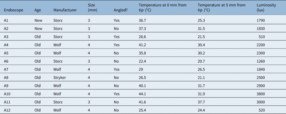
In total, 12 different endoscopes were assessed. These varied in terms of: manufacturer (Wolf (n = 6), Storz (n = 5) or Stryker (n = 1)), size (3 mm (n = 5) or 4 mm (n = 7)), age (new (n = 2) or used (n = 10)) and angulation (0-degree (n = 7) or angled (n = 5)) (Table 1). In addition, three different light bulb types were trialled, comparing LED (Stryker L9000 LED and Storz Power LED 175), halogen (Storz 201133 20 and Olympus CLK-4) and xenon (Stryker X8000) light sources.
Endoscopes utilised in study

The temperature and luminosity measurements shown were recorded at 100 per cent light source intensity with a light-emitting diode source.
Light source
Surprisingly, in our study, LED light sources generated higher temperatures in comparison to halogen or xenon bulbs. When testing the light cable without an endoscope, at maximum light intensity and following 180 seconds of exposure, LED light sources generated temperatures between 116°C and 174.3°C; in comparison, halogen light sources generated temperatures between 61.9 and 98°C, and xenon light sources generated a temperature of 62°C.
Reassuringly, the temperatures at the tip of the endoscope were significantly lower: when assessing temperatures and luminosity with a range of endoscopes of different sizes, angulation, age and model, at maximum light intensity and following 180 seconds of exposure. The LED light sources generated temperatures of 22.4–44.1°C and luminosity of 510–3800 lumens. Halogen light sources generated temperatures of 26.5–41.6°C and luminosity of 520–3000 lumens. Xenon light sources generated temperatures of 21.4–25.4°C and luminosity of 168–620 lumens. The Storz LED and halogen light sources produced higher luminosity and temperatures compared with their respective Stryker and Olympus counterparts.
Old versus new endoscopes
Two new endoscopes were sourced from Storz and directly compared with used models of the same endoscope. A new angled endoscope (Stryker 3 mm, 30-degree) reached a luminosity of 1790 lumens and a highest temperature of 36.7°C, while a used model of the same angled endoscope reached a luminosity of 510 lumens with the highest temperature 26.6°C. Similarly, a new 0-degree endoscope (Stryker 3 mm, 0-degree) reached a luminosity of 1830 lumens and a temperature of 37.3°C, while a used 0-degree endoscope of the same model reached a luminosity of 1260 lumens and a temperature of 22.4°C.
Zero-degree versus angled endoscopes
The luminosity and temperature production of 0-degree endoscopes were compared with those of angled endoscopes. The mean maximal tip temperature was 32.7°C for 0-degree endoscopes compared with 35.5°C for angled endoscopes, without a significant difference between groups (t = 0.62, df = 10, p = 0.549). Similarly, the mean luminosity in the 0-degree endoscopes (2044 lumens) in comparison to the mean luminosity in the angled endoscope group (2028 lumens) was also not significantly different (t = 0.027, df = 10, p = 0.979).
Size 3 mm versus 4 mm endoscopes
Temperature and luminosity were compared between 4 mm and 3 mm endoscopes. Higher mean temperatures and mean luminosity levels were noted in 4 mm endoscopes (34.6°C and 2294 lumens, respectively) in comparison to 3 mm endoscopes (32.9°C and 1678 lumens, respectively). However, neither of these differences was statistically significant.
Temperature at tip versus 5 mm from tip
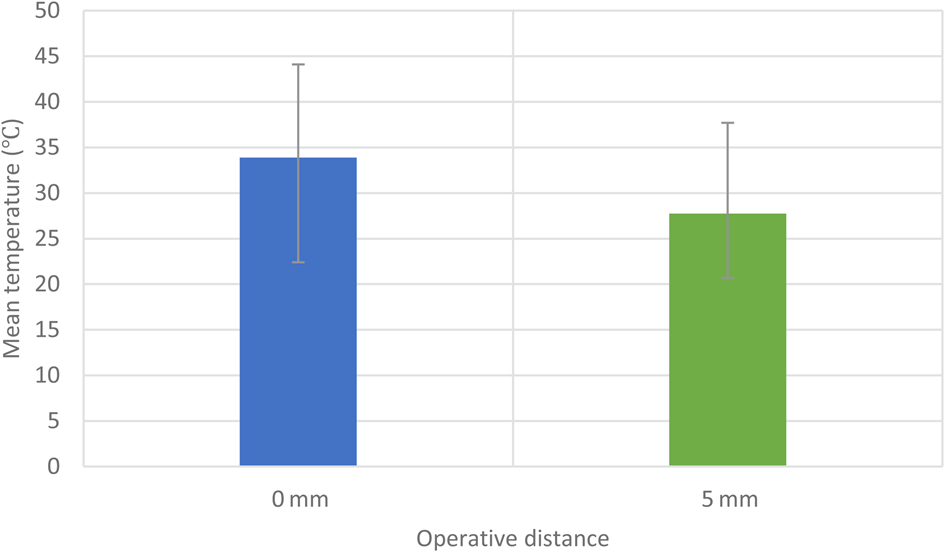
When comparing temperatures produced by the endoscopes, measurements were taken with the thermometer touching the tip and with the thermometer placed 5 mm from the tip. There was a statistically significant difference between temperature measurements when taken at the tip of the endoscope (mean = 33.9°C) and when measured 5 mm from the tip (mean = 27.7°C) (t = 5.63, df = 11, p < 0.005) (Figure 2).
Bar chart demonstrating mean temperatures for all endoscopes when recorded either at the endoscope tip (0 mm) or at 5 mm from the endoscope tip. Error bars represent the minimum and maximum values recorded.

Light intensity versus temperature
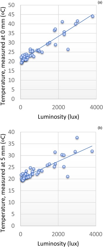
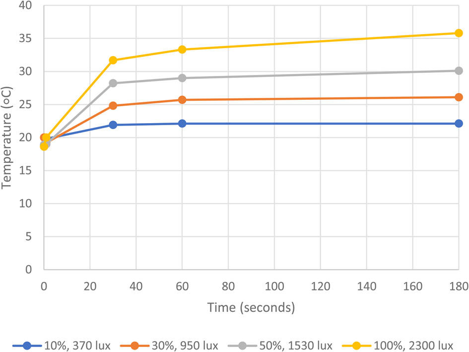
There was a strong positive correlation between luminosity and temperature, whereby the greater the luminosity, the higher the temperature noted at the tip and 5 mm from the endoscope tip. These correlations were statistically significant for measurements taken at the tip of the endoscope (R = 0.77, p = 0.004) and for those taken at 5 mm from the endoscope tip (R = 0.67, p = 0.016) (Figure 3). Temperature rises also demonstrated a logarithmic pattern of increase over time (Figure 4), with a gradual reduction in the rate of temperature increase between 0 and 180 seconds. This was most evident at higher light intensities.
Line graph demonstrating a positive linear correlation between temperature and luminosity.

Example line graph demonstrating the logarithmic relationship between temperature increase (recorded at the endoscope tip (0 mm)) and time, for an old, 0-degree, Wolf endoscope.

Discussion
This study has taken a unique approach by measuring luminosity using a lux meter rather than solely relying on light source settings. Additionally, this study has considered an extensive list of variables, which have previously not been considered together in a succinct experimental study.
We have demonstrated that thermal energy transfer from operating endoscopes is greatest when the light intensity is high and when the endoscope is touching the surface. Furthermore, new endoscopes appear to generate more heat in comparison to older endoscopes, and, in contrast to some other studies, LED light sources generate the greatest temperature and luminosity.
The thermal energy generated by operating endoscopes has been linked to the ignition of drapes and damage to skin,Reference Sandhu, Turner and Pozo13,Reference Bellina and Haas14 and altering of the workings of the inner ear.Reference Kozin, Lehmann, Carter, Hight, Cohen and Nakajima4,Reference Kahana, Rosenblith and Galambos6–Reference Bottrill, Perrault and Poe8,Reference Das, Mitra, Agarwal and Sengupta11 However, the maximal temperature achieved has varied greatly amongst the different papers studied. For example, Hensman et al. noted temperatures as high as 225°C within 10 minutes of switching on the light source.Reference Hensman, Hanna, Drew, Moseley and Cuschieri15 Conversely, Kozin et al. demonstrated temperatures of 46°C at 0.5–1 mm from the tip of the endoscope.Reference Kozin, Lehmann, Carter, Hight, Cohen and Nakajima4 Our study therefore aimed to determine what factors led to these variances in temperature.
The effects of high temperatures on body tissues has been assessed in several studies. Guinea pig models, exposed to high temperatures produced by a xenon light source for 5 minutes, showed lower distortion product otoacoustic emissions and an increased auditory brainstem response threshold.Reference Aksoy, Dogan, Ozturan, Eren, Veyseller and Gedik7 Similarly, Kahana et al. noted changes to multiple aspects of cochlear microphonics in the hamster cochlea when temperatures rose only to 39°C.Reference Kahana, Rosenblith and Galambos6 Vestibular dysfunction was reported by Bottrill et al. when a temperature rise in the ear caused caloric stimulation.Reference Bottrill, Perrault and Poe8 Various studies have specifically investigated the propensity for tissue damage and damage to the inner workings of the ear associated with the thermal energy generated by endoscopes used in ENT.Reference Kozin, Lehmann, Carter, Hight, Cohen and Nakajima4,Reference Aksoy, Dogan, Ozturan, Eren, Veyseller and Gedik7,Reference Bottrill, Perrault and Poe8,Reference Tomazic, Hammer, Gerstenberger, Koele and Stammberger10,Reference MacKeith, Frampton and Pothier16–Reference Lewis, Levin and Sommer18 However, thus far, only one paper has reported thermal-related injury in a patient undergoing otology surgery.Reference Das, Mitra, Agarwal and Sengupta11 Das et al. demonstrated audiometric change in higher frequencies, which was greater with xenon light sources. Vomiting, vertigo and tinnitus were also noted in patients exposed to either xenon or LED light sources.Reference Das, Mitra, Agarwal and Sengupta11
While the maximal temperature recorded in the study, from an exposed light cable, was 174.3°C, the maximal temperature at the tip of the endoscope, in any experimental condition, was 44.1°C. Furthermore, temperatures recorded at lower light intensities were significantly decreased. This trend has been demonstrated in previous studies. Tomazic et al. reported temperatures as high as 91.4°C at 100 per cent light intensity with a xenon light source, which decreased to 44.3°C at 33 per cent light intensity.Reference Tomazic, Hammer, Gerstenberger, Koele and Stammberger10 Ito et al. measured the safety of heat generated by endoscopes with xenon and LED light sources in a 3D model of a human temporal bone.Reference Ito, Kubota, Takagi, Watanabe, Futai and Furukawa5 It was noted that a xenon light source generated more thermal energy at 100 per cent light intensity than at 30 per cent intensity.Reference Ito, Kubota, Takagi, Watanabe, Futai and Furukawa5 Importantly, the use of a lower light intensity intra-operatively is feasible: McCallum et al. demonstrated no detriment to image quality as a result of the very low light levels used, as low as 10 per cent, which was in turn associated with lower heat generation.Reference McCallum, McColl and Iyer12
This study also demonstrated a significant increase in temperature recordings when the endoscope tip was in contact with the thermometer. This highlights the importance of avoiding resting the endoscope tip on operative surfaces, particularly in areas that may be sensitive to thermal damage. Other factors studied included: endoscope diameter, angled endoscopes versus 0-degree endoscopes, and whether the endoscope was new or used. Larger-diameter endoscopes, angled endoscopes and new endoscopes appeared to generate greater heat; however, none of these factors achieved significance. While some studies have demonstrated higher thermal energy production from endoscopes with larger diameters,Reference Aksoy, Dogan, Ozturan, Eren, Veyseller and Gedik7,Reference MacKeith, Frampton and Pothier16 because of the minimal temperature difference in larger-diameter endoscopes measured in this study, the authors would not advocate this as a reason for selecting smaller endoscopes.
Our study investigated the maximum temperatures reached at 0, 30, 60 and 180 seconds after switching on different light sources at 100 per cent intensity. Previous studies have investigated temperatures reached up to 30 minutes after the light source was switched on, although a number of studies showed that maximum temperatures were reached within 60 seconds of switching on the light source.Reference Kozin, Lehmann, Carter, Hight, Cohen and Nakajima4,Reference Aksoy, Dogan, Ozturan, Eren, Veyseller and Gedik7,Reference Tomazic, Hammer, Gerstenberger, Koele and Stammberger10,Reference MacKeith, Frampton and Pothier16 This appeared to be the case in our study, with temperature change over time demonstrating a logarithmic pattern of increase, with the greatest temperature rise within the first 60 seconds (Figure 4). These findings suggest that operative pauses, while rapidly cooling the endoscope,Reference Kozin, Lehmann, Carter, Hight, Cohen and Nakajima4 are unlikely to be of benefit overall to the patient because of the rapid return to close to maximal temperatures. One additional method to avoid excessive heat exposure within the ear is frequent aspiration and irrigation, and this could be adopted intra-operatively.Reference Ozturan, Dogan, Eren and Aksoy19
• Temperature rise in ear surgery has been linked to: cochlear microphonic alteration, reduced otoacoustic emissions and auditory brainstem responses, caloric effect, and direct tissue damage
• This study found a strong positive correlation between luminosity and temperature, whereby greater luminosity was associated with higher temperature
• Light-emitting diode sources generated higher temperatures and luminosity, compared to halogen or xenon bulbs
• Higher temperatures were recorded from endoscopes with larger diameters, angled endoscopes and new endoscopes, but differences were not significant
• It is recommended that operative light intensity is maintained at the lowest possible level, and that surgeons avoid contact between patient tissues and the endoscope tip
While there were attempts to optimise the experimental design, this study does have some limitations. It was only possible to assess 12 endoscopes, as these were the only otological endoscopes available for study in the department. This led to a small sample size, which could have affected statistical outcomes. The authors argue, however, that if the influence of a particular factor on intra-operative temperatures would only be significant with a large sample size, its relevance clinically would be minimal. In addition, the plastic tubing and matt black surface will not have accurately simulated the middle-ear environment. However, it was not feasible to use animal or cadaveric tissue with the luminosity meter or in the operating theatre environment; furthermore, it would not have been possible to test as many endoscopes without this simulation.
Conclusion
This study has demonstrated that light intensity and contact with the temperature sensors significantly elevated operative temperatures. In addition, new endoscopes and LED light sources appeared to generate greater heat. The authors of this study would therefore recommend that operative light intensity is maintained as low as possible, and that the surgeon avoids contact between patient tissues and the endoscope tip. For maximal assurance, the surgeon could consider testing the maximal temperature of any new endoscope to ensure it is a safe operative tool.
Acknowledgement
Funding for equipment was provided by ENT Scotland.
Competing interests
None declared