Introduction
The Heterostraci is a subclass of jawless vertebrates in which the cephalothorax was completely sheathed in bony armor. They were diverse, with more than 300 species described to date, and inhabited shallow marine and brackish environments around the Old Red Sandstone Continent (now North America, the Canadian Arctic, and Western Europe) from the middle Silurian (Wenlock) to the Late Devonian (late Frasnian) (Janvier, Reference Janvier1996). Due to the common occurrence of heterostracan plates and scales in sediments that often yield little other organic remains, they have been used extensively in biostratigraphic schemes for the Early Devonian (Janvier and Blieck, Reference Janvier, Blieck and Long1993; Elliott and Johnson, Reference Elliott, Johnson, Klapper, Murphy and Talent1997; Blieck et al., Reference Blieck, Cloutier, Elliott, Goujet, Loboziak, Reed, Rodina, Steemans, Valiukevicius, V'yushkova, Yolkin, Young, Blieck and Turner2000).
One of the main taxa, the Pteraspidiformes (Blieck, Reference Blieck1984), has a complex arrangement of the armor in which a number of plates grew separately and, in some taxa, fused at maturity to form larger elements. The dorsal shield was composed of a median dorsal plate and median rostral, pineal, and spinal plates, surrounded by paired orbital, branchial, and cornual plates. The earliest members of this taxon appear in the Pridoli of the Canadian Arctic (Elliott and Dineley, Reference Elliott and Dineley1983), after which they spread around the coast of the Old Red Sandstone Continent. The last members of this taxon occur in channels of Middle Devonian age in Idaho and Alberta (Denison, Reference Denison1968; Elliott et al., Reference Elliott, Dineley and Johnson2000a), and in the Water Canyon Formation of northern Utah (Ørvig, Reference Ørvig1961). Although the fauna from the Yahatinda Formation has been described (Elliott et al., Reference Elliott, Dineley and Johnson2000a), and the heterostracans from Idaho also (Denison, Reference Denison1968), new material from both localities prompts a review and redescription of the pteraspidiforms. In addition, the use of the same genus name for the material from the Water Canyon Formation (Ørvig, Reference Ørvig1961) and from Idaho (Denison, Reference Denison1968) is considered to be incorrect because the Water Canyon material consists only of incomplete ventral shields that are not usually considered distinctive enough for taxonomic definition.
The Middle Devonian taxa show most similarity to the protaspids, which are common pteraspids of the Early Devonian of the western United States (Denison, Reference Denison1970). Characteristically, they are moderate sized to large pteraspids with broad dorsal shields, absent or poorly developed cornual plates, and branchial plates that extend to the posterior margin of the dorsal median plate (Denison, Reference Denison1970). None shows an ornament of separated tubercles, but in several species the ridges are composed of rows of oval tubercles.
Geologic setting
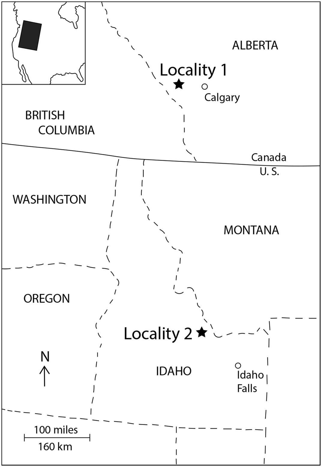
The vertebrate localities in Alberta and Idaho occur in channel-fill deposits (Aitken, Reference Aitken1966; Denison, Reference Denison1968; Fig. 1), while the locality in the Water Canyon Formation of northern Utah (Williams and Taylor, Reference Williams and Taylor1964) occurs in sediments that represent an environment of deposition in tidally influenced deltaic and estuarine conditions in a restricted embayment (Reed, Reference Reed1997).
Location map for the Middle Devonian pteraspid localities. Locality 1 indicates the Yahatinda Formation, north of “End Mountain” and Bowfort Creek, east of Banff, Alberta. Locality 2 indicates the Spring Mountain Beds at the head of Spring Mountain Canyon, Lemhi Range, Idaho.

Channel deposits of Middle Devonian age were described from the Banff area of southern Alberta by Aitken (Reference Aitken1966) as part of a study of the succession of strata named the Ghost River Formation by Walcott (Reference Walcott1921) (Fig. 1.1). The channel-fills together with the more continuous underlying and westerly sediments related to them were named the Yahatinda Formation and divided by Aitken (Reference Aitken1966) into two facies that are laterally and vertically distinct from each other. The “channel” facies occupies the deeper parts of the more pronounced sub-Devonian channels and consists mostly of purple-red to pink dolomite and dolosiltite. Conglomerate and breccias are also common. Within the dolomitic siltstones are a number of localities where plant and fish fragments have been found. Aitken (Reference Aitken1966) concluded that deposition must have been alluvial or marine to brackish deltaic based on the absence of marine fauna in the “channel” facies, the presence of mud-cracks, and the fact that deposition initially took place only in the lower parts of erosional channels and valleys. Vertebrates were collected from this formation and a preliminary identification by D.L. Dineley (Aitken, Reference Aitken1966) identified these as tuberculate pteraspids together with an antiarch placoderm, the arthrodire Holonema, large indeterminate arthrodires, osteolepiforms, and porolepiforms—a fauna that was subsequently described by Elliott et al. (Reference Elliott, Dineley and Johnson2000a). This fauna is similar in composition to that found in channel deposits in the Lemhi Range of Idaho by Denison (Reference Denison1968), and dated by him as Middle Devonian, and to that reported from the upper part of the Grassy Flat Member of the Water Canyon Formation in northern Utah (Ørvig, Reference Ørvig1961; Elliott and Johnson, Reference Elliott, Johnson, Klapper, Murphy and Talent1997). Work on plant megafossils and spores from the Yahatinda Formation (Greggs et al., Reference Greggs, McGregor and Rouse1962; McGregor, Reference McGregor1963, Reference McGregor1964) and later reassessed (C. McGregor, personal communication, 1992) indicated a late middle Givetian age.
Further collections of vertebrates were made at several localities in the Yahatinda Formation both by the Geological Survey of Canada and by D.L. Dineley during the 1960s, and by a party from the University of Alberta, Edmonton in 2006. This most recent material was collected from the NE Spur of End Mountain and one specimen is the subject of description here.
A channel in the Lemhi Range, central Idaho (the Spring Mountain Channel; Fig. 1. Locality 2) is 700 m wide and cut into the Ordovician Fish Haven Dolomite and Silurian Laketown Dolomite (Dehler, Reference Dehler1995). Although originally referred to the basal unit of the Jefferson Formation by Denison (Reference Denison1968) and to the Beartooth Butte Formation by Johnson and Sandberg (Reference Johnson, Sandberg, Murphy, Berry and Sandberg1977) because of the presence of pteraspids, at that time considered to be purely Early Devonian, Dehler (Reference Dehler1995) distinguished them as the Spring Mountain Beds. Denison (Reference Denison1968) described a vertebrate fauna from this channel that included Psephaspis idahoensis, Holonema haiti, and Asterolepis sp., which shows strong similarities to that from the Yahatinda Formation at the generic level. However, the presence of the brachiopod Stringocephalus in the base of the overlying Jefferson Formation (Dehler, Reference Dehler1995) constrains the age of the Spring Mountain Beds to the Eifelian. Additional collections were made in 1994 by Elliott and Dehler, and further material was borrowed from the collections of Brigham Young University-Idaho. This material shows the presence of a new pteraspid, which is described here.
The upper or Grassy Flat Member of the Water Canyon Formation in northern Utah has also been correlated with the Lower Devonian Beartooth Butte Formation in the past (Poole et al., Reference Poole, Sandberg, Boucot, Stewart, Stevens and Fritsche1977), and this is confirmed by the vertebrate fauna in the lower part of the member. However, revision of the faunal lists (Elliott et al., Reference Elliott, Johnson, Cloutier, Carr, Daeschler, Blieck and Turner2000b) shows that the upper part of the Grassy Flat Member contains a fauna similar to that of the Yahatinda Formation and the Spring Mountain Beds. Ørvig (Reference Ørvig1961) described Psephaspis williamsi from the upper part of the Grassy Flat Member and noted the presence of euarthrodires, antiarchs, and sarcopterygians in his collections. The presence of antiarchs is supported by the collection of an Asterolepis anterior dorsolateral plate in the uppermost Grassy Flat Member (C.A. Sandberg in Elliott et al., Reference Elliott, Johnson, Cloutier, Carr, Daeschler, Blieck and Turner2000b). This part of the formation is clearly Middle Devonian in age, and the age is constrained by the overlying Hyrum and Jefferson formations, which are dated as middle to late Givetian based on their invertebrate fauna (Sandberg et al., Reference Sandberg, Poole, Johnson, McMillan, Embry and Glass1988; Johnson et al., Reference Johnson, Sandberg, Poole, McMillan, Embry and Glass1989). Two large plates were collected from this level near Logan, Utah by Ørvig (Reference Ørvig1961), named by him Psephaspis idahoensis and described by him as belonging to the Drepanaspididae, a group of mostly Late Devonian heterostracans, hitherto unknown from North America. This genus name was used by Denison (Reference Denison1968) in his description of pteraspids from the Spring Mountain Beds in Idaho, however, new material indicates the presence of a distinctive new tuberculate pteraspid at this locality and thus casts doubt on the use of that genus name beyond the Water Canyon Formation.
Materials and methods
The material from the Yahatinda Formation is preserved in indurated dolosiltstone that proved resistant to preparation and so no preparation was carried out on it beyond taking latex peels of two specimens. The material from the Spring Mountain beds in Idaho is preserved in dolosiltite and in some cases was prepared mechanically with a vibrotool and by using the transfer method of Toombs and Rixon (Reference Toombs and Rixon1959), in which specimens are embedded in clear plastic and the matrix removed with a dilute solution of acetic acid.
Repositories and institutional abbreviations
The specimens are deposited in the collections of the University of Warsaw (DUW); Field Museum of Natural History, Chicago (FMNH-PF); the Idaho Museum of Natural History (IMNH); and the Vertebrate Paleontology Collections of the University of Alberta (UALVP).
Systematic paleontology
Class Pteraspidomorphi Goodrich, Reference Goodrich and Lankester1909
Subclass Heterostraci Lankester, Reference Lankester1868
Order Pteraspidiformes Berg, Reference Berg1937
Family Pteraspididae Claypole, Reference Claypole1885, sensu Denison, Reference Denison1970
Remarks
In the original description of this species from the Yahatinda Formation (Elliott et al., Reference Elliott, Dineley and Johnson2000a) the genus name Helaspis was created. However, it has since come to our attention that this name was already preoccupied by the modern pterygotid insect Helaspis mexicana McKenzie, Reference McKenzie1963, thus invalidating its use for this heterostracan. We are rectifying this here by creating a new genus name for this organism.
Genus Clavulaspis new genus
- Reference Elliott, Dineley and Johnson2000a
Helaspis Elliott et al., p. 124.
Type species
Helaspis finis Elliott, Dineley, and Johnson, Reference Elliott, Dineley and Johnson2000a
Diagnosis
As for species by monotypy.
Etymology
Latin clavus, diminutive clavulus, a nail, and Greek aspis, a shield, referring to the appearance of the tuberculate ornament.
Remarks
As noted above, this genus is renamed due to the fact that the original name is preoccupied and the description is expanded by the addition of new material.
Clavulaspis finis (Elliott, Dineley, and Johnson, Reference Elliott, Dineley and Johnson2000a) new combination
Figures 2, 3.1
- Reference Elliott, Dineley and Johnson2000a
Helaspis finis Elliott, Dineley, and Johnson, p. 124, figs. 2.1, 2.2a, b, 2.3–2.5, 3, 4.2.
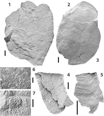
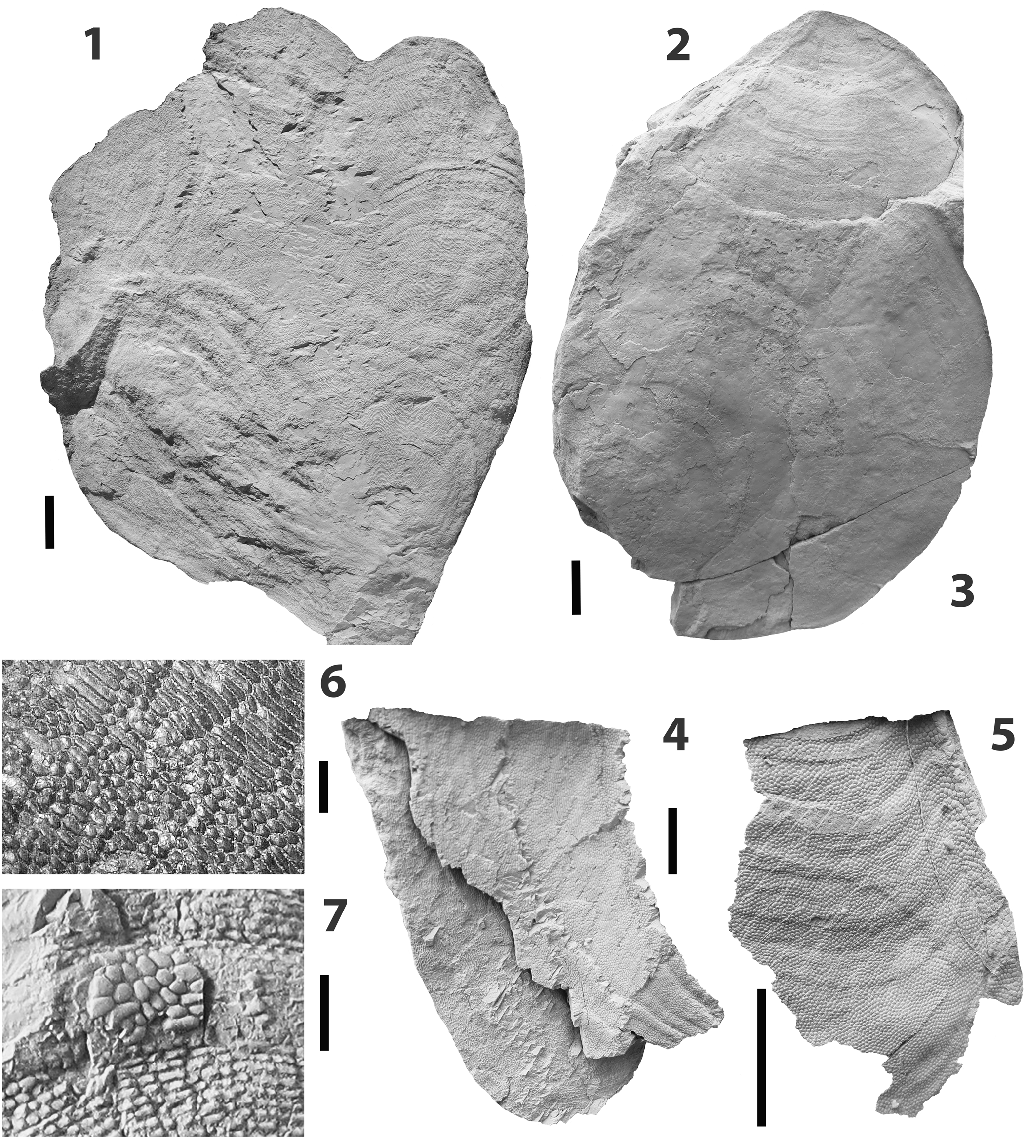
Clavulaspis finis (Elliott et al., Reference Elliott, Dineley and Johnson2000a) n. comb. from the Middle Devonian “Channel” facies of the Yahatinda Formation, north face of “End Mountain” and head of Bowfort Creek, east of Banff, Alberta. All specimens dusted with ammonium chloride. (1) UALVP 52814 latex cast of median dorsal plate; (2) GSC 118224 rostral plate; (3) GSC 118223 ventral plate; (4) GSC 118225 posterior part of branchial plate; (5) GSC 118226c latex cast of right lateral part of rostral plate; (6) UALVP 52814 detail of ornament; (7) UALVP 52814 secondary ‘blister’ covering primary ornament. (1–5) Scale bars = 10 mm; (6) scale bar = 1 mm; (7) scale bar = 2 mm.
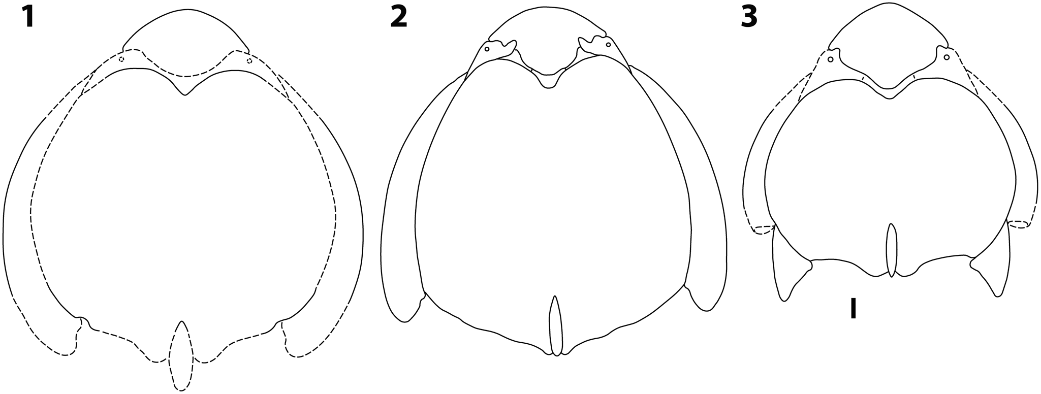
(1) Clavulaspis finis (Elliott et al., Reference Elliott, Dineley and Johnson2000a) n. comb., reconstruction based on UALVP 52814, GSC 118226, and GSC 118237. (2) Scutellaspis wilsoni n. gen. n. sp., reconstruction based on the holotype, FMNH-PF14551, IMNH 2536/5392, and FMNH-PF14556. (3) Ecphymaspis idahoensis (Denison, Reference Denison1968) n. comb., reconstruction based on the holotype, FMNH-PF5680, FMNH-PF5684, and FMNH-PF14559. Scale bar = 10 mm.

Holotype
GSC 118222a, 118222b a partial dorsal plate.
Emended diagnosis
Large pteraspid with broad dorsal shield. Rostrum broadly rounded; dorsal plate broadly convex rising to postero-median eminence. Branchial plates elongated and curved; branchial openings located at posterolateral corners of shield; cornual plates absent. Ornament of small, polygonal, smoothly convex or bluntly pointed tubercles with denticulate margins over most of the shield, but grading into patches of sharp-crested, discontinuous ridges with crenulate margins within the dorsal plate and on the lateral margins of the dorsal and lateral plates.
Occurrence
“Channel” facies of the Yahatinda Formation, north face of “End Mountain” and head of Bowfort Creek, east of Banff, Alberta (Fig.1.1).
Description
A new specimen of the dorsal plate (UALVP 52814, Fig. 2.1) provides additional information about the size, outline, and proportions of the plate. A reinterpretation of some of the original material also enables a more complete description of the species to be made.
The new dorsal plate is incomplete, but preserves the central part of the anterior margin and the left lateral part of the posterior margin (Fig. 2.1). The lateral margins are broken and incomplete, but this plate is clearly large, broad, and gently convex with a length of 122 mm and breadth of at least 106 mm. Lateral margins appear to have been slightly convex (based on growth lines on the plate), median anterior margin laterally convex with a deep median concavity to accommodate the pineal plate. Surface gently convex posteriorly, rising to rounded median posterior prominence. Base of dorsal spine projects as a convex median point posteriorly, dorsal spine not preserved. Ornament is variable across dorsal plate. Small, convex polygonal tubercles with crenulate margins and flat to convex or bluntly pointed tops are dispersed in patches across the plate and grade laterally into discontinuous, sharp-crested ridges (Fig. 2.6). The ridges are arranged parallel to the plate margins and are more common along plate margins (extending uninterrupted for 25 mm along upper plate margin) and along the midline. Several patches of what appear to be second generation tubercles form ‘blisters’ on the plate (Fig. 2.7). These consist of tubercles that are larger and rounder than those on the rest of the plate. These ‘blisters’ form as rounded patches or, in several cases, elongated bands situated on raised growth lines on the plate.
Distinct growth lines are present, paralleling the plate margins. Because this plate does not have the pineal, rostral, or lateral plates attached, it is assumed to be from a juvenile, suggesting that adults were somewhat larger.
The previously described partial branchial plate (GSC 118225; Fig. 2.4), also shows the same gradation of ornament, but much of the rest of the material used in the original description unfortunately shows little or no surface ornament. GSC 118226 (Fig. 2.5) was identified in the original description as being part of a branchial plate. However, further study suggests that it is in fact the right posterolateral part of a rostral plate (Fig. 2.5).
The specimens described as Helaspis sp. in the original description (Elliott et al., Reference Elliott, Dineley and Johnson2000a) have been restudied and are now attributed to Clavulaspis finis n. comb. These consist of a small juvenile dorsal plate (GSC 118236a, b) and a juvenile branchial plate (GSC 118237). Study of the ornament on these plates shows that it is also composed of small denticulate tubercles grading into sharp-crested ridges, with the ridges predominating on the lateral margins of the dorsal disc and on the branchial plate, and is therefore similar to that seen on the larger plates.
The additional material enables a reconstruction of Clavulaspis finis n. comb. to be made and shows it to have been a medium to large pteraspid of Protaspis type (Fig. 3.1).
Materials
GSC 118223, GSC 118227–118230, partial dorsal median plates; GSC 118236a, b, juvenile dorsal median plate; GSC 118224, GSC 118226, rostral plates; GSC118225, GSC 118226a–c, GSC 118231–118234, branchial plates; GSC 118237, juvenile branchial plate; GSC 118235, ?ventral plate.
Remarks
All the material attributed to Clavulaspis finis n. comb. shows an ornament in which patches of small, polygonal, crenulate, gently rounded to flat-topped tubercles are interspersed with crenulate, sharp-crested ridges. This is an ornament that has not been reported for any other pteraspid and serves to distinguish this species. Several patches of what appear to be second generation tubercles form ‘blisters’ on the dorsal plate. These consist of tubercles that are larger and rounder than those on the rest of the plate (Fig. 2.7), and it is noticeable that these ‘blisters’ are preferentially developed on raised growth lines. Ørvig (Reference Ørvig1961, Reference Ørvig1976) noted the presence of this phenomenon in his material from the Water Canyon Formation and Denison (Reference Denison1968) also noted it on specimens from Idaho. They have only otherwise been reported from the pteraspids Protaspis mcgrewi Denison, Reference Denison1970 and P. brevispina Denison, Reference Denison1970 (Denison, Reference Denison1973) from the Early Devonian of Wyoming, but are a common feature of the related drepanaspids (Ørvig, Reference Ørvig1976), prompting Ørvig (Reference Ørvig1961) to place the Water Canyon material within the Drepanaspididae. Tarlo (Reference Tarlo1964) suggested that the blisters were developed in response to an irritant, such as surface abrasion of the plates, however, Ørvig (Reference Ørvig1976) noted that development seemed to occur despite no indication of abrasion, and certainly the specimens described here show no abrasion of the plate surface. Denison (Reference Denison1973) showed that wear on dorsal plates of Protaspis mcgrewi and P. brevispina had occurred during the life of the animals because ‘blisters’ of secondary denticles were found in the worn areas and, more commonly, the spaces in the spongy reticular layer exposed at the surface had been infilled by dentine. No conclusion was reached as to how these animals developed wear on the dorsal surface (Denison, Reference Denison1973), and it is difficult to understand what behavior might have caused it to happen. The new material does not provide any additional information towards solving why the denticles formed.
Tuberculate pteraspids are otherwise only known from the Early and Middle Devonian of the western United States. Tuberculaspis elyensis Ilyes and Elliott, Reference Ilyes and Elliott1994 and Pirumaspis lancasteri Ilyes and Elliott, Reference Ilyes and Elliott1994 occur in the Early Devonian of Nevada (Ilyes and Elliott, Reference Ilyes and Elliott1994); however, their overall morphology is different from that of Clavulaspis finis as are the details of their ornament. In both, the tubercles are domed and the high point is usually to the posterior of the tubercle, and sharp-crested ridges are not present. The very similar ornament means that the ornament alone cannot be used as a diagnostic feature to separate these two Early Devonian species despite their very different gross morphology (Ilyes and Elliott, Reference Ilyes and Elliott1994).
Psephaspis has been reported from the Middle Devonian Water Canyon Formation in Utah and the Spring Mountain Beds at the Spring Mountain channel in Idaho (Elliott et al., Reference Elliott, Johnson, Cloutier, Carr, Daeschler, Blieck and Turner2000b). In addition, a new species from the Spring Mountain channel is described here. In neither species is the ornament similar to that of Clavulaspis finis n. comb.
Genus Scutellaspis new genus
Type species
Scutellaspis wilsoni new species.
Diagnosis
As for the type species by monotypy.
Etymology
Latin scutella, a little plate, and Greek aspis, a shield in reference to the small additional plates between the pineal and orbital plates.
Remarks
Scutellaspis n. gen. is based on new material from the Spring Mountain Beds, which is distinct from the previously described Psephaspis idahoensis from the same locality.
Scutellaspis wilsoni new species
Figures 3.2, 4
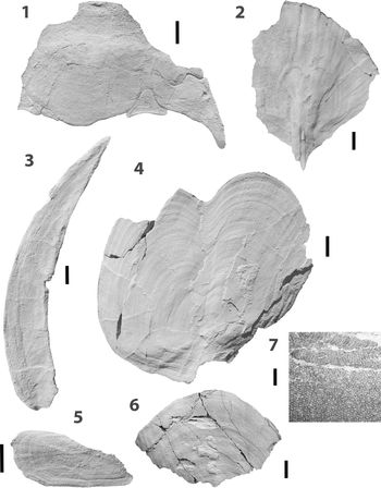
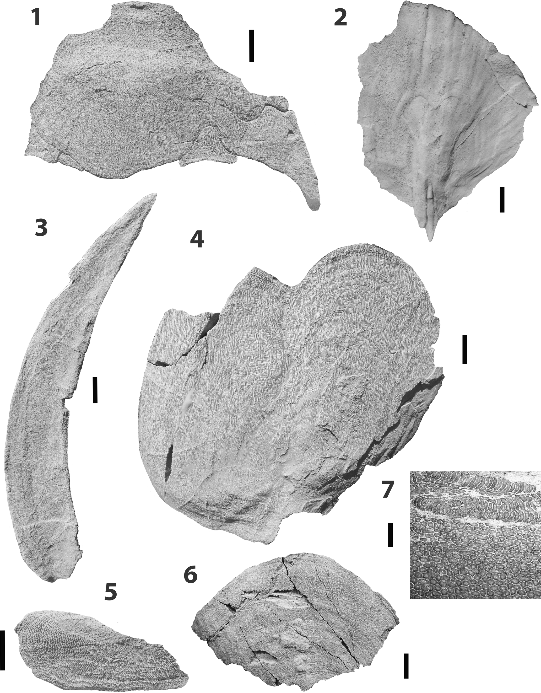
Scutellaspis wilsoni n. gen. n. sp., from the Middle Devonian Spring Mountain Beds of Idaho. All specimens dusted with ammonium chloride. (1) FMNH-PF14551 holotype rostral, orbital, and anterior accessory plates; (2) FMNH-PF14552 posterior part of dorsal plate, latex peel; (3) FMNH-PF14556 branchial plate; (4) INMH 2536/5392 median dorsal plate; (5) FMNH-PF14557 small plate or scale; (6) FMNH-PF14554 anterior margin of ventral plate; (7) FMNH-PF14554 detail of ornament and secondary ‘blister’ overgrowing primary tubercles. (1–6) Scale bars = 10 mm, (7) scale bar = 1 mm.
Holotype
FMNH-PF14551 partial rostral plate with attached right orbital plate and right and partial left anterior platelets.
Diagnosis
Large pteraspid with very broad dorsal shield. Rostrum broadly rounded; dorsal disc convex rising to postero-median eminence. Branchial plates elongated and curved; branchial openings located at posterolateral corners of shield. Additional triangular paired platelets present mediad to the orbital plates and projecting into the rostral plate; anterior margin of the orbital plate showing two anterior projections: one broad projection is placed laterally on the anterior margin, the second is more medially placed and elongated. Ornament of very small, round, smoothly convex, crenulated tubercles over most of the shield, but marginally becoming elongated and oriented parallel to the plate margins in the orbital and ventral plates and anterior platelets.
Occurrence
A channel at the head of Spring Mountain Canyon, just northeast of Big Windy Peak in the Lemhi Range. Located in the Gilmore 7.5’ topographic quadrangle at T11N, R22E, Section 29.
Description
A large species with a broad dorsal shield (estimated to be 272 mm long and 290 mm wide). Dorsal surface almost flat anteriorly, convex posteriorly, and rising to a steep peak where the dorsal spine is inserted. Rostrum broad and rounded anteriorly; laterally the shield margin is convex, posteriorly almost transverse and deeply notched for insertion of the spinal plate.
The rostral plate (FMNH-PF14551; Fig. 4.1) appears to have been broadly rounded anteriorly with a slightly convex surface. Posteriorly the border is notched for the anterior projection of the orbital plates and the anterior platelets, and is bounded centrally by the pineal plate. It does not appear to contact the anterior border of the dorsal median plate.
The only orbital plate is almost complete and has an anterior margin in which a broad, rounded, anterior projection is followed medially by an additional narrow anterior projection, 12.7 mm long, narrowing from 10.4 mm at the base to 3.2 mm distally (Fig. 4.1). Posteriorly the plate is bounded by the anterior margin of the dorsal plate and laterally by an additional anterior platelet. The posterior projection is incomplete, but appears to narrow rapidly to a point. The orbit is small (diameter 3.0 mm) and placed dorso-laterally.
Mediad to the anterior projection of the orbital plate is an additional small plate that is not part of the normal complement of plates in the dorsal shield of pteraspids. The entire right plate and most of the left are present in a symmetrical arrangement (Fig. 4.1), indicating that this is not just a growth anomaly in this individual. We refer to it here as the anterior platelet. The right platelet is triangular with concave margins; the anterior projection is 10.0 mm long and narrows from 12.2 mm wide at the base to 5.2 mm near the termination, which is bluntly rounded. Narrow, tapering, medial and lateral projections are present; the medial projection is 17.3 mm long measured from the apparent growth center on the posterior margin of the plate, the lateral projection is broken but is estimated to have been 20.0 mm long. The posterior margin of the plate contacts the anterior margin of the dorsal plate, and the medial projection presumably contacts the pineal plate. The left anterior platelet is incomplete but shows a medial projection that narrows to a natural truncated point, suggesting that it contacted a narrow lateral projection of the pineal plate (Fig. 4.1).
There are no specimens of the pineal plate. However, its boundaries with the dorsal, rostral, and orbital plates, and the anterior platelets show its shape (Fig. 3.2). Posteriorly it is very convex at its boundary with the dorsal plate and anteriorly very concave at its boundary with the rostral plate. The lateral projections are extremely narrow and appear to have contacted the medial extensions of the anterior platelets.
One almost complete dorsal median plate is present (IMNH 2536/5392; Fig. 4.4). This is clearly from a juvenile individual because there are no anterior or lateral plates fused to it. It is broad with convex lateral margins; anteriorly there is a deep, broad, rounded notch for reception of the pineal plate. The posterior margin is almost transverse, with a deep, narrow notch for the dorsal spine. The plate is crushed slightly but the surface is convex posteriorly, becoming almost flat anteriorly. It is 104 mm long and 113 mm wide (although the width may be enlarged due to crushing). No pores of the sensory canal system can be seen. Growth lines parallel the margins of the plate. One uncrushed example of the posterior part of an adult plate shows that the plate is steeply peaked at the posterior and that the spine was long, narrow, and recumbent (FMNH-PF14552; Fig. 4.2). The spine base in this specimen is 30.3 mm long and 2.5 mm wide.
The single available branchial plate is preserved in dorsal aspect and is complete except for the posterior margin (FMNH PF-14556; Fig. 4.3). It is an elongated curved plate 204 mm long, broadening steadily to a width of 30 mm at the posterior margin. The inner, concave margin appears to have a broken edge suggesting detachment from an adult dorsal shield. The plate surface is convex at the outer edge, concave medially, and then slightly convex at the inner edge. The posterior margin is broken and incomplete so that the branchial opening cannot be discerned.
Two partial ventral plates are known. One shows the anterior margin, which is broadly curved and reaches a width of 90.1 mm as preserved (FMNH-PF14554; Fig. 4.6). The other is 99.3 mm long, but is incomplete laterally and posteriorly. Both specimens show only a slight surface convexity.
A small plate (Fig. 4.5) may be a lateral scale. It is triangular, 38 mm long and 16 mm wide, rounded at one end and coming to a narrow point at the other. It is almost complete with part of one long margin missing and a longitudinal crest is present dividing it into a narrower and a broader side. Growth lines follow the margins of the plate and the ornament consists of small, closely packed tubercles. This is not a median scale, but may be a lateral scale, although the arrangement of the ornament is unlike that usually found on them.
No sensory pores have been recognized anywhere on the dorsal or ventral shield, so nothing is known of the sensory canal system.
The ornament is uniform across the dorsal plate and consists of very small, round, round-topped tubercles 0.2 mm in diameter and with crenulations at the base (Fig. 4.7). They are regularly arranged, but do not appear to indicate rows and they do not change towards the margins of the plate. The same is true of the rostral plate, but on the anterior platelet and the orbital plate the tubercles become elongated and oriented in rows and in the anterior platelets delineate a center of growth close to the posterior margin and centrally placed. On the ventral plate elongation of the tubercles is present in a band 5 mm deep along the plate margin. They reach 0.6 mm in length and are oriented in rows parallel to the margin. On the margin of one ventral plate there is a ‘blister’ of secondary tubercles (Fig. 4.7). A patch of secondary tubercles is also present on FMNH-PF14553, the posterior part of a dorsal plate.
Etymology
Named after Dr. Mark Wilson of the University of Alberta, Edmonton, in recognition of his important contributions to the study of early vertebrates.
Materials
IMNH 2536/5392 median dorsal plate; FMNH-PF14552 posterior part of median dorsal plate; FNMH-PF14554, FMNH-PF14555 partial ventral plates; FMNH-PF14556 branchial plate; FMNH-PF14557 lateral scale?.
Remarks
This species (Fig. 3.2) bears most resemblance to the protaspids from the western United States (Denison, Reference Denison1970), which are rather flat, broad pteraspids with long, curved branchial plates and reduced or missing cornual plates. Cosmaspis transversa Denison, Reference Denison1970 is a typical example in which there are no cornual plates and the branchial opening is a slit at the posterior end of the branchial plate, a situation that is most likely to be the case also with Scutellaspis wilson n. gen. n. sp. The presence of the anterior platelets and the configuration of the anterior margin of the orbital plates are unique to Scutellaspis n. gen.
External platelets have been described previously in two species. Tuberculaspis elyensis (Ilyes and Elliott, Reference Ilyes and Elliott1994, figs. 2.4, 2.5, 3.1–3.3) has a series of small platelets (termed accessory plates) described as being, small, thin, and loosely attached, that occur along the margin of the dorsal plate from behind the orbit to the base of the dorsal spine, and cover the suture between the median dorsal and branchial plates. A similar set of small plates in the same position can be seen in an undescribed species from the Lochkovian of the Canadian Arctic (Blieck, Reference Blieck1984, fig. 58). However, the anterior platelets in Scutellaspis n. gen. are different from these in that they are separate plates that appear to have developed by subdivision from the orbital plates or possibly the rostral plate, and are sutured to the plates surrounding them and so are not loosely attached surface features. The center of growth, centrally placed close to the posterior margin, shows that the anterior platelets developed as separate plates. The reason for development of these platelets or configuration of the anterior margin of the orbital plates remains unknown at present.
Genus Ecphymaspis new genus
- Reference Tarlo1965
Schizosteus (part); Tarlo, p. 55, 56.
- Reference Denison1970
Psephaspis (part); Denison, p. 36.
- Reference Blieck1984
Psephaspis (part); Blieck, p. 71, 103, 104.
Type species
Psephaspis idahoensis Denison, Reference Denison1968.
Diagnosis
As for the type species by monotypy.
Etymology
Greek ekphyma, an eruption of pimples, and Greek aspis, a shield, in reference to the tuberculated plate ornament.
Remarks
Because this species is described partly to resolve some taxonomic problems concerning these pteraspids, these problems are reviewed here before its description.
Ørvig (Reference Ørvig1961) reported the collection of fragments of large heterostracan plates with tuberculate ornament from the upper or Grassy Flat Member of the Water Canyon Formation at Blacksmith Fork in the Bear River Range of northeastern Utah. The heterostracans occurred together with “Euarthrodires (or Antiarchi?) with coarsely tuberculated dermal bones, Acanthodians and Crossopterygians” (Ørvig, Reference Ørvig1961, p. 533). Ørvig identified the heterostracan as a drepanaspid, based on the tuberculate ornament and the presence of ‘blisters’ or areas of overgrowth, both of which were features known only in drepanaspids at that time. He named the species Psephaspis williamsi and designated the largest (although fragmentary) plate, identified by him as a median ventral, as the holotype (Ørvig, Reference Ørvig1961, fig. 6). Subsequently, Tarlo (Reference Tarlo1965, p. 63, 64) attributed a plate with tuberculate ornament from the Spring Mountain channel in the Lemhi Range to the psammosteid Schizosteus wellsi Tarlo, Reference Tarlo1964. Denison (Reference Denison1968, p. 281, 282) criticized this on the basis that the type specimen of Schizosteus wellsi had tubercles that were different in appearance and spacing from the Lemhi material. In 1968 Denison described a tuberculate pteraspid from the same channel in the Lemhi Range of eastern Idaho. The morphology of the dorsal shield indicated quite clearly that this was a pteraspid and not a drepanaspid, and so Denison (Reference Denison1968) removed Psephaspis from the Drepanaspididae and named the new species Psephaspis idahoensis. The dorsal shield of this species is short and broad, and the type specimen is clearly adult because both dorsal spine and pineal plate are attached, so it can be compared directly with Ørvig's material. However, it is considerably smaller than the ventral plate reported by Ørvig and differently proportioned, indicating that it could not be the same species and that the morphology of the two species might have been different. The ornament is similar, however, both in size and morphology, and shows areas of overgrowth, as in P. williamsi. At the time that Denison (Reference Denison1968) was describing Psephaspis idahoensis, it was only the second species of tuberculate pteraspid known, and it appeared logical to place it in the same genus as P. williamsi. However, the genus was based on what was later recognized as a fragmentary ventral plate (Denison, Reference Denison1968), which has none of the characters necessary for a satisfactory diagnosis, and this genus is therefore considered to be insufficiently diagnosed and the name a nomen dubium. In addition, Ilyes and Elliott (Reference Ilyes and Elliott1994, figs. 3.11, 5.1) showed that similar tuberculate ornament is present in two morphologically very different pteraspid genera from the Lower Devonian (Emsian) of the western United States, and is therefore not a reliable criterion absent morphological information. Other new, large, tuberculate species are also present in collections made by Denison from the Water Canyon Formation, but never described (DKE, personal observation). It is not clear therefore, that the genus Psephaspis is sufficiently well diagnosed, because it is based on an incomplete ventral shield, or that P. idahoensis is correctly attributed to Psephaspis, because similar tuberculate ornament is now known to occur on morphologically very different species. For these reasons we have not used the genus Psephaspis for the new species from the Yahatinda Formation and the Spring Mountain channel. However, this also raises the question of whether the material described by Denison (Reference Denison1968) from the Spring Mountain channel should have been attributed to Psephaspis. Denison (Reference Denison1968, fig. 2) did provide a new reconstruction of the fragmentary ventral plate used to characterize the genus (Ørvig, Reference Ørvig1961, fig. 6), which shows a plate 245 mm wide and 261 mm long. This is far larger than the ventral plate of P. idahoensis (Denison Reference Denison1968, fig. 3), which is 155 mm wide and accords well with the width of 154 mm for the holotype dorsal shield (Denison, Reference Denison1968, fig. 2). Denison (Reference Denison1968, fig. 7) also shows the ornament of both species at the same scale and they are certainly very similar, however, as we have noted previously, we do not consider that tubercular ornament alone is enough to distinguish taxa. It is our opinion therefore, that Psephaspis is inadequately diagnosed as a genus and that P. idahoensis cannot be attributed to that genus. We are therefore designating the Water Canyon material described by Ørvig (Reference Ørvig1961) as P. williamsi as a nomen dubium, but renaming (below) the material described by Denison (Reference Denison1968) as P. idahoensis, while adding some additional material to its description.
Ilyes and Elliott (Reference Ilyes and Elliott1994) were the first authors to use a computerized cladistic analysis of some Pteraspidiformes, however, they only analyzed 11 taxa limited to the Early and Middle Devonian of the western United States, and they restricted the characters to those that were not considered to be adaptive. This did place Psephaspis (as then understood) as the most derived form, with the Early Devonian tuberculated taxa Tuberculaspis and Pirumaspis forming a sister clade.
Ecphymaspis idahoensis (Denison, Reference Denison1968) new combination
Figures 3.3, 5, 6
- Reference Tarlo1965
Schizosteus wellsi (part); Tarlo, p. 63, 64, pl. 14, fig. 5.
- Reference Denison1968
Psephaspis idahoensis; Denison, p. 270, 281, figs. 1, 7B, 8.
- Reference Blieck1984
Psephaspis idahoensis; Blieck, fig. 56B.
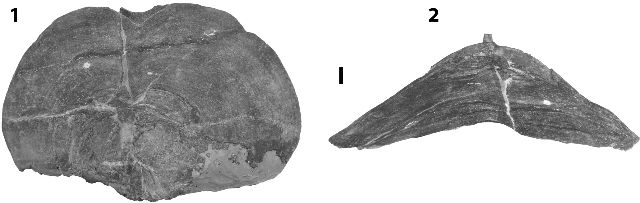
Ecphymaspis idahoensis (Denison, Reference Denison1968) n. comb. from the Middle Devonian Spring Mountain Beds of Idaho. FMNH-PF5680 holotype median dorsal plate (1) in dorsal view, and (2) in anterior view. Scale bar = 10 mm.

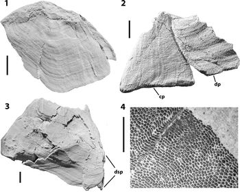
Ecphymaspis idahoensis (Denison, Reference Denison1968) n. comb. from the Middle Devonian Spring Mountain Beds of Idaho. All specimens dusted with ammonium chloride. (1) FMNH-PF5684 rostral plate; (2) FMNH-PF14559 left cornual plate with posterolateral part of dorsal median plate; (3) FMNH-PF5682 left posterior part of median dorsal plate showing the posterolateral margin; (4) FMNH-PF5684 detail of ornament on rostral plate. (1–3) Scale bars = 10 mm, (4) scale bar = 5 mm. cp = cornual plate; dp = median dorsal plate; dsp = dorsal spine.

Holotype
FMNH-PF5680, a dorsal shield deposited in the Field Museum of Natural History, Chicago. Recovered from a channel at the head of Spring Mountain Canyon on the crest of the Lemhi Range, east-central Idaho.
Emended diagnosis
Medium sized pteraspid with broad dorsal shield. Rostrum broadly rounded with postero-lateral embayments for the anterior margin of the orbital plates; dorsal disc convex, rising very steeply to postero-median eminence. Small triangular cornual plates. Branchial opening placed at the posterior margin of the branchial plate. Ornament of small, round, crenulated denticles, with a surface that is relatively flat and sloping in one direction over most of the shield but marginally becoming elongated and oriented parallel to plate margins.
Occurrence
A channel at the head of Spring Mountain Canyon, just northeast of Big Windy Peak in the Lemhi Range. Located in the Gilmore 7.5’ topographic quadrangle at T11N, R22E, Section 29.
Description
The type specimen is a median dorsal plate (FMNH-PF5680) that has the posterior dorsal spine and pineal plate attached (Denison, Reference Denison1968, fig. 1; Fig. 5). The anterior and lateral margins are present, but the posterior margin is incomplete. However, specimens FMNH-PF5681 and FMNH-PF5682 (Fig. 6.3) preserve this margin and show that it is concave laterally, but extended into a median projection that is deeply notched to house the dorsal spine. The dorsal plate is broad with a width of 155 mm and a length of 104 mm, it is also extremely steeply vaulted, almost conical, with the sides sloping at an internal angle of 120° posteriorly (Fig. 5.2). The anterior margin is deeply notched for the pineal plate, which is short and broad, and extended laterally into narrow projections (Denison, Reference Denison1968, fig. 1; Fig. 5.1). However, because the lateral projections are incomplete, it is unclear if they connected to the medial projections of the orbital plates. The lateral margins of the dorsal plate are convex, but the posterior part is missing on both sides of the holotype so it is unclear if there were indentations for the branchial openings at the posterolateral corners of the shield.
Several rostral plates have been identified (Fig. 6.1; Denison Reference Denison1968, fig. 4). They show considerable variation in proportions, some being broad and short, others being narrow and long. Some of this is due to tectonic deformation, as seen in Denison (Reference Denison1968, fig. 4A; Fig. 6.1), which has clearly been distorted. In all cases, there is a small indentation in the lateral part of the posterior margin indicating the position of the anterior projection of the orbital plate.
A single small triangular plate that is attached to a fragment of the posterolateral part of a median dorsal plate appears to be a cornual plate (Fig. 7.2). The ornament is not that of Scutellaspis wilsoni and it is similar to that on the other specimens of Ecphymaspis idahoensis n. comb. and we are, therefore, attributing it to the new genus. The plate is 25 mm long along the medial edge where it attaches to the dorsal plate, 23 mm along the posterior margin, and 29 mm along the lateral margin. The medial and posterior edges are concave, the lateral edge convex. The lateral and median edges are undamaged and show a rounded edge with ornament. The plate is complete except for a small part missing at the anterior corner.
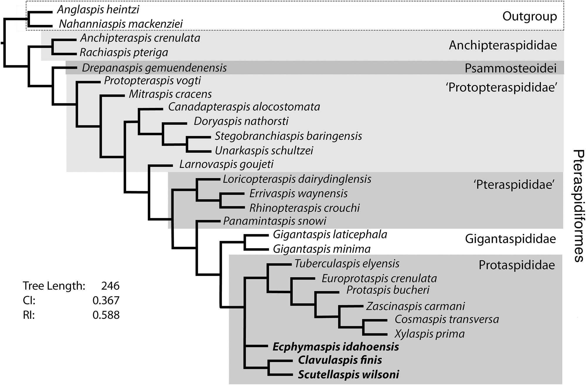
Pteraspidiform phylogenetic analysis results for 27 taxa (two outgroup taxa) and 54 characters. The three new Middle Devonian taxa added to the Pernègre and Elliott (Reference Pernègre and Elliott2008) data matrix are bolded. This is a strict consensus tree from five most parsimonious trees generated using PAUP* and TNT (Tree length 246; CI 0.367; RI 0.588).

Denison (Reference Denison1968, fig. 5) illustrated a possible fragment of a branchial plate that he identified as an incomplete posterior end. A similar fragment has been collected (FMNH-PF14558), but in neither case is the specimen complete enough to show the actual configuration of the branchial opening, although parts of the dorsal edge are preserved. Two additional specimens mentioned by Denison (Reference Denison1968, p. 275) appear to have come from the central part of a branchial plate, but are only slightly curved and so would not have fitted against the lateral margin of the type dorsal plate.
The ornament consists of small, round, crenulated tubercles with a surface that is relatively flat and sloping in one direction (Fig. 7.4). These show a tendency to elongate at the margins of plates (Fig. 7.4). As noted by Denison (Reference Denison1968, p. 277, fig. 8), patches of larger tubercles forming overgrowths or ‘blisters’ can be seen in several places on the plates.
Pores of the sensory canal system are only seen on one specimen, which is an incomplete median dorsal plate (FMNH-PF5681). In this specimen, eight longitudinal slits, each ~2 mm long, mark the position of the first lateral commissure.
Materials
FMNH-PF5671, 5674, 5675, 5681–5683, incomplete dorsal plates; D. 77. U.W. partial dorsal median plate; FMNH-PF5661 incomplete ventral plate; FMNH-PF5673, 5684, rostral plates; FMNH-PF5677, 14558, fragments of branchial plate; FMNH-PF14559, cornual plate.
Remarks
The reconstruction (Fig. 3.3) shows this to be a medium sized pteraspid that is unusually broad because it has an estimated length of 156 mm and a width of 170 mm.
Although cornual plates are unknown in the other Middle Devonian pteraspids, both of which appear to have elongated branchial plates with the branchial opening close to the posterolateral corner of the dorsal shield, Ecphymaspis idahoensis n. comb. has a small plate attached to the dorsal shield behind the branchial opening. A similar arrangement is seen in Oreaspis dunklei Denison, Reference Denison1970, however, in that case there is some confusion as to the naming of the plate. Oreaspis dunklei was originally described as Protaspis (Protaspis) cf. bucheri by Denison (Reference Denison1953), and the projecting plate behind the branchial opening was termed the “posterior scale-like process of the cornual plate” (Denison, Reference Denison1953, figs. 72, 79). His illustrations show a plate that is attached to a posterior process of the branchial plate and partly to an internal projection that he terms the “anterior lamina of the cornual plate” (Denison, Reference Denison1953, figs. 72, 79). Although there is some discussion of the relationship between the branchial opening and the cornual plate, the reason for separating the cornual plate into two parts is not explained (Denison, Reference Denison1953, p. 320–322). In a revision of the Pteraspididae in 1970, Denison described the same specimen as Oreaspis dunklei and described the external projection as the “postbranchial plate” and the internal part as the “? cornual plate” (Denison, Reference Denison1970, fig. 20). No explanation is given of this change in identification. In E. idahoensis, the plate appears to be attached directly to the dorsal plate and does not have an internal projection, and so we are referring to it here as the cornual plate.
Phylogenetic analysis
Background
Although heterostracans are a numerous and taxonomically diverse group, little phylogenetic analysis has been carried out on them until recently. Currently the Cyathaspididae (Lundgren and Blom, Reference Lundgren and Blom2013), the Psammosteoidei (Glinskiy, Reference Glinskiy2017), and the Pteraspidiformes (Pernègre and Elliott, Reference Pernègre and Elliott2008; Randle and Sansom, Reference Randle and Sansom2016, Reference Randle and Sansom2017) have been analyzed. The first computerized analysis of the Pteraspidiformes was published by Ilyes and Elliott (Reference Ilyes and Elliott1994), but was limited to a small number of taxa from the western USA. Limited computerized analysis to define the position of Doryaspis was published by Pernègre (Reference Pernègre2002) and Gigantaspis by Pernègre and Goujet (Reference Pernègre and Goujet2007). The first full analysis, published by Pernègre and Elliott (Reference Pernègre and Elliott2008), showed that Pteraspidiformes could be separated into five families: Anchipteraspididae, ‘Protopteraspididae,’ Pteraspididae, Gigantaspididae, and Protaspididae, while also placing the psammosteid Drepanaspis between the Anchipteraspididae and the ‘Protopteraspididae,’ thus supporting the view expressed by Blieck (Reference Blieck1984). Most recently, Randle and Sansom (Reference Randle and Sansom2017) were the first to attempt a combined analysis of the Pteraspidiformes and Cyathaspididae, producing trees that provided little insight into the relationships within the main taxa although confirming the place of the Anchipteraspididae as the sister group of all other pteraspidiformes.
Methods
This analysis uses the phylogeny published by Pernègre and Elliott (Reference Pernègre and Elliott2008) to evaluate the three new taxa described here (Clavulaspis finis n. comb., Ecphymaspis idahoensis n. comb., and Scutellaspis wilsoni n. gen. n. sp.). It also includes Mitraspis cracens Elliott, Schultze, and Blieck, Reference Elliott, Schultze and Blieck2015, which was described since the original analysis. Of the baseline data matrix (26 taxa and 59 characters) of Pernègre and Elliott (Reference Pernègre and Elliott2008), 24 taxa and 54 characters were retained for this analysis (Supplementary data). The original study had additional species of Doryaspis and Gigantaspis because their phylogenetic position was the primary purpose of that study. For this analysis, Doryaspis nathorsti (Lankester), Reference Lankester1884 alone was considered to be sufficient to reflect the relationship of doryaspids within the Pteraspidiformes. Two Gigantaspis species, G. laticephala (Blieck and Goujet, Reference Blieck and Goujet1983) and G. minima Pernègre and Goujet, Reference Pernègre and Goujet2007, were used to retain the family Gigantaspididae. These taxa were also used in the recent, more comprehensive analysis of the psammosteids (Glinskiy, Reference Glinskiy2017), in which Drepanaspis gemuendenensis Schlüter, Reference Schlüter1887 was selected as a basal and relatively complete taxon to represent the Psammosteoidei. Extensive recoding of the character states for Drepanaspis was required because the published matrix (Pernègre and Elliott, Reference Pernègre and Elliott2008) showed a considerable number of errors. The original characters included several that were considered uninformative or not valid for this analysis and were therefore not used. This included the use of arbitrary bins for continuous data, such as width/length ratios or ornament density. Although data of this type were used in an earlier study (Randle and Sansom, Reference Randle and Sansom2017), the raw data were not detailed and so could not be analyzed. Weighting to resolve polytomies was also not used (see Congreve and Lamsdell, Reference Congreve and Lamsdell2016 for discussion of issues). The spreadsheet data were copied into a text file (Microsoft Notepad) and converted into NEXUS (Maddison et al., Reference Maddison, Swofford and Maddison1997) and TNT (Goloboff and Catalano, Reference Goloboff and Catalano2016), formatted versions using Mesquite (version 3.2) (Maddison and Maddison, Reference Maddison and Maddison2017). The analysis was carried out using PAUP* and TNT (Swofford, Reference Swofford2003; Goloboff and Catalano, Reference Goloboff and Catalano2016). A replica of the original matrix (Pernègre and Elliott, Reference Pernègre and Elliott2008, appendix 3) was built into a Microsoft® Excel® spreadsheet to verify the original phylogeny result (Pernègre and Elliott, Reference Pernègre and Elliott2008, fig. 5). For TNT (Goloboff and Catalano, Reference Goloboff and Catalano2016) the analysis used the traditional search option for 10,000 replications, tree bisection reconnection saving 25 trees per replication with space for 10,000 trees in memory. TNT was also used to estimate bootstrap support by bootstrap sampling with replacement for 10,000 replicates, also using the traditional search option. Analysis was repeated in PAUP* version 4.01b (Swofford, Reference Swofford2003) in a heuristic parsimony search with 10,000 replicates and 100 random additional replicates using TBR and ACCTRAN optimization. Bootstrap analysis was also conducted in PAUP* to verify TNT results and for tree display tools. PAUP* options to keep trees of different scores (tree length) was used to evaluate Bremer support values. Both software tools have distinctive advantages and were used for fast analyses (TNT) and output documentation (PAUP*).
Results
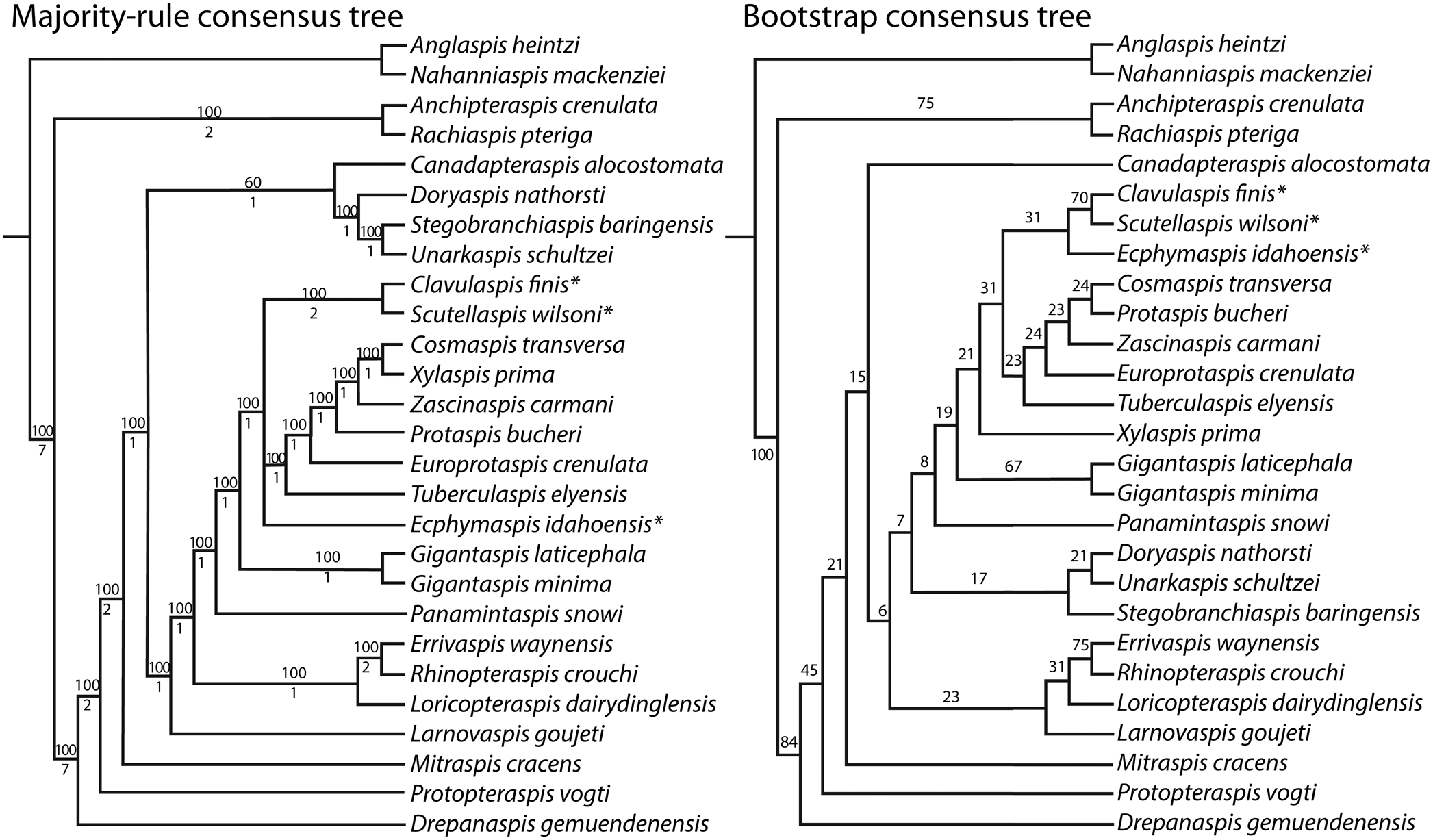
The phylogenetic analyses produced five most parsimonious trees with two taxa (Anglaspis heintzi Kiaer, Reference Kiaer1932, Nahanniaspis mackenziei Dineley and Loeffler, Reference Dineley and Loeffler1976) from the Cyathaspidiformes used as the outgroup. The strict consensus of these trees (Fig. 7) is well resolved, although the Bremer support values are low (1–2) for most clades within the ingroup. This has been typical of other analyses for this group and may reflect issues in the assumptions and definitions of the characters that were not evaluated for this kind of support in the past. Bootstrapping support is also weak, with only the division between the Cyathaspidiformes and Pteraspidiformes as a whole producing good support between these major groups (Fig. 8.1). Otherwise, bootstrap results show the strongest support for the relationships within the Gigantaspis genera, two of the new pteraspids (Clavulaspis finis n. comb. and Scutellaspis wilsoni n. gen. n. sp.) and two other pteraspids (Errivaspis waynesis [White, 1935] and Rhinopteraspis crouchi [Lankester, 1865]).
Additional versions of the analysis from parsimony (majority-rule consensus tree) and bootstrap (bootstrap consensus tree) with the three new taxa indicated by an asterisk. Majority rule consensus tree using PAUP* and TNT analyses showing the percentage of the five most-parsimonious trees that display branched groups as the upper number and Bremer support value as the lower number. Bootstrap values provided for tree support from 10,000 replicates using parsimony analysis.

The analysis supported the tree produced previously by Pernègre and Elliott (Reference Pernègre and Elliott2008) with Drepanaspis gemuendenensis positioned between the Anchipteraspididae and the ‘Protopteraspididae’ (Fig. 7). The recently described taxon Mitraspis cracens is resolved as a basal member of the ‘Protopteraspididae,’ as suggested by Elliott et al. (Reference Elliott, Schultze and Blieck2015), and the three new Middle Devonian taxa form part of the Protaspididae, which is the most derived clade.
Conclusions
The description of three new taxa of pteraspids from the Middle Devonian of western North America expands the known diversity and range of Middle Devonian heterostracans. The genus Psephaspis is shown to be a nomen dubium and is restricted to the original material from Utah, while the species from the Spring Mountain channel in Idaho is renamed Ecphymaspis idahoensis n. comb., and additional material is used here to provide a more complete reconstruction. A new species from the same locality is described and named as Scutellaspis wilsoni n. gen. n. sp. This species exhibits a feature new to pteraspids with the addition of a small platelet between the orbital, rostral, pineal and median plates. A previously described species from the Yahatinda Formation in Alberta is renamed Clavulaspis finis n. comb., due to the preoccupation of the original generic name, and new material adds to the known features of this taxon. The phylogenetic analysis of these taxa shows them to be part of the derived clade Protaspididae.
The Middle Devonian species are mostly broad and dorso-ventrally compressed, and exhibit tuberculate surface ornament instead of the crenulated ridge ornament commonly seen in Early Devonian species. These late-appearing taxa all have elongated branchial plates with terminal branchial openings, and the cornual plates are reduced or lost. These trends are also seen in the Psammosteoidei and continue in that lineage to the extinction of the Pteraspidiformes in the Late Devonian (Glinskiy, Reference Glinskiy2017). The similarity among these features in the pteraspids and psammosteids caused the original Middle Devonian pteraspids to be identified as psammosteids, however, this new analysis confirms previous studies in placing the Psammosteoidei in a basal position within the Pteraspidiformes.
Acknowledgments
We are indebted to F. Gahn, Brigham Young University-Idaho for making the Lemhi median dorsal plate of Scutellaspis wilsoni n. gen. n. sp. available, the Paleontology Program of the Park Service for the collecting permit, and M. Thompson of the IMNH for curating the specimen. C. Dehler, Utah State University, Logan, is thanked for assisting during the collection of the Lemhi material and for collecting the new Yahatinda material, and J. Bruner, University of Alberta, Edmonton, made it available for study. A. Stroup helped with the curation of material at the FMNH Chicago, and very kindly provided photographs of the type median dorsal plate of Ecphymaspis idahoensis n. comb. H. Davis drafted Figure 3. We also acknowledge the help of M. Serio in the initial part of this study. Finally, we would like to thank V. Glinskiy and two unknown reviewers for comments and suggestions that substantially improved the manuscript.
Accessibility of supplemental data
The final character list, data matrix, and NEXUS (Maddison et al., Reference Maddison, Swofford and Maddison1997) and TNT files used in this analysis are available from the Dryad Digital Repository: https://doi.org/10.5061/dryad.dncjsxkvt