I. INTRODUCTION
Veatchite polytypes have been said to have X-ray powder diffraction (XRPD) patterns that are too similar to allow for proper identification via this method (Stewart et al., Reference Stewart, Chalmers and Phillips1954; Clark and Mrose, Reference Clark and Mrose1960; Clark and Christ, Reference Clark and Christ1971; Kumbasar, Reference Kumbasar1979; Grice and Pring, Reference Grice and Pring2012). Until now, the polytypes were studied and discussed mainly on the basis of their crystal structures. One of the authors (Rémy Poulin, supervised by J.D. Grice) in his bachelor's thesis at the University of Ottawa (Poulin, Reference Poulin2012), came to the conclusion that the polytypes should be distinguishable by XRPD. This project confirms this by using all of the available veatchite specimens in the mineral collection of the Canadian Museum of Nature in addition to a sample of type material provided by the Smithsonian Institution. Another objective here is to outline a simple step-by-step method that would allow a better use of XRPD data when studying polytypes and minerals with similar structures. The usefulness of micro-powder-diffraction for such a project is extremely advantageous because of its low cost, easy availability, quick sample preparation and analysis time compared to a single crystal diffractometer. It also requires a lot less material than a standard powder diffractometer, which makes it more suitable for microscopic crystals.
The structural relationships of the three veatchite polytypes are discussed in detail by Grice and Pring (Reference Grice and Pring2012). Typical of polytypes, the structures are layered. There are two different layer types, labeled A and B. In the triclinic polytype, veatchite-1A, the layer sequence is AABBAABB. In the two monoclinic polytypes veatchites-1M and -2M, the layer sequence is ABABAB. The doubling of one cell dimension in veatchite-2M is due to a slight lateral shift in the layer stacking. The slight differences in crystal symmetry and cell parameters induced by different stacking sequences are expected to slightly modify the measured diffraction patterns, which are the basis of the investigation for this project.
II. MATERIALS
Initially, veatchite-1A from Emet, Turkey (type material NMNH 105697) and veatchite-2M for Sussex, NB (sample # F97-35) was available for XRPD. Although the authors had veatchite-1M from Reyershausen, Germany, it was a single crystal of type material from the Smithsonian Institution and hence not available because of the destructive nature of XRPD analysis, which consists of crushing the mineral. Veatchite from Billie Mine, Inyo County, California (CMNMC 57568 and 57569) and the Sterling Borax Mine (Lang), California (CMNMC 57620) were also examined to insure that our approach yielded reliable results.
Veatchite occurs as white fuzzy powder and rare rhombic plates in Emet, Turkey and as orange rhombohedra in the Sussex, New Brunswick material. On both samples from Billie Mine, veatchite was found in two distinct crystal forms; larger rhombic plates and elongated blades that often form sprays. The two habits are often intergrown and difficult to separate. The Sterling Borax Mine veatchite specimen has radiating rhombic plates. The identification method uses a profile fitting approach when comparing polytype calculated diffraction scans with any of the measured patterns acquired from the specimens.
III. METHOD
All the analyses were performed using a Bruker D8 Discover micro-diffractometer with the Hi-Star detector calibrated according to Rowe (Reference Rowe2009). Factors that could influence the experiment are the shape, size and crystallite size of the powder mount and the crystallinity of the minerals. The grid lines of the alignment camera were used to estimate if the powder mounts were of similar size.
The first problem that was noted when comparing polytypes using popular search/match software is the abundance of XRPD data available and how much variance there is for each polytype. Important elements that need to be taken into account and were identified as issues to tackle are as follows:
-
– The nature of the data in the available databases often differs (calculated or measured)
-
– Experimental error or limits associated with published patterns (instrumentation used, software used, data manipulation involved, etc.). As an example; Jenkins et al. (Reference Jenkins, Edmonds, Cooper and Post1990) identify intensity approximation of XRPD films from a Gandolfi camera as a source of error.
-
– The point form layout of X-ray diffraction data (a single line for each measured peak or calculated reflections depending on the nature of the data). The user is often left wondering if some lesser but useful reflections were not reported.
-
– The limits in resolution of an X-ray diffractometer; some adjacent reflections will merge into a single peak if the sample is too close to the detector (Rowe, Reference Rowe2009).
The best approach to limit introduced experimental error is to calculate a continuous diffraction scan (2θ calc/I calc profile) using the initial structure data for each polytype; for this project, the structure data from Grice and Pring (Reference Grice and Pring2012) was used. The software POWDER CELL (Kraus and Nolze, Reference Kraus and Nolze1996) was used to calculate the XRPD using the experimental conditions for the instrument and this program offers the option of exporting the data as a full scan in a .RAW file that can be imported in Bruker EVA Search/Match software (Bruker, 2010). A comparative method of visual profile fitting using MEASURED SCAN VS CALCULATED SCAN is believed to be the most appropriate to compare polytypes. The advantages of the approach are as follows:
-
– The qualitative nature of a scan contains useful features not usually found in a diffraction table (peak shoulders, skewness and broadness). These shapes can be hints of diagnostic reflections hidden by the limits in resolution of the instrument.
-
– Visual profile fitting allows for quick results, plus it is more evident than comparing d-spacing values and relative intensities in an XRD table.
-
– Allows comparing broad 2θ areas, therefore identifying diagnostic diffraction ranges rather than single peaks. The 2θ calc/I calc profile calculated from the matching polytype would ideally fit under the broader features found in the measured pattern. Shifting peak positions and intensities will help in differentiating the polytypes.
-
– Shows how the various calculated patterns (ideal) translate into measured patterns (real) considering the limited resolution of a micro powder-diffractometer.
These advantages will become more apparent as this paper unfolds since the objective is to confirm which polytype occurs at different localities with a high confidence level.
XRPD patterns were calculated using POWDER CELL software (Kraus and Nolze, Reference Kraus and Nolze1996) for veatchite-1M, -2M and -1A using the data of Grice and Pring (Reference Grice and Pring2012) structure determinations using the .INS files created in SHELX-97 (Sheldrick, Reference Sheldrick1997). The Inorganic Crystal Structure Database (ICSD) can be used as a valuable source for the structural data to import in POWDER CELL. The only issue is that POWDER CELL cannot import recent ICSD text files. To overcome this issue, the modern ICSD text file needs modification to look more like the text files fit for POWDER CELL. The supplementary material (available online at http://www.journals.cambridge.org/PDJ) includes an example for a modified ICSD text file with necessary data and parameters for POWDER CELL to calculate and draw the diffraction pattern properly. It was noted that veatchite (ICSD #2942) used as an example in the supplementary material was mislabeled as veatchite-1M in the ICSD database, but the reference Clark and Christ (Reference Clark and Christ1971) is indeed for polytype-2M. For other minerals, there are many structures available from ICSD. It is important to carefully select the material from which the structure was described. An ideal candidate has to be described from a naturally occurring specimen that was not involved in any heating, doping, cation-exchange or any other experiments that could alter its naturally occurring structural arrangement.
IV. POLYTYPE IDENTIFICATION RESULTS
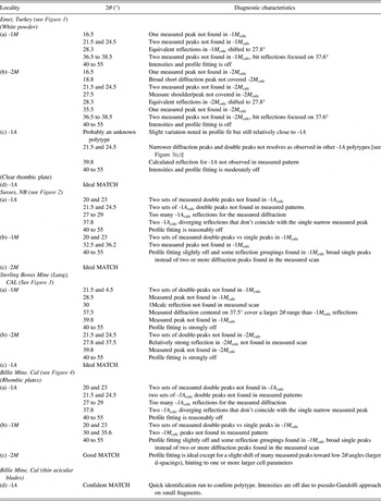
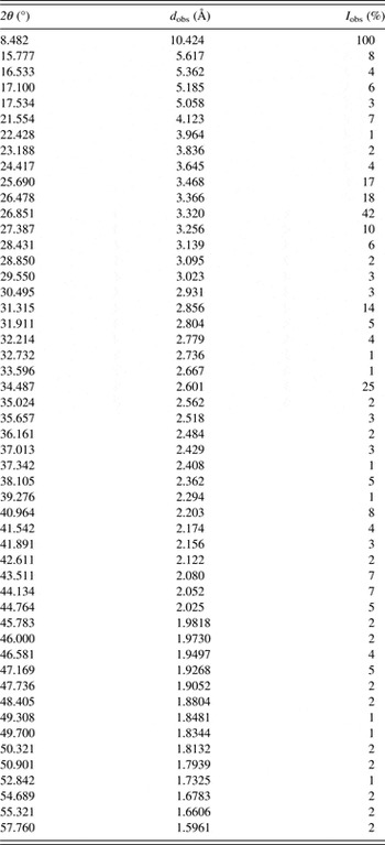
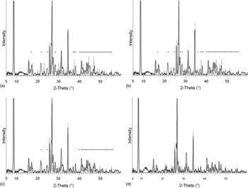
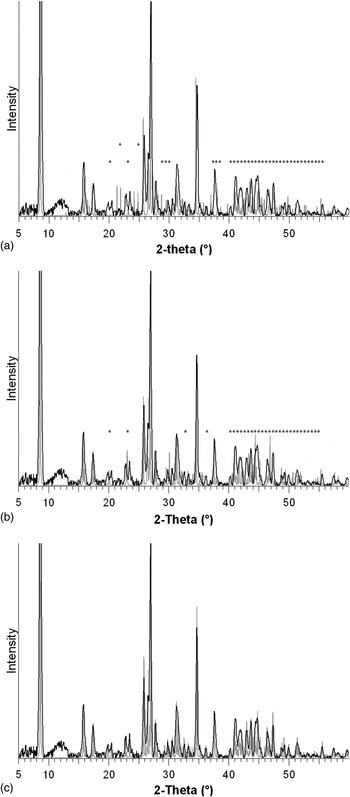
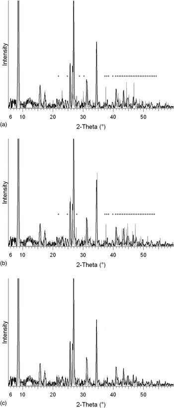
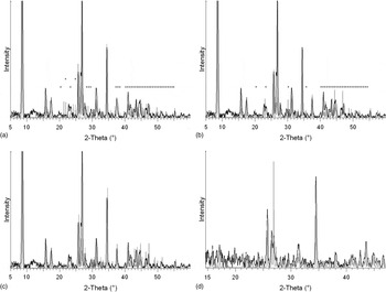
The current research paper has two distinct objectives: first, to find a quick method for identifying polytypes using X-ray diffraction data, which is achieve by a visual comparison of every measured diffraction scan, for all habits, from each of the localities, to the calculated scans for polytypes -1A, -1M and -2M in Figures 1–4. The matching calculated polytype profile matches almost ideally the measured profile. All the diagnostic differences and results observed in Figures 1–4 are recorded in Table I, which is in fact a summary of the observations leading to proper polytype identification for each of the studied veatchites. Table I also serves our second objective, which is to identify diagnostic diffraction areas for an even more simplified comparison of veatchite polytypes. This could be used for subsequent polytype identification for new veatchite occurrences.
Detailed comparison of veatchite measured diffraction scans from each locality with the calculated scan of veatchite-1A, -1M and -2M.

A few interesting discoveries resulted from this polytype identification project. It was found that most of the white silky and powdery veatchite from Emet, Turkey is actually a different, unknown polytype, with a diffraction pattern that differs slightly from veatchite-1A, which was described for the rare rhombic plates found on the same specimen, which is confirmed by this study [Figure 1(d)]. Unfortunately, the new polytype is not suitable for structure work because of the lack of a suitable single crystal, but its measured diffraction pattern is reported in Table II for future reference. Only veatchite-2M was analyzed from Penobsquis near Sussex, NB. The sprays of rhombic plates from the Sterling Borax Mine (Lang) were confirmed as veatchite-1A. At first the analyses from Billie Mine gave a mixture of veatchites -1A and -2M, but a careful selection of crystals of different habit confirmed that the clear rhombic plates were veatchite-2M and the flat elongated blades, veatchite-1A.
Unknown veatchite polytype: X-ray powder diffraction data (CuKᾱ).

The profile fitting method made good use of the differences observed within the 5–60°2θ range studied (Table I and Figures 1 to 4), but it could be narrowed down to a diagnostic area between 20 to 25°. In this range, veatchite-1A has two unique sets of double peaks at approximately 21.5 and 24.5°2θ (−103, 114 and 1–14, 105), veatchite-2M has two unique set of double peaks at approximately 20 and 23°2θ (merged −113, 023; 113; merged 114, 024; and 114) whereas veatchite-1M only has single peaks at 20 and 23°2θ (merged 130, 031, −131 and merged 140, 041, −141). The unknown polytype from Emet, Turkey has single peaks at approximately 21.5 and 24.5° instead of double peaks for -1A.
Profile fitting for veatchite from Emet, Turkey. (a) white powdery vs -1M (b) white powdery vs -2M (c) white powdery 1A (d) clear rhombic plates vs -1A.

Profile fitting for veatchite from Penobsquis, near Sussex, New Brunswick, Canada. (a) vs -1M (b) vs -2M (c) 1A

Profile fitting for veatchite from the Sterling Borax Mine (Lang), California, USA. (a) vs -1M (b) vs -2M (c) 1A

Profile fitting for veatchite from the Billie Mine, California, USA. (a) rhombic plates vs -1M (b) rhombic plates vs -2M (c) rhombic plates 1A (d) thin acicular blades vs -1A.

V. DISCUSSION
This project is in no way a full description of the veatchite polytypes occurring at each locality, but at least a clear confirmation for the ones observed. Only one or two hand specimens for each locality were studied, but hopefully the method will allow for a clarification of the worldwide distribution of veatchites -1A, -1M and -2M, which seems to be debated. Clark and Christ (Reference Clark and Christ1971) listed some hypotheses from the literature that were not conclusive due to the inability to easily distinguish veatchite polytypes, but which could now be verified with this approach. They thought, probably unjustified, that Murdoch and Webb (Reference Murdoch and Webb1964) and Pemberton (Reference Pemberton1968) suggested that both veatchites -2M and 1M were found at the Sterling Borax mine (Lang), but maybe the presence of veatchite-1A confirmed in [Figure 3(c)] could explain this assumption that two different veatchite polytypes occurred there. Polytype-1A was described only in 1979. They also mentioned the fact that Clark et al. (Reference Clark, Konnert and Christ1969) suggested veatchite-1M to be from marine evaporates and veatchite-2M to be from continental saline environment. Also it could help validate or discredit veatchite -1M (labeled veatchite-p) from Billie Mine, which is sold and, or reported on various websites (e.g., mindat.org). Nothing confirming veatchite-1M (veatchite-p) from Billie Mine seems to be available in the scientific literature. Grice and Pring (Reference Grice and Pring2012) concluded that “it appears that temperature and pressure control the occurrence of each (veatchite) polytypes.” With improved identification of polytypes using these techniques mineralogists should be able to better interpret the genesis of the associated geological formations.
VI. CONCLUSION
The identification results obtained using calculated scans from the structure data proved to be extremely accurate and in fact gives almost undisputable validation of veatchite polytypes at different localities. The approach might not be suitable for structures that are too similar, but its potential can be assessed by comparing calculated scans of polytypes or polymorphs and identifying possible diagnostic reflections. The method has also been used to distinguish quasi-isostructural minerals with different chemistry, but as previously mentioned, its potential should be assessed on a case-to-case basis. It is understood that this method does not provide the same certainty as a full structure characterization, but it allows expanding on the usefulness of data from XRPD. (The calculated .RAW files for veatchites -1A, -1M and -2M are available in the supplementary material found with the online publication.)
SUPPLEMENTARY MATERIALS AND METHODS
The supplementary material for this article can be found at http://www.journals.cambridge.org/PDJ