Introduction
Mollusc-parasitic nematodes (MPNs) comprise eight families – namely, Agfidae, Alaninematidae, Alloionematidae, Angiostomatidae, Cosmocercidae, Diplogasteridae, Mermithidae and Rhabditidae (Pieterse et al., Reference Pieterse, Malan and Ross2017a). Phasmarhabditis hermaphrodita (Schneider) Andrássy is the only MPN to be successfully developed as a biological molluscicide (Rae et al., Reference Rae, Verdun, Grewal, Robertson and Wilson2007). The nematode is commercially available from BASF (formerly Becker Underwood) and Dudutech under the trade names Nemaslug® and SlugTech®, respectively. Like other rhabditid nematodes, P. hermaphrodita responds to depleting food supply and to poor environmental conditions with the formation of a metabolically suppressed, third juvenile stage, which is known as the infective juvenile (IJ), or the dauer juvenile. IJs, which are adapted for long-term survival, actively search out molluscs, entering through natural openings (Wilson et al., Reference Wilson, Burch, Tourna, Aalders and Barker2012). The IJs then recover and develop into self-fertilizing hermaphrodites, whereupon they start to reproduce. The host usually dies within 4–21 days following infection, after which the nematodes colonize the entire slug cadaver, feeding and reproducing until the food source is depleted. The IJs are then produced, which move into the soil in search of new mollusc hosts (Wilson et al., Reference Wilson, Glen and George1993; Tan & Grewal, Reference Tan and Grewal2001). Phasmarhabditis hermaphrodita is capable of infecting a number of mollusc species, including the snail species Monacha cantiana (Montagu), Cepaea hortensis (Müller), Theba pisana (Müller), Cochlicella acuta (Müller), Cernuella vigata (Da Costa) and Lymnaea stagnalis (Linnaeus), as well as the slug species Deroceras reticulatum (Müller), Deroceras panormitanum (Lessona & Pollonera), Deroceras laeve (Müller), Arion silvaticus Lohmander, Arion intermedius Normand, Arion distinctus Mabile, Tandonia sowerbyi (Férussac), Tandonia budapestensis (Hazay) and Leidyula floridana (Leidy) (Rae et al., Reference Rae, Verdun, Grewal, Robertson and Wilson2007). It also has a necromenic life cycle (Mengert, Reference Mengert1953), as well as being able to reproduce on such organic substrates as slug faeces, dead animal tissue and leaf compost (Tan & Grewal, Reference Tan and Grewal2001; MacMillan et al., Reference MacMillan, Haukeland, Rae, Young, Crawford, Hapca and Wilson2009; Nermut’ et al., Reference Nermut’, Půža and Mráček2014). Although P. hermaphrodita has been the most studied MPN to date, very little is known about its biology and life cycle. This information would be of importance when considering improved mass production conditions.
There is a total of 16 described species within Phasmarhabditis; however, little to no information is available on their life cycles. One such nematode is Phasmarhabditis papillosa (Schneider) Andrássy, which was first described in 1866 based on the minimal amount of information regarding the female body length, as well as basic morphometric data relating to the males and females (Schneider, Reference Schneider1866). Schneider (Reference Schneider1871) later reported that the males and females of P. papillosa occur in roughly equal numbers, whereas the males are relatively rare in cultures of P. hermaphrodita (Andrássy, Reference Andrássy1983; Tandingan De Ley et al., Reference Tandingan De Ley, Holovachov, McDonnell, Bert, Paine and De Ley2016). In 2016, P. papillosa was redescribed after being isolated for the first time in California, USA (Andrássy, Reference Andrássy1983; Tandingan De Ley et al., Reference Tandingan De Ley, Holovachov, McDonnell, Bert, Paine and De Ley2016), and then in 2017, the description was further updated following the nematode's isolation in South Africa (Pieterse et al., Reference Pieterse, Malan, Kruitbos, Sirgel and Ross2017b). Pieterse et al. (Reference Pieterse, Tiedt, Malan and Ross2017c) demonstrated that P. papillosa can cause mortality in the European slug species, Deroceras invadens Reise, Hutchinson, Schunack & Schlitt (Agriolimacidae) (Pieterse et al., Reference Pieterse, Tiedt, Malan and Ross2017c).
Phasmarhabditis bohemica Nermuť, Půža, Mekete & Mráček was described in 2017 from D. reticulatum in the Czech Republic (Nermuť et al., Reference Nermut’, Půža, Mekete and Mráček2017). The ability of this species to cause mortality in its hosts requires further investigation as no apparent mortality was caused under natural conditions, but high mortality of D. reticulatum was observed under laboratory conditions (Nermut’ et al., Reference Nermut’, Půža, Mekete and Mráček2017). In the description of this nematode it was reported that P. bohemica can complete its life cycle on decaying animal matter within several days when kept at 15°C (Nermut’ et al., Reference Nermut’, Půža, Mekete and Mráček2017).
Phasmarhabditis kenyaensis Pieterse, Rowson, Tiedt, Malan, Haukeland & Ross was isolated from the slug Polytoxon robustum (Simroth) (Urocyclidae), collected in Nairobi, Kenya. The most notable characteristic of P. kenyaensis is that the IJ stage has the longest body length in the genus; however, its pathogenicity to molluscs requires further investigation (Pieterse et al., Reference Pieterse, Rowson, Tiedt, Malan, Haukeland and Ross2020).
In the current study, the biology and development of inoculated (parental) IJs to the next generation of IJs of P. hermaphrodita, P. papillosa, P. bohemica and P. kenyaensis from ex vivo cultures with freeze-killed slug cadavers were studied. This study contributes to the knowledge of the biology of the genus and will help to establish the in vitro liquid cultures of different species of the genus.
Materials and methods
Source of nematodes
The following nematode species were used in the study: P. hermaphrodita, P. papillosa, P. bohemica and P. kenyaensis. The P. papillosa isolate was obtained from a collection of the Department of Conservation Ecology and Entomology at Stellenbosch University (Pieterse et al., Reference Pieterse, Malan, Kruitbos, Sirgel and Ross2017b). The P. bohemica and P. hermaphrodita isolates were supplied by Vladimir Půža from the Biology Centre CAS, Institute of Entomology ASCR, Laboratory of Entomopathogenic Nematodes, Czech Republic. The P. kenyaensis was provided by Solveig Haukeland from the International Centre of Insect Physiology and Ecology (icipe) in Nairobi, Kenya. Inoculum was prepared by recycling through a defrosted slug and the IJs harvested were used within two weeks of storage.
Nematode preparation
Deroceras invadens Reise, Hutchinson, Schunack & Schlitt was used as the host species, as it was previously shown to be susceptible to Phasmarhabditis species (Pieterse et al., Reference Pieterse, Tiedt, Malan and Ross2017c). These slugs were also available to be collected in large quantities by hand and frozen for future use. The life cycles of the four Phasmarhabditis spp. were studied by adding 100 IJs (rinsed with distilled water) of each species to 1 g of the freeze-killed homogenized D. invadens, placed on modified White traps, along with a piece of damp filter paper in a 90-mm-diameter Petri dish. The cultures were sealed with Parafilm® and kept in darkness at 20°C. Every 24 h thereafter, the development of each species was recorded by taking one Petri dish of each species and washing with 0.9% saline and heat-killing (~85°C) the nematodes with triethanolamine-formalin fixative (Courtney et al., Reference Courtney, Polley and Miller1955). The nematodes were then classified into the different life stages and counted using a compound microscope (Leica DM200, Leica Microsystems, Wetzlar, Germany), to determine the daily population structure development. The experiment was repeated on a different test date with a fresh batch of the different nematode species.
Nematode biological observations and measurements
Fifty nematodes of each life stage were measured for each of the Phasmarhabditis spp. studied. The nematodes were placed in a drop of water on a microscope slide, covered with a coverslip and killed with gentle heat on a hot plate. They were then measured using a compound microscope, fitted with a digital camera and with the software Leica Application Suite version 3.5.0, (https://www.leica-microsystems.com/products/microscope-software/p/leica-application-suite/)with live measurement capability. The body length and width, as well as the life stage of the nematodes, were recorded. The experiment was repeated twice, providing an average measurement of each life stage, based on 100 nematodes.
Statistical analysis
Statistical analyses were performed using Statistica version 13.3 (StatSoft Europe, 2021). Results of the measurements of the different life stages (females, males and IJs) were analysed using a one-way analysis of variance, followed by a non-parametric post-hoc Games–Howel multiple comparison.
Results
Development of different life stages
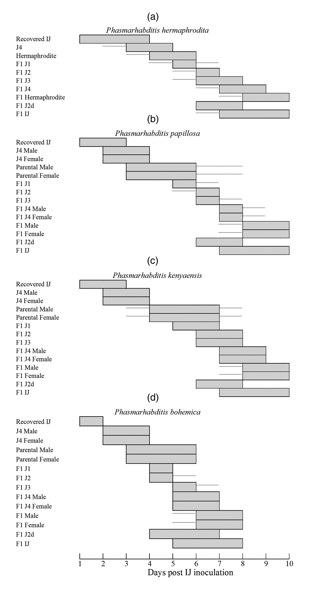
The ex vivo development of the four Phasmarhabditis species is illustrated in fig. 1, with bars demonstrating the development of inoculated IJ (100) over a period of 8–10 days on 1 g of frozen homogenized D. invadens, by which point the food source was depleted. The lines in the bars indicate when the population of each stage was less than 10% of the maximum (fig. 1).
The ex vivo development of 100 IJs on 1 g of homogenized Deroceras invadens over a period of ten days for (a) Phasmarhabditis hermaphrodita, (b) Phasmarhabditis papillosa, (c) Phasmarhabditis kenyaensis and (d) Phasmarhabditis bohemica. The lines indicate when the population of each stage was less than 10% of the maximum.

The recovery of IJs of P. hermaphrodita occurred from one to four days after inoculation (DAI), with J4 visible from days three to five. Hermaphrodites developed from four to six DAI, and by day six, most of the population consisted of parental hermaphrodites, with eggs starting to hatch and first-stage juveniles (J1) of the first generation (F1) starting to appear from five to six DAI. By day six, the development of the second-stage juvenile (J2) and third-stage juvenile (J3) phases was observed, until day eight for J3. The fourth-stage juveniles (J4) appear after seven days and were visible until day nine. F1 hermaphrodites started developing at eight DAI, reaching maturity by day ten and showing fertilized eggs by the end of day eight. A new cohort of IJs start to develop by day six. In some hermaphrodites, endotokia matricida was observed eight DAI (fig. 1a).
In the development of the inoculated IJs of P. papillosa, the IJs started recovering from one to three DAI, and the J4 (with male and female primordial developing genitals visible) were distinguishable on two–four DAI. By day three, the parental males and females reached adulthood, by which time female egg development had also started. By five–six DAI, the F1 J1s appear, with them mostly developed into J2s by seven DAI, and into J3s by six–eight DAI. The F1 males and females started developing on seven DAI, while some juveniles developed into F1 pre-dauer second stage juveniles J2d (pre-IJs) and IJs at the same time. Males and females reached adulthood over a period of eight–ten DAI, with fertilized eggs developing by day ten. Endotokia matricida was observed at day ten in some females (fig. 1b).
Phasmarhabditis kenyaensis inoculated IJs started to recover from one to three DAI, with male and female characteristics showing by two DAI. The parental males and females were fully developed by day 4 and started producing J1 larvae by day five. By six DAI, most of the F1 larvae had developed into J2, J3 or J2d stages, and by seven DAI, J4 males, J4 females and IJs had formed. On eight DAI, the males and females had completed their development, and by ten DAI, the females had fertilized and developed their eggs, which were either released or had hatched out inside the adult female's body (fig. 1c).
With P. bohemica, the IJs started recovering from one to two DAI, with many parental males and females formed after three DAI. By five DAI, the first F1 J1 larvae started to appear and develop into J2 and J3, or into J2d six DAI. By five DAI, the majority of the nematodes in the population consisted of F1 J4 males, J4 females, J3 larvae, J2d larvae or IJs. On six DAI, many of the males and females had reached maturity. By eight DAI, the females contained fertilized and developed eggs, which were ready for release, or had hatched inside the body of the adult female nematode as IJs (fig. 1d).
Difference in body length and body width
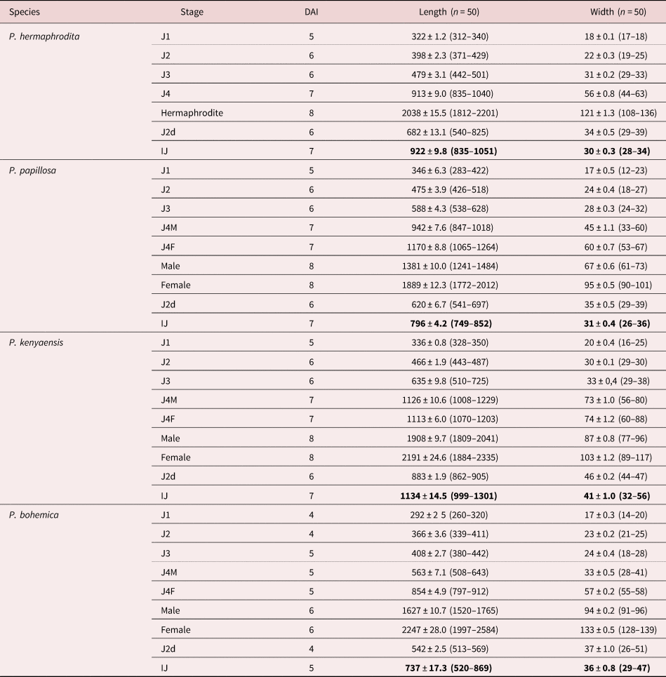
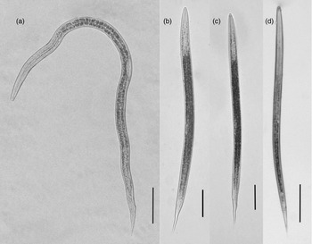
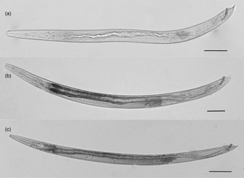
Analysis of the results of the measurements of the mean body length and width of the different life stages of the four species showed significant differences (P < 0.01) between all stages and species. The only exception was in the case of the body width of the IJ of P. hermaphrodita and P. papillosa, which did not differ significantly (P = 0.57) from each other. The IJs of P. hermaphrodita (fig. 2a) are longer and thinner than those of P. papillosa (fig. 2b) and P. bohemica (fig. 2c). The IJs of P. hermaphrodita are, however, generally shorter and thinner than those of P. kenyaensis (fig. 2d), which has the largest IJ body size, measuring 1134 μm in length and 41 μm in width (table 1 and illustrated in figs 2–4).
Mean body length and width (μm) of the different life stages of Phasmarhabditis hermaphrodita, Phasmarhabditis papillosa, Phasmarhabditis bohemica and Phasmarhabditis kenyaensis at specific days after inoculation (DAI) with infective juveniles (IJs), for the duration of the development of 100 IJs on 1 g of Deroceras invadens homogenized tissue.

All measurements are in μm and in the form: mean ± standard error (range).
IJs of (a) Phasmarhabditis hermaphrodita, (b) Phasmarhabditis papillosa, (c) Phasmarhabditis bohemica and (d) Phasmarhabditis kenyaensis. Scale bars: (a–c) 100 μm; (d) 200 μm.

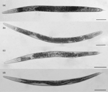
Images of (a) hermaphrodite of Phasmarhabditis hermaphrodita, (b) female Phasmarhabditis papillosa, (c) female Phasmarhabditis bohemica and (d) female Phasmarhabditis kenyaensis. Scale bars: 200 μm.

Images of males of (a) Phasmarhabditis papillosa, (b) Phasmarhabditis bohemica and (c) Phasmarhabditis kenyaensis. Scale bars: (a, b) 100 μm; (c) 200 μm.

The adult hermaphrodites of P. hermaphrodita (fig. 3a) are longer (2038 μm) and thicker (121 μm) than the females of P. papillosa (fig. 3b) (1889 μm length, 95 μm width), but both are shorter than the adult females of P. kenyaensis (fig. 3d), which measure 2191 μm in length and 103 μm in width. The adult females of P. bohemica (fig. 3c) were the largest of the Phasmarhabditis species studied, measuring 2247 μm in length and 133 μm in width.
Of the three species that had males present, P. papillosa (fig. 4a) had the smallest males, measuring 1381 μm in length and 67 μm in width. The males of P. bohemica (fig. 4b) were larger, measuring 1627 μm in length and 94 μm in width, with the males of P. kenyaensis (Fig 4c) being the largest of the species studied, with a length of 1908 μm and a width of 87 μm.
Development took approximately 48 h longer for P. hermaphrodita, P. papillosa and P. kenyaensis (day eight) compared to P. bohemica, which developed into F1 IJ, adult males and females by day six.
The most important difference between the four species is the mode of reproduction. Phasmarhabditis papillosa, P. bohemica and P. kenyaensis formed males and females that required copulation to reproduce, whereas P. hermaphrodita only formed self-fertilizing hermaphrodites, meaning that each nematode could reproduce on its own.
Discussion
Of the many nematode species associated with terrestrial molluscs, P. hermaphrodita is the only species, to date, to have been successfully formulated into a commercial biological molluscicide (Rae et al., Reference Rae, Verdun, Grewal, Robertson and Wilson2007; Barua et al., Reference Barua, Williams and Ross2021). Its success as a biocontrol agent can be attributed to its wide host range, to its capacity to form self-fertilizing hermaphrodites, as well as to its ability to cause mortality to its mollusc hosts within 4–21 days after infection (Rae et al., Reference Rae, Verdun, Grewal, Robertson and Wilson2007). The success of the product, alongside the regulatory restrictions associated with the use of P. hermaphrodita in countries where it is non-native, has led to a boom in research within the genus. To date, there are 16 species in the genus, many of which have been described over the last decade.
Aside from the work conducted on P. hermaphrodita, very little has yet been published on the mass culture and virulence of other species within the Phasmarhabditis genus (Pieterse et al., Reference Pieterse, Tiedt, Malan and Ross2017c). The potential of a nematode candidate to be considered for biocontrol development depends on its virulence, as well as on the ease with which it can be mass cultured. An important part of nematode mass production is to understand, and the ability to manage, the nematode population dynamics. The current study provides new insights into the life cycles of the four phasmarhabditids used in this study associated with slugs. Of the four nematode species used, P. bohemica completed its development the quickest, completing it within eight days, while P. hermaphrodita, P. papillosa and P. kenyaensis took ten days. This means that P. bohemica has an advantage over the other Phasmarhabditis species, in that it develops into the adult stage two days earlier, but due to the inability of P. bohemica to produce self-fertilizing hermaphrodites, it can be regarded as a more complex biocontrol candidate. The above means that the culturing would take longer, and that the conditions within the flasks or bioreactors would have to be altered to allow copulation between the males and females. When mass-culturing nematodes, the goal is to produce the largest amount of IJs in the least amount of time. Even a day difference in the lifecycle length of the nematodes will have a significant impact on production efficiency and economics associated with mass production.
This means that the reproduction of P. hermaphrodita may be easier in liquid culture, with the use of less inoculum compared to that of P. papillosa, P. bohemica and P. kenyaensis. The results indicated that despite the promising pathogenicity data noted by Pieterse et al. (Reference Pieterse, Tiedt, Malan and Ross2017c) demonstrating the ability of P. papillosa to control D. invadens, based on the lifecycle results of the current study, it would not necessarily be a better biocontrol candidate compared to P. hermaphrodita.
Previous research has shown that growth substrate and the bacterial associates affect the amount and quality of IJs produced in P. hermaphrodita (Tan & Grewal, Reference Tan and Grewal2001; Rae et al., Reference Rae, Robertson and Wilson2009; Nermuť et al., Reference Nermut’, Půža and Mráček2014). As it is a facultative parasite, it can reproduce on a variety of organic materials (Maupas, Reference Maupas1900; Mengert, Reference Mengert1953; Tan & Grewal, Reference Tan and Grewal2001; Rae et al., Reference Rae, Robertson and Wilson2006; Nermuť et al., Reference Nermut’, Půža and Mráček2014). Rae et al. (Reference Rae, Robertson and Wilson2009) analysed the number of infected juveniles produced by P. hermaphrodita when grown on homogenized snail, slug or earthworm, and found that higher numbers of IJs were achieved when it was grown on the slug species D. panormitanum (Rae et al., Reference Rae, Robertson and Wilson2009). Nermuť et al. (Reference Nermut’, Půža and Mráček2014) studied the influence of growth substrates on the development time and size of three different P. hermaphrodita strains that were grown on freeze-killed slug cadavers or grown in monoxenic production, and found that monoxenic strains had a shorter development time, longer length and a higher yield (Nermuť et al., Reference Nermut’, Půža and Mráček2014). As the nematodes in our study were grown with a variety of unknown bacteria from the homogenized slug substrate, development time, length and yield would, therefore, improve with mass-culturing, as this is done in monoxenic conditions. When selecting a nematode species for mass-culturing, the next step after analysing the life stage would, therefore, be to determine the best growth substrate and bacterial associate.
To optimize the conditions in liquid cultures for the development of nematode species, it is crucial to understand their life cycles and to have a basic knowledge of the different life stages. The results of the study regarding ex vivo development and the body size of the different species can be used for future improvements on the culturing of the nematodes and on the optimizing of yields for possible commercial production. To enable the recommendation of nematode species for, or their eliminating from, consideration for development as possible biocontrol agents, the pathogenicity and the life cycles of the other species in the Phasmarhabditis genus should also be investigated.
Financial support
This work was supported by the National Research Foundation of South Africa (NRF-THRIP TP14062571871) and the Council for Scientific and Industrial Research.
Conflicts of interest
None.
Ethical standards
The authors assert that all procedures contributing to this work comply with the ethical standards of the relevant national and institutional guides on the care and use of laboratory animals.