Implications
The dietary calcium level is a critical factor in poultry production, which plays an essential structural and metabolic function in bone formation, eggshell quality and egg production. The present study determined the dietary calcium requirement for optimal egg production, eggshell quality, bone mineralization and ovarian morphology in laying duck breeders. The obtained results have a significant role, for feed formulators and producers, in enhancing the egg production profits, and health of laying duck breeders.
Introduction
Calcium (Ca) is the critical nutritional factor for egg production, eggshell formation and bone quality of laying birds. Much is known about the Ca requirements of laying hens, but little information is available on the Ca requirements for duck breeders. The U.S. National Research Council (1994) recommended a Ca level of 2.75% for white Pekin duck breeders, whereas 3.0% Ca for breeder ducks was suggested in Commercial Poultry Nutrition (2004). Usually, obtaining good egg production with an acceptable shell quality in the laying hens requires sufficient dietary Ca and bones’ Ca reservoir. The laying performance, eggshell and tibial quality of hens were affected by the level, source and particle size of dietary Ca (Cufadar et al., Reference Cufadar, Olgun and Yildiz2011; Ganjigohari et al., Reference Ganjigohari, Ziaei, Ramzani and Tasharrofi2018; Manangi et al., Reference Manangi, Maharjan and Coon2018). Wang et al. (Reference Wang, Chen, Zhang, Ruan and Lin2014) found that using the large-particle size limestone (0.85 to 2.0 mm) to provide 3.49% dietary Ca facilitates superior productive performance, egg quality and bone characteristics of laying ducks. Castillo et al. (Reference Castillo, Cuca, Pro, Gonzalez and Morales2004) showed that the optimal biological level for maximal egg production and egg-specific gravity in Hy-Line W-98 hens are 4.34% and 4.62% Ca, respectively. Roland et al. (Reference Roland, Bryant, Rabon and Self1996) found that 5% dietary Ca increased bone quality without any adverse effect on the egg production of commercial Leghorns.
The Longyan duck breed is one of the most important egg-type duck breeds, which lays huge amounts of eggs. In southern China, more than 300 million birds are raised from such breed, producing around 80 billion eggs annually (Ruan et al., Reference Ruan, Fouad, Fan, Xia, Wang, Chen, Lin, Wang, Yang and Zheng2018). In a previous study, we found that diets containing 3.2% to 3.6% Ca exhibited superior egg production and bone quality of Longyan laying ducks during their peak laying period (Xia et al., Reference Xia, Zhang, Lin and Zheng2015). The established levels of Ca requirements for duck layers are not adequate to meet the requirements of duck breeders, which have a different genetic capacity for egg production, egg size, egg quality, fertility and hatchability. It is hypothesized that determining the optimal dietary Ca level for the breeding ducks is essential to maximize their reproductive performance. The objective of the current study, therefore, was to estimate the dietary Ca requirements for optimal productive and reproductive performance as well as eggshell and tibial quality for Longyan duck breeders.
Materials and methods
Animals and treatments
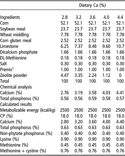
In total, 450 egg-laying Longyan-duck breeders with the same genetic background and comparable BW at 21 weeks of age were allotted randomly into five treatments, each with six replicates of 15 birds. The experimental groups had a similar initial BW (1.39 ± 0.02 kg) according to ANOVA test (P >0.05). The data collection started from 23 weeks of age and continued over the following 35 weeks. The experimental ducks were housed in individual galvanized battery cages (having the dimensions: length 27.8 cm × width 40 cm × height 55 cm, purchased from Guangzhou Huanan Poultry Equipment Co., Ltd, Guangzhou, China). The ducks were purchased at the age of 17 weeks, and from 17 to 20 weeks of age, they were fed a diet containing 2600 kcal metabolizable energy/kg, 16.0% CP, 0.90% Ca and 0.40% available phosphorus (P). Each battery cage was equipped with a feeder and nipple drinker. The different groups corresponded to five dietary treatments containing 2.8%, 3.2%, 3.6%, 4.0% or 4.4% Ca. The Ca content was adjusted using finely ground (<0.1 mm) limestone and zeolite as ‘filler’, and 0.40% available P (0.63% total P) was provided by the inclusion of calcium hydrogen phosphate. The other dietary nutrient levels were covered according to our previous findings for laying ducks (Ruan et al., Reference Ruan, Fouad, Fan, Xia, Wang, Chen, Lin, Wang, Yang and Zheng2018). Chemical and calculated analyses of the experimental diets are shown in Table 1. Water was available ad libitum and 85 g of feed was introduced twice daily at 0700 h and 1500 h throughout the experimental period. In addition to ambient daylight, 4 h of artificial light (15 lx per m2) was provided from 1830 h to 2230 h to give in total 16 h light: 8 h dark a day.
Composition and nutrient levels in the experimental diets of laying duck breeders (%, as fed basis)
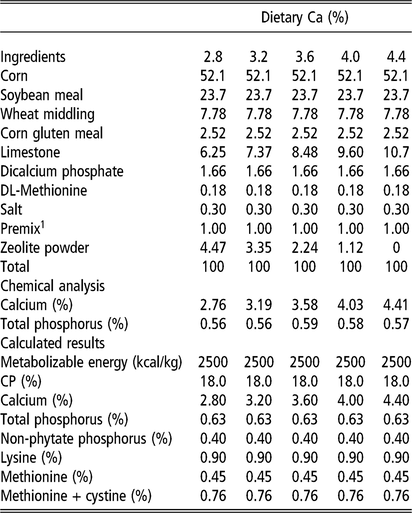
1 The premix provided the following per kilogram of diet: vitamin A 12 000 IU, vitamin D3 1800 IU, vitamin E 26 IU, vitamin K 1.0 mg, vitamin B1 3.0 mg, vitamin B2 9.6 mg, vitamin B6 6.0 mg, vitamin B12 0.03 mg, choline 500 mg, D-calcium pantothenate 28.5 mg, folic acid 0.6 mg, biotin 0.15 mg, Fe 50 mg, Cu 10 mg, Mn 90 mg, Zn 90 mg, I 0.50 mg and Se 0.40 mg.
Tissue sampling and storage
At the termination of the study, two birds were randomly selected from each replicate and blood samples were collected from the left-wing vein using 5 ml vacutainer tubes at 1000 h after an overnight fast for 12 h. Within 30 min of collection, the plasma was separated by centrifugation (1200×g for 10 min) and stored in 0.5 ml Eppendorf tubes at −20°C until analysis. The birds were then euthanized by cervical dislocation and exsanguinated, and the uteruses, ovaries and tibiae were collected. In addition, samples from the uteruses and ovaries were washed with phosphate-buffered saline, frozen by direct bluning into liquid nitrogen, and stored at −80°C until analysis.
Productive performance
Throughout the experimental period, the numbers of total broken and shell-less eggs were recorded daily on a replicate basis. All daily oviposited eggs were weighed on an individual basis. The feed intake (the difference between the added and refused feed amounts), egg number, egg weight, egg mass (egg weight/bird/day) and feed conversion ratio (FCR, daily feed intake/daily egg mass) were calculated on a per replicate basis, and then expressed as averages for their corresponding laying periods: early laying period (18 weeks, average daily egg production by all ducks ranged from 50% to 80%), peak laying period (17 weeks, average daily egg production by all ducks >80%) and the entire experimental period (35 weeks).
Eggshell quality
In total, 90 eggs were used for evaluating the eggshell quality (three eggs/replicate/four weeks). In each collection time, the treatment average (N = 18 eggs) was calculated to determine the eggshell quality traits. The breaking strength of the shell was determined by an Egg Force Reader (model EFR-01, ORKA Food Technology, Ramat HaSharon, Israel). The shell thickness was measured at three different points of the shell without membranes from the blunt, mid-length and pointed ends using a digital micrometer, and the obtained values were then averaged. To calculate the ash content, shells without membranes were washed under a gentle flow of water to remove any adhering albumen, dried in an oven at 65°C to a constant weight, and then ashed overnight in a muffle furnace between 550 and 600°C.
Incubation indices
During the peak laying period, all birds were artificially inseminated twice (with a three-day interval) with 100 μl of diluted fresh semen (diluted with 0.9% saline solution in a proportion of 1:1 volume/volume). The semen samples were collected from drakes belonging to the same breed. Fifty eggs per replicate (egg weight >63 g, with no soft shells, cracks, dirtiness or double yolks) were collected from the third through tenth day following the first insemination. All eggs were incubated in the same incubator (JXB2000; Dezhou Jingxiang Technology Co., Ltd, China) from 36.5°C to 38.4°C and from 45% to 65% relative humidity for 1 to 28 days. Eggs were turned (16 turns/day) throughout the incubation period and sprayed with water once daily from the 15th day of incubation until they hatched. The egg fertility was checked by candling at the seventh day of incubation. After 28 days, the healthy hatched ducklings were counted and recorded, and eggs that failed to hatch were counted. Hatchability was calculated as hatchability of fertile eggs. Healthy ducklings (clean and dry, free of deformities and with bright eyes) were determined macroscopically as described by Tona et al. (Reference Tona, Onagbesan, De Ketelaere, Decuypere and Bruggeman2004).
Ovary-related indices
The collected ovaries, small yellow follicles (SYF; 3 mm < diameter < 8 mm) and large yellow follicles (LYF; diameter ≥ 8 mm) were dissected and weighed. The numbers of SYF and LYF were recorded, and their weights were each expressed as proportions of ovarian weight. The relative ovary weight was calculated on the basis of live BW.
Tibial quality
The collected tibiae were trimmed of muscles and tendons, and weighed. Left tibiae then were boiled in water for 6 min and de-fatted by soaking in diethyl-ether for 96 h. Thereafter, they were oven dried to a constant weight and ashed using a muffle furnace (550 to 600°C for 24 h) to determine the tibial ash, which was expressed on the basis of dry-defatted weight (Lin and Shen, Reference Lin and Shen1979). In addition, Ca and P levels in the tibial ash were estimated. Besides, the right tibiae were used in examining the bone mineral density at the Guangzhou Overseas Chinese Hospital with an X-ray osteodensitometer (Lunar Prodigy, General Electric Company, Fairfield, CT, USA). The breaking strength was carried out at half of the tibia length using a materials tester (Instron 4411, Instron Corporation, Grove City, PA, USA). The tibia bone was held by two supporters, spaced 30 mm apart, and the physical power was applied to the midpoint of the bone by a static load cell with a crosshead speed of 30 mm/min. The breaking strength measured the failure point of each loading curve according to Fleming et al. (Reference Fleming, McCormack and Whitehead1998).
Chemical analysis of plasma, uterus, eggshells and tibiae
Ca and P concentrations of plasma and uterus were determined spectrophotometrically in duplicates using kits purchased from Jiancheng Bioengineering Institute, Nanjing, China. Approximately 1 g of shell or ash from tibiae was mixed in a solution comprising 15 ml of nitric acid and 2 ml of perchloric acid. The mixture was boiled until white fumes appeared, which was then diluted with distilled water to 100 ml. Ca cotent was determined by titration with EDTA, while P was titrated by ammonium molybdenate according to the procedures of Yoshida et al. (Reference Yoshida, Forno, Cock and Gomez1976). Plasma concentrations of progesterone, estradiol-17β (E2), FSH, LH, calcitonin (CT) and parathyrin (PTH) were determined following the procedure of Yang et al. (Reference Yang, Medan, Arai, Watanabe and Taya2005) with radio-immunoassays, using kits purchased from Beijing North Institute of Biological Technology, Beijing, China.
Transcript abundance of matrix protein genes in the uterus
To extract the RNA samples from the frozen uteruses, an extraction kit (Invitrogen, Carlsbad, CA, USA) was used. The extracted RNA was then treated with DNAase (Takara, Biotechnology Co. Ltd, Dalian, China) and evaluated with gel electrophoresis. The quality of the test was confirmed via the optical density at 260 and 280 nm (1.7 < OD260:280 < 2.0).
Thereafter, complementary DNA (cDNA) was obtained by reverse transcription using 1.0 μg of RNA in a final volume of 20 μl of reaction mixtures according to the manufacturer’s instructions (Promega, Madison, WI, USA). The primer’s design was prepared according to the GenBank sequences using Primer Premier 6.0, which was manufactured by Shanghai Shenggong Biological Company (Shanghai, China). The primer sequences for the examined genes were as follows:
carbonic anhydrase 2 (CA2, GenBank: XM_013096806.2),
5′-CGAGACTATTGGACATACCCTGGCT-3′ and 5’-GTGGCACACGGGCTCATTCTC-3′;
calbindin 1 (CALB1, GenBank: XM_013108520.2),
5′-TGGTATTTGTGAATGAGTCTCTGCC-3′ and 5′-CCTGCCTTCTTCCTCGCCT-3′;
secreted phosphoprotein 1 (SPP1, GenBank: XM_005012779.3),
5′-CAGTTCTTTGCTTATGCCTTATCAG-3′ and 5′-GCCAGGTCATTCTGCGGGT-3′;
ovocalyxin-32 (OCX-32, GenBank: XM_005027579.3),
5′-CCTTTGCGTTTGCTGTTCG-3′ and 5’-CAGGTAATACTTCTGACCCATGCC-3′;
ovocalyxin-116 (OC-116, GenBank: XM_005012815.1),
5′-GAGGAACCAGACGCAGATAAAGAAG-3′ and 5’-GTTTTCAGGCTTGGGGCTGTA-3′; and β-actin (GenBank: EF667345.1),
5′-GCTATGTCGCCCTGGATTT-3′ and 5′-GGATGCCACAGGACTCCATAC-3′.
Amplified volumes of cDNA were generated by PCR standard protocol, which started with an initial denaturation at 94°C for 5 min, followed by 35 cycles at 94°C for 30 s, 30 s at 59°C or 60°C and 30 s at 72°C, with a final extension for 10 min at 72°C. The evaluation of PCR products was performed using electrophoresis on 1.5% agarose gel. These products were then excised from the gels and sequenced in order to verify their authenticity.
Quantitative real-time PCR was done by MXPro 3500 system (Stratagene, La Jolla, CA, USA), using 1 μl of the cDNA product in a total volume of 20 μl, which contained 10 μl of SYBR-green PCR master Mix (Takara, Biotechnology Co. Ltd, Dalian, China) and 0.5 μL (10 mmol) of each primer. The reaction accuracy was examined by evaluating the melting curve of the product. The following method was used: denaturation for 30 s at 95°C, followed by 35 cycles of 20 s at 95°C, 30 s at 60°C and 20 s at 72°C. The transcript quantification was assayed based on a standard curve derived from tenfold serial dilutions of cDNA. The samples were measured in triplicates with a standard deviation lower than 0.5.
The ΔCt method (R = 2−ΔΔ Ct), described previously by Chen et al. (Reference Chen, Zhao, Tian, Zhang, Ruan, Li, Wang, Zheng and Lin2015), was used to determine the relative abundance of each target gene, where R represents the relative expression level of the target gene and ΔCt reflects the value resulting from subtracting the Ct of β-actin mRNA from the Ct value of the target mRNA.
Statistical analysis
Data were statistically analyzed following the general linear model method, and using the statistical software SAS version 9.1 (Copyright (c) 2002–2012, SAS Institute Inc., Cary, NC, USA). Polynomial contrasts were used to identify the linear and quadratic responses to the dietary Ca treatments. A quadratic regression equation, predicted on 95% of the maximum replication, was employed to identify the optimal Ca requirement whenever the quadratic response (P <0.05) was significant (Corzo et al., Reference Corzo, Dozier and Kidd2006).
Results
Productive performance
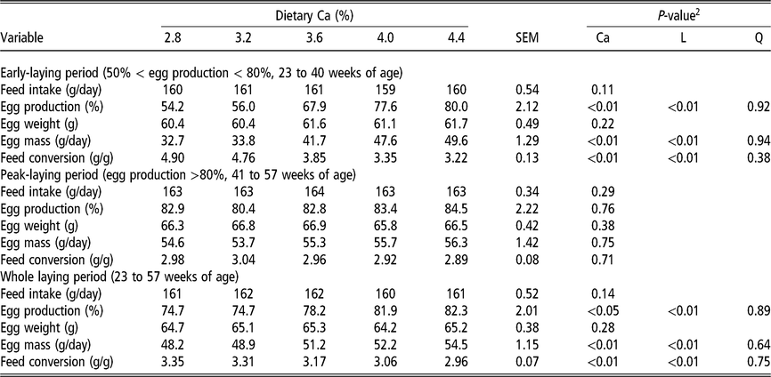
As shown in Table 2, the egg production and egg mass of breeder ducks linearly increased as dietary Ca levels increased from 2.8% to 4.4%, and a linear (P <0.01) improvement in FCR was obtained during the early- and entire laying periods. There were no differences in the laying performance of laying duck breeders during the peak laying period. In addition, the different Ca levels had no significant effects on the feed intake throughout the experimental periods.
Effects of dietary Ca levels on laying performance of duck breeders aged 23 to 57 weeks1

SEM = standard error of the mean.
1 Data are means for n = 6 replicates (15 birds/replicate).
2 Ca = treatment effect; L = linear; Q = quadratic.
Eggshell quality
The effects of dietary Ca levels on the eggshell quality of duck breeders are shown in Table 3. Eggshell thickness and eggshell content increased (linear, P <0.01) as the dietary Ca levels increased, and a quadratic response (P <0.05) was observed in the eggshell thickness. According to the regression model of eggshell thickness in relation to dietary Ca levels, the optimal Ca level of 3.86% was reached.
Effects of dietary Ca levels on eggshell quality of duck breeders aged from 23 to 57 weeks1

SEM = standard error of the mean.
1 Data are means for n = 6 replicates (3 eggs/replicate).
2 Ca = treatment effect; L = linear; Q = quadratic.
3 Regression equation based on dietary Ca level (%); quadratic equation: Y (eggshell thickness) = 0.17 + 0.08 (Ca) – 0.01 (Ca)2; R 2 = 0.58; P-value <0.01 yielded the optimized dietary Ca value of 3.86%.
Incubation indices
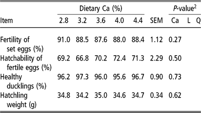
The incubation indices of Longyan-duck breeders were unaffected by the tested dietary Ca levels (Table 4).
Effects of dietary Ca levels on the incubation indices of duck breeders aged 41 weeks1

SEM = standard error of the mean.
1 Data are means for n = 6 replicates (50 eggs/replicate).
2 Ca = treatment effect; L = linear; Q = quadratic.
Ovary-related indices
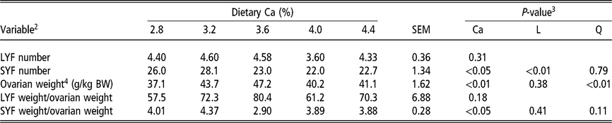
As shown in Table 5, a quadratic effect of ovarian weight was observed in response to the graded dietary Ca levels, and the highest ovarian weight was obtained with 3.48% Ca according to the regression model. The SYF number and SYF weight/ovarian weight were affected by dietary Ca levels, and the linear response was significant in relation to the SYF number.
Effects of dietary Ca on reproductive organ indices for duck breeders aged 57 weeks1

SEM = standard error of the mean.
1 Data are means for n = 6 replicates (2 birds/replicate).
2 LYF = large yellow follicles, diameter > 8 mm; SYF = small yellow follicles, diameter = 3 to 8 mm.
3 Ca = treatment effect; L = linear; Q = quadratic.
4 Regression equation based on dietary Ca level (%); quadratic equation: Y (ovarian weight) = −86.1 + 71.7 (Ca) – 9.80 (Ca)2; R 2 = 0.29; P-value = 0.01 yielded the optimized dietary Ca value of 3.48%.
Tibial quality
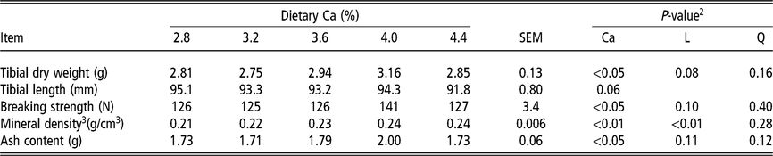
The tibial quality indices are summarized in Table 6. Of the examined indices, the tibial dry weight, breaking strength, and ash content were affected by dietary Ca levels, and the highest values were obtained with the level 4.0% Ca. The tibial mineral density linearly (P <0.01) increased as dietary Ca levels increased, and the optimal Ca level for tibial mineral density was obtained with 4.00% according to the regression model.
Effects of dietary Ca levels on tibial quality of duck breeders aged 57 weeks1

SEM = standard error of the mean.
1 Data are means for n = 6 replicates (2 birds/replicate).
2 Ca = treatment effect; L = linear; Q = quadratic.
3 Regression equation based on dietary Ca level (%); broken linear equation: Y = 0.24 – 0.025 × (4.00 – Ca), Ca ≤4.00; Y = 0.24, Ca >4.00; R 2 = 0.99; P-value <0.01 yielded the optimized dietary Ca value of 4.00%.
Chemical analysis of plasma, uterus, eggshells and tibiae
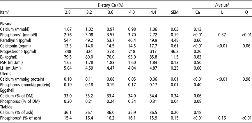
Plasma P concentration and tibial P content exhibited a quadratic (P <0.01) response to dietary Ca levels. According to the regression models, the highest plasma and tibial P concentrations were obtained with 3.47% and 3.49% Ca, respectively. The Ca concentration in the uterus linearly (P <0.01) decreased as dietary Ca levels increased. In addition, the plasma CT concentration linearly (P <0.01) increased as dietary Ca levels increased (Table 7).
Effects of dietary Ca levels on chemical analysis of plasma, uterus, eggshell and tibiae of duck breeders aged 57 weeks1

SEM = standard error of the mean.
1 Data are means for n = 6 replicates (2 birds/replicate).
2 E2 = estradiol-17β; FSH = follicle-stimulating hormone; LH = luteinizing hormone.
3 Ca = treatment effect; L = linear; Q = quadratic.
4 Regression equation based on dietary Ca level (%); quadratic equation: Y (plasma phosphorus) = −14.0 + 9.62 (Ca) – 1.32 (Ca)2, R 2 = 0.29, P-value <0.01; Y (tiabial phosphorus) = 2.16 + 7.72 (Ca) – 1.05 (Ca)2, R 2 = 0.28, P-value <0.01 yielded the optimized dietary Ca values of 3.47% and 3.49%, respectively.
Transcript abundance of matrix protein genes in the uterus
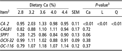
With the exception of CA 2 relative expression in the uterus, no differences were observed in the matrix protein gene expression indices due to the dietary Ca levels. There were both linear and quadratic effects of CA 2 relative expression in response to dietary Ca levels, and the highest CA 2 relative expression was obtained with 3.2% Ca.
Discussion
The findings of the present study confirm that the tested dietary Ca levels affect egg production, eggshell quality, ovary development and tibial quality of duck breeders. Certainly, numerous studies demonstrated that Ca-deficient diets impaired the reproductive organs and decreased the egg production of hens (de Bernard et al., Reference de Bernard, Stagni, Camerotto, Vittur, Zanetti, Zallone and Teti1980; Abdallah et al., Reference Abdallah, Harms and EL-Husseiny1993). Chen et al. (Reference Chen, Zhao, Tian, Zhang, Ruan, Li, Wang, Zheng and Lin2015) showed that the egg production and the egg mass of laying ducks were reduced to 84% and 47% of control (3.6% Ca) for 1.8% and 0.38% Ca, respectively. In laying hens, Safaa et al. (Reference Safaa, Serrano, Valencia, Frikha, Jimenez-Moreno and Mateos2008) found that Lohmann Brown laying hens (58 weeks of age) that were fed a diet containing 4.0% Ca had higher egg production, egg mass and better FCR than those fed 3.5% Ca. In addition, similar to our results (Table 2), the latter authors found that the daily feed intake was not affected by 3.5% to 4% dietary Ca levels. The findings of Cufadar et al. (Reference Cufadar, Olgun and Yildiz2011) suggest that molted brown laying hens required 3.6% Ca in the diet to maintain their normal reproductive performance. These latter studies indicate that the optimal dietary Ca level is well established for the maximum productive performance of laying birds. In the current study, the results of egg-type duck breeders showed that increasing the dietary Ca levels from 2.8% to 4.4%, which was accompanied with a linear increase in plasma CT concentration, linearly increased the egg production, egg mass and SYF number, with linearly decreased FCR (Table 2, Table 5 and Table 7). This result, therefore, suggests that the diets with <4.4% Ca level yielded an inferior productive performance in duck breeders. However, the maximal ovarian weight, which is one of the most important indices of the reproductive system, was obtained with 3.48% Ca according to the regression model (Table 5), thereby indicating that 3.48% Ca facilitates follicles development in duck breeders. This goes in line with the recommendation of 3.5% Ca for breeder hens in Ministry of Agriculture of the People’s Republic of China (2004) and exceeds the US National Research Council (1994) suggestion with a Ca level of 2.75% for breeder Pekin ducks.
In the 60 g shelled egg of laying hens, approx. 60% to 75% of Ca is obtained from the diet and the remainder Ca (25% to 40%) stems from the body reserves (Comar and Driggers, Reference Comar and Driggers1949; Mueller et al., Reference Mueller, Schraer and Schraer1964). It is known that medullary bone is a woven bone that acts as a labile source of Ca for eggshell formation (Whitehead, Reference Whitehead, McCormack, McTeir and Fleming2004). In laying hens, the results of Roland et al. (Reference Roland, Bryant, Rabon and Self1996) indicated that increasing the dietary Ca level up to 5% increased tibial density and breaking strength without any adverse effect on egg quality indices. A similar result was obtained in the present study, where the highest tibial dry weight, breaking strength and ash content were achieved when duck breeders fed a diet containing 4.0% Ca (Table 6). Ca and P are important for bone development and mineralization in the form of hydroxyapatite (99% Ca, 80% P) (Veum, Reference Veum, Vitti and Kebreab2010). Bone mobilization occurs and Ca ion releases to fulfill Ca requirement when Ca supplied from feed is insufficient, with increasing concentration of plasma P during the time of shell formation (Hurwitz and Bar, Reference Hurwitz and Bar1965). In the present study, the highest plasma and tibial P concentrations were reached when the birds fed the diets containing 3.47% and 3.49% Ca according to the regression model (Table 7), respectively. This finding suggests that using 3.47% Ca is insufficient for maximum eggshell formation in egg-type duck breeders. Furthermore, the results indicated that the dietary Ca level (3.86%) is optimal to maximize eggshell thickness. The best relative eggshell weight was reached with 4.0% and 4.4% dietary Ca, but there were no beneficial effects of the levels higher than 4.0% Ca on the other shell quality indices. When the Ca supply is excessive and concentrations in the gut exceed 1 mmol/L, the homeostatic mechanism is reversed by the secretion of CT; this inhibits the intestinal activation of VD3, which lead to reduction in Ca absorption in the intestine (Beckman et al., Reference Beckman, Goff, Reinhardt, Beitz and Horst1994). This is consistent with the remarkable increase of plasma CT with 4.4% Ca in the current study; however, the high egg mass obtained in this group (4.4% Ca) implies that higher amounts of Ca and P, either from dietary supplementation or bone mobilization, have been used in egg and eggshell formation versus the other groups. In addition, the increased tibial ash content could be associated with increased contents of other minerals (e.g. Mn, Mg, Zn). Significant linear improvements in eggshell strength and thickness were observed with increasing dietary Ca levels from 3.5% to 4.7% in 70-week-old Hy-Line Brown layers (An et al., Reference An, Kim and An2016). In molted laying hens, Cufadar et al. (Reference Cufadar, Olgun and Yildiz2011) noted that the level of dietary Ca had no significant effect either on eggshell breaking strength or on eggshell thickness. Much amount of Ca ion is usually excreted from the shell gland combined with the bicarbonate ion in the form of Ca carbonates, which are deposited in the shell matrix (Hunton, Reference Hunton2005). This could imply why increasing the dietary Ca level from 2.8% to 4.4% linearly decreased free Ca ion concentrations in the uterus (Table 7).
The formation of CaCO3 predominantly occurs in the presence of carbonic anhydrase (CA) enzyme, where the bicarbonate ions are mainly formed in the glandular cells from metabolic CO2 in a reaction catalyzed by CA: CO2 + H2O ↔ HCO3− + H+ (Nys et al., Reference Nys, Hincke, Arias, Garcia-Ruiz and Solomon1999). In the present study with egg-type duck breeders, a quadratic effect was observed on the CA 2 relative mRNA expression in the uterus in response to increased dietary Ca levels (Table 8). In a recent study, Chen et al. (Reference Chen, Zhao, Tian, Zhang, Ruan, Li, Wang, Zheng and Lin2015) found that using calcium-deficient diets, containing 1.8% or 0.38% Ca, decreased the relative expression of CA 2 in the uteruses of laying ducks. Considering the latter result along with the increasing tendency of Ca content in eggshell, this indicates that the deposition of CaCO3 crystallites in eggshell is dependent on the optimal dietary Ca level, which goes in harmony with the obtained results of eggshell thickness (Table 3). SPP1 (also named osteopontin) is a glycosylated and highly phosphorylated protein that is characterized by Ca ion transport in bone, kidney and uterus. As one of the major phosphorylated proteins of the avian eggshell matrix localized specifically in the palisade and mammillae layers (Mann et al., Reference Mann, Olsen, Macek, Gnad and Mann2007; Fernandez et al., Reference Fernandez, Escobar, Lavelin, Pines and Arias2003), SPP1 expression in chicken uterus is stimulated by the presence of the egg (Brionne et al., Reference Brionne, Nys, Hennequet-Antier and Gautron2014) and mechanical distension of the uterine wall (Lavelin et al., Reference Lavelin, Yarden, Ben-Bassat, Bar and Pines1998). The results of the current study point out that the levels of SPP1 relative mRNA expression in the uterus could be associated with the plasma LH levels, where both of them showed a tendency to decrease with an increase in the dietary Ca level (Table 8).
Effects of dietary Ca levels on the mRNA expression levels of matrix protein genes in the uterus of duck breeders aged 57 weeks1

SEM = standard error of the mean.
1 Data are means for n = 6 replicates (2 birds/replicate).
2 CA 2 = carbonic anhydrase 2; CALB1 = calbindin 1; SPP1 = secreted phosphoprotein 1; OCX-32 = ovocalyxin-32; OC-116 = ovocalyxin-116.
3 Ca = treatment effect; L = linear; Q = quadratic.
It is concluded that, in egg-type duck breeders, the egg production and egg mass linearly increased as dietary Ca levels increased from 2.8% to 4.4%. In addition, 4.4% Ca seems to be high for the duck breeders, where the birds fed a diet with 4.0% Ca showed superior eggshell and tibial quality. The birds fed a diet with 3.6% Ca obtained the heaviest ovarian weight. The regression analysis revealed that the optimal dietary Ca requirements for maximum eggshell thickness, ovarian weight and tibial mineral density were 3.86%, 3.48% and 4.00%, respectively.
Acknowledgements
This work was supported by the Earmarked Fund for the National Key Research and Development Program (Grant No. 2018YFD0501504), the China Agriculture Research System (Grant No. CARS-42-13), the Science and Technology Program for Pearl River Nova of Guangzhou City (Grant No. 201710010159), Key Project of the Science and Technology Program of Guangzhou City (Grant No. 201804020091).
Declaration of interest
No potential conflict of interest declared.
Ethics statement
All of the procedures employed in this study were approved by the Animal Care and Use Committee of Guangdong Academy of Agricultural Sciences.
Software and data repository resources
None of the data were deposited in an official repository.