Introduction
Partial arrhinia (hyporhinia) is a rare congenital nasal anomaly with only a few cases reported in the literature.Reference Baruah, Dubey, Gupta and Kumar1 The cause of its occurrence is still unknown with a few cases being familial and a few sporadic.Reference Hou2 It is often found to be associated with other malformations of the craniofacial area, central nervous system anomalies, cleft palate and ear anomalies, which in turn can lead to problems with feeding, airway obstruction and phonetics.
Case report
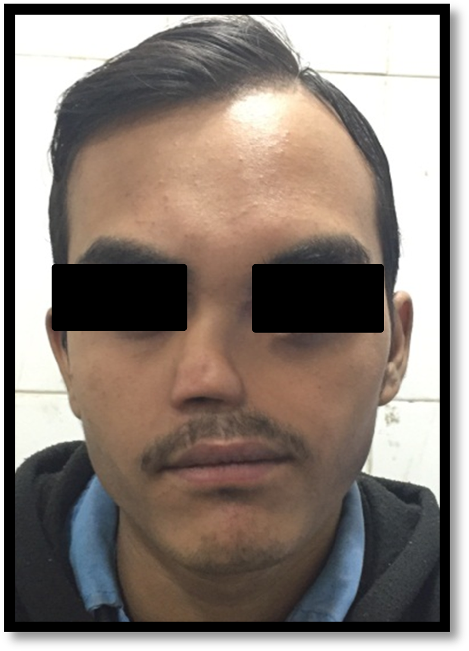
We report a rare case of a 20-year-old male presenting with left-sided facial deformity, left-sided epiphora and intermittently discharging fistula at the medial canthus of the left eye (Figure 1). There were no vision-related, hearing-related, feeding or breathing problems. There was no family history of any congenital malformation. He was born at term by normal vaginal delivery out of a non-consanguineous marriage. The mother does not recall any antenatal problems or neonatal problems in the child except facial deformity. Clinical examination showed a depressed nasal bridge with single anterior nasal nare and flattening of the facial contour on the left side.
A 20-year-old male with left-sided facial deformity.

Imaging was done using multi-detector computed tomography (CT) using thin 0.9 mm sections for evaluation of the nasal cavity and paranasal sinuses followed by CT dacryocystography. The external opening of discharging fistula at medial canthus of the left eye was cannulated, and multi-detector CT was performed after introduction of contrast to delineate the fistulous tract.
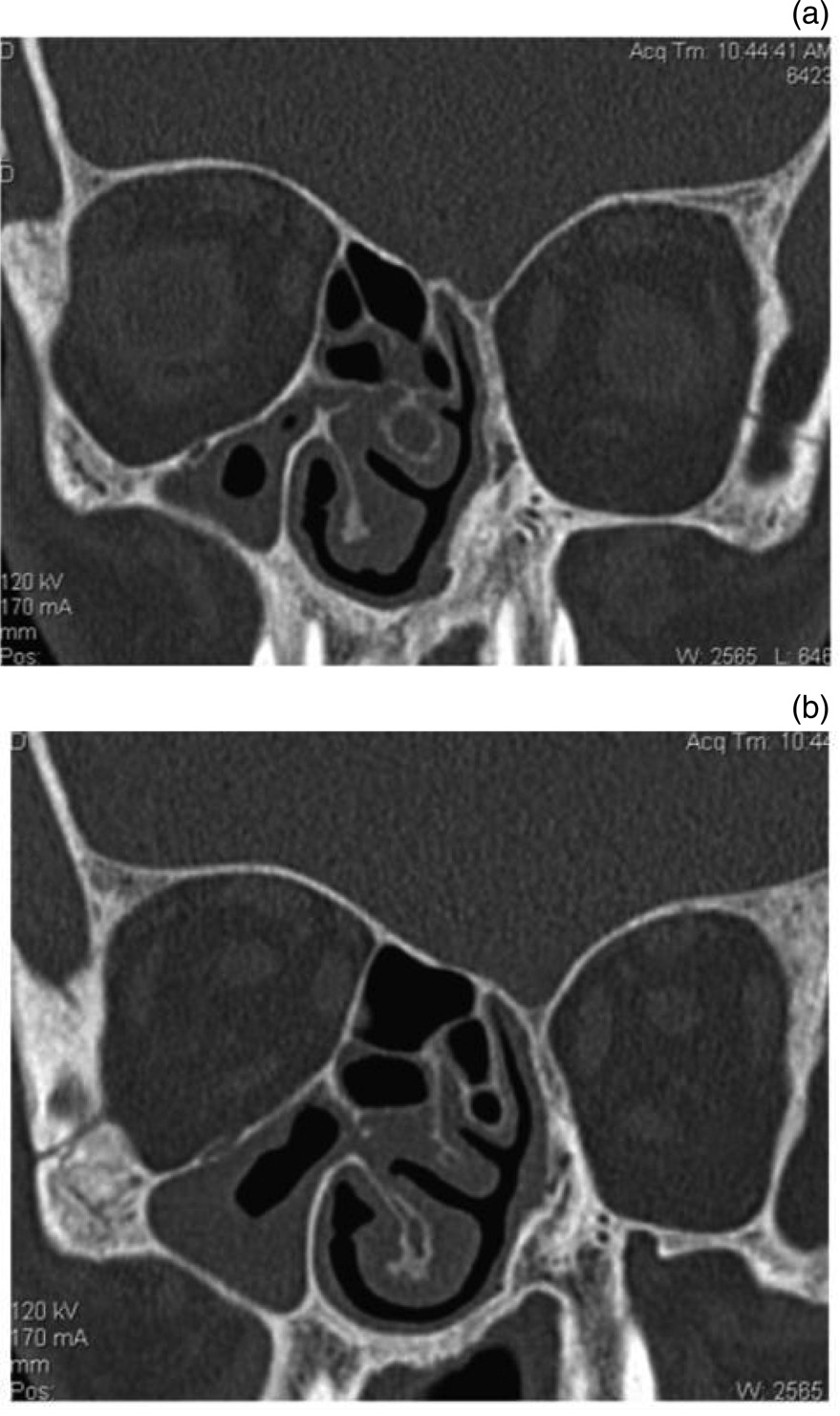
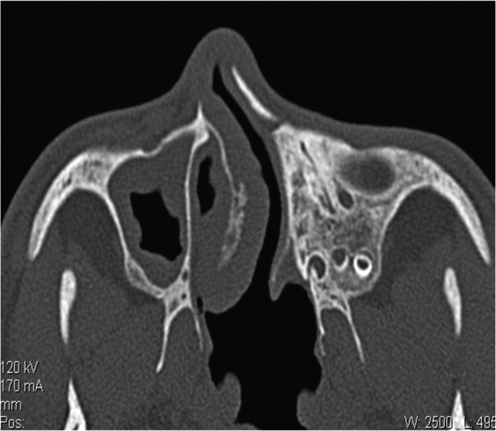
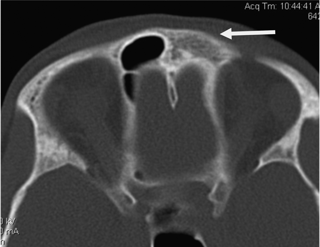
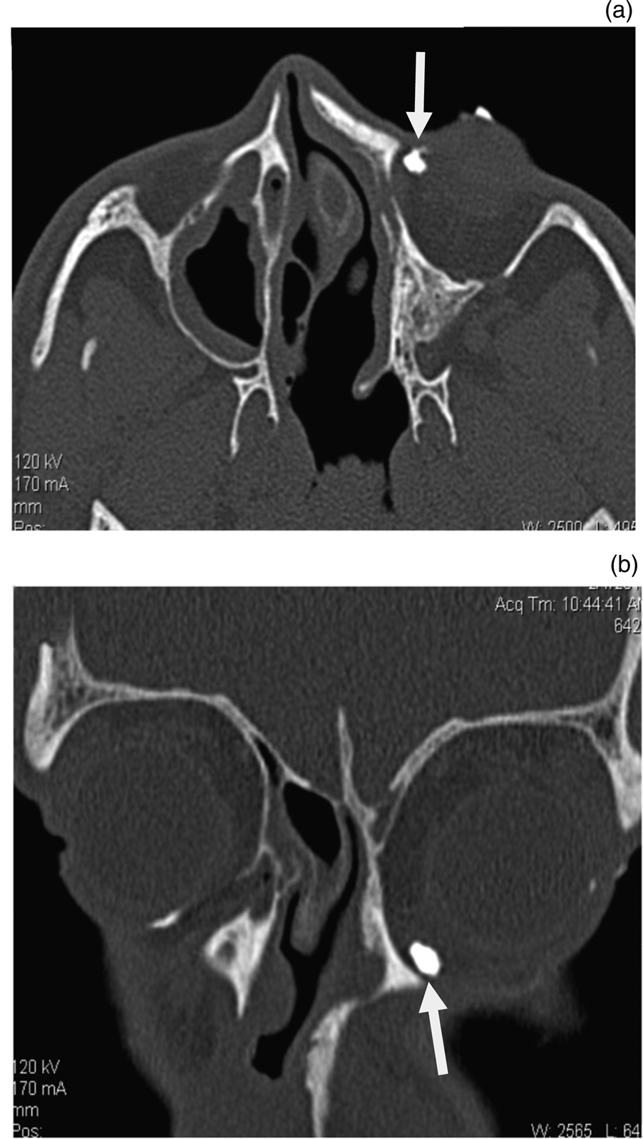
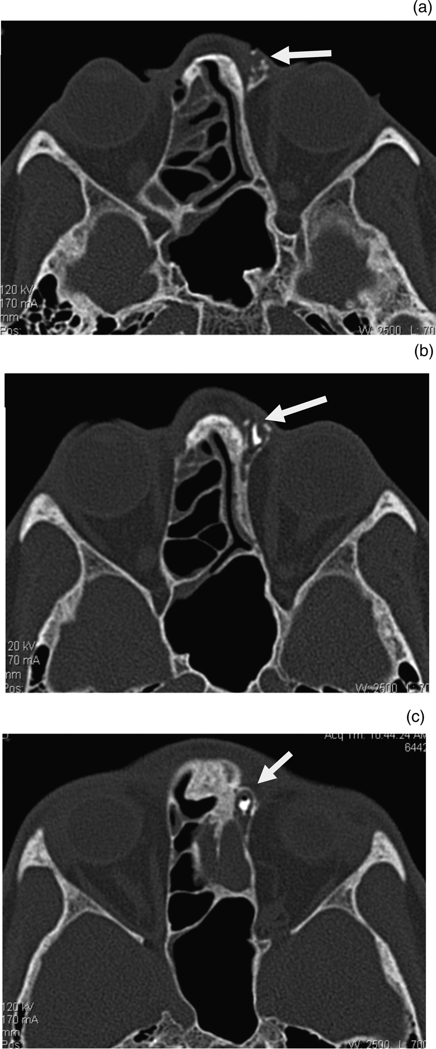
Multi-detector CT showed left-sided nasal atresia and atretic ipsilateral paranasal sinuses (Figures 2, 3 and 4). Singular right nasal cavity with patent posterior choana communicating with the nasopharynx was seen (Figure 3). On CT dacryocystography, there was non-opacification of the left nasolacrimal duct (Figure 5). A fistulous tract was also seen at the left medial canthus communicating with the ipsilateral frontal recess (Figure 6).
Multi-detector computed tomography coronal reformats of paranasal sinuses show (a) non-development of left nasal cavity along with (b) atretic left maxillary sinus.

Multi-detector computed tomography axial section of paranasal sinuses shows non-development of the left nasal cavity (singular nasal cavity seen) along with atretic left maxillary sinus.

Multi-detector computed tomography axial section of paranasal sinuses shows atretic left frontal sinus. The arrow indicates the atretic left frontal sinus, which ideally should have been filled with air (black); however, there was also bone in its place.

(a) Axial plane image (arrow indicates proximal end of the nasolacrimal duct) and (b) coronal plane image (arrow indicates proximal end of the nasolacrimal duct) from computed tomography dacryocystography show non-passage of contrast beyond left lacrimal sac.

Multi-detector computed tomography axial sections of paranasal sinuses after cannulation of external opening show opacification of fistulous communication with left frontal recess. (a) Image shows contrast in left frontal recess with arrow indicating opening of frontal recess, (b) image shows contrast in external opening of left medial canthus with arrow indicating opening of frontal recess near medial canthus of left eye and (c) image shows contrast in external opening of left medial canthus with arrow indicating the external opening with pooling of dense contrast in it. Note the single enlarged sphenoid sinus.

Discussion
The embryological process of facial development includes cranial neural crest cells migrating from the trigeminal nerve region to the face, which occurs between the third and tenth weeks of gestation.Reference Lee and Lee3 Nasal placode forms as a focal thickening of the surface of the ectoderm and develops from the frontal process advancing laterally between the medial and lateral nasal processes. The nasal nuclei are formed at the fifth week by invagination of nasal placode. Nostrils develop from the nasal nuclei, which migrate posteriorly to form nasal cavities. The nasal septum develops at the ninth week when the palate and inferior septum unite and form the secondary palate. Hard palate develops by the eighth or ninth week, and the soft palate formation finishes by eleventh or twelfth week.
Various causal mechanisms for arrhinia are described including underlying chromosomal abnormalities, perinatal insult like maternal drug abuse and environmental factors; however, exact aetiology is still elusive. According to the proposed theory, arrhinia could develop because of a defect in the medial and lateral nasal processes or overdevelopment and early fusion in the medial nasal processes.Reference Albernaz, Castillo, Mukherji and Ihmeidan4 Abnormal migration of neural crest epithelial cells or arrest of absorption of the nasal epithelial plates during weeks 13–15 are other possible mechanisms leading to arrhinia.Reference Akkuzu, Akkuzu, Aydin, Derbent and Ozluoglu5
Congenital arrhinia is very rare with most cases being sporadic, although a few familial cases have also been reported. A variety of anomalies such as coloboma of olfactory bulbs, microphthalmia, coloboma of the iris, absence of the paranasal sinuses, absent or occluded nasolacrimal duct, high arched palate, cleft palate, hypotelorism, hypertelorism, microtia, midline central nervous system defects, umbilical hernia, hypospadias and syndactyly may accompany arrhinia.Reference Olsen, Gjelland, Reigstad and Rosendahl6,Reference McGlone7 Imaging is essential for planning further surgical management. Computed tomography of the nose and paranasal structures is required for detecting associated structural abnormalities. Magnetic resonance imaging is required for evaluating intracranial midline central nervous system defects. In symptomatic neonates, treatment aims at providing nursing care for breathing and feeding problems with surgical correction of arrhinia. Placement of a tracheostomy tube for severe airway obstruction and nasogastric or gastrostomy tube for feeding is performed. A multidisciplinary team consisting of a radiologist, otorhinolaryngologist, plastic surgeon and prosthodontist is required for planning and reconstruction of arrhinia.
• Congenital arrhinia is very rare with most cases being sporadic
• A variety of anomalies may accompany arrhinia
• Our case was a sporadic variety of congenital partial arrhinia with ipsilateral atresia of the paranasal sinuses and nasolacrimal duct
• A multidisciplinary team consisting of a radiologist, otorhinolaryngologist, plastic surgeon and prosthodontist is required for management of arrhinia
Our case was of a sporadic variety of congenital partial arrhinia with ipsilateral atresia of paranasal sinuses and nasolacrimal duct. We considered our patient as having partial arrhinia because there were some remnants of the external nose present on the affected side. The uniqueness of this case lies in the absence of breathing or feeding problems leading to delay in seeking treatment by the patient and adult presentation of a rare face-deforming congenital anomaly.
Competing interests
None declared