Introduction
The diagnosis of cholesteatoma is a clinical one generally made on history and otoscopy, or confirmed at operation. Once a cholesteatoma is removed surgically, simple observation may not detect residual disease in some situations, for instance after canal wall up tympanomastoidectomy or mastoid cavity obliteration procedures.
Canal wall up mastoidectomy has grown in popularity, partly as it does not leave the patient with a cavity requiring subsequent care and water exclusion. After primary surgery, the general practice is to perform second-look surgery to exclude residual disease, usually after approximately one year. The residual cholesteatoma rate certainly varies amongst surgeons, but is often quoted as being in the region of 10–20 per cent.Reference Robinson1 This means that a large proportion of patients undergoing second-look procedures could in fact have avoided this procedure if it were possible to accurately detect cholesteatoma pre-operatively. Extra surgery brings additional risks to the patient, and further health care costs. Cholesteatoma removal may also be followed by mastoid cavity obliteration in the same surgical sitting; here, the concern is that residual skin may be trapped in a site that cannot be observed in the clinic, or easily viewed in a second-stage procedure.
Both of these situations call for an imaging technique that can reliably detect even small pearls of residual cholesteatoma. Single-shot, turbo spin-echo, diffusion-weighted magnetic resonance imaging (MRI), pioneered by a Belgian group, shows potential as the imaging modality of choice in this situation.Reference De Foer, Vercruysse, Bernaerts, Maes, Deckers and Michiels2 Our centre, like many others, has begun to use similar sequencing (in our case, a propeller, diffusion-weighted image sequence on a GE Signa scanner) to determine its accuracy in detecting residual cholesteatoma, in patients who are already committed to undergoing surgery. This enables retrospective analysis of whether the imaging findings correctly predicted those found at surgery, taking the latter as the ‘gold standard’. The question that remains is how accurate the technique needs to be in order to negate the need for exploratory surgery, and what are the implications of false negative results?
Case report
A 41-year-old woman presented with long-standing chronic otitis media. By the time of initial presentation to our tertiary referral clinic, she had undergone surgery on both ears. On the right side, there was a modified radical mastoid cavity with tympanic membrane perforation. On the left, there was a deep attic defect and grossly retracted tympanic membrane. Both ears were discharging intermittently and had severe to profound mixed hearing loss.
The senior author (BW) subsequently cleared the right cavity of residual cholesteatoma and partially obliterated the cavity with bone pâté and a Palva flap (at the time of writing, this had remained dry for five years). The left cavity was further modified, elsewhere, with removal of cholesteatoma, but it was still discharging on an intermittent and frequent basis when the patient re-presented to our clinic approximately four years later.
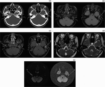
Due to the patient's concurrent hearing deficit, she was assessed for cochlear implantation candidacy. Both ears met the audiological criteria for implantation. The patient reported still receiving a small benefit from a conventional hearing aid on the right side, without aggravating that cavity. For this reason, and due to continued discharge from the left side, the latter ear was chosen for revision tympanomastoidectomy and cochlear implantation. A pre-operative computed tomography (CT) scan demonstrated a soft tissue mass and bony erosion, in keeping with cholesteatoma (Figure 1a). At surgery, extensive residual cholesteatoma was discovered and removed, and a Fisch closure of the ear canal with fat obliteration of the cavity was performed. A section of the descending mastoid portion of the facial nerve was dehiscent, so a Silastic® sheet was left in situ to aid subsequent safe access to the round window niche.
(a) Pre-operative, axial computed tomography scan (bone algorithm, slice thickness 0.625 mm): a soft tissue mass is seen in the right middle ear, corresponding to the sinus tympani, where residual cholesteatoma was found (arrow 1); on the left side, the anterior limb of the superior semicircular canal is dehiscent with some loss of attenuation (arrow 2), corresponding to the location of the subsequently discovered residual cholesteatoma, possibly represented by an adjacent soft tissue mass visible in the middle-ear space. (b) Axial, T1-weighted, fat-saturated magnetic resonance imaging (MRI) scan (echo train = 3, repetition time = 500 ms, echo time = 31.12 ms; 2 mm contiguous slices): the right ear lesion shows some hyperintensity with patches of more central isointensity (arrow 1); on the left side, hyperintense signal is seen lateral to the void left by the retained Silastic sheet which represents the pus and infected tissue found at re-exploration (arrow 2), while the soft tissue adjacent to the superior semicircular canal is isointense. (c) Axial, T1-weighted, post-gadolinium, fat-saturated MRI: the soft tissue mass in the right ear does not enhance, excluding granulation tissue (arrow 1), but does show rim enhancement, suggestive of cholesteatoma; on the left side, the soft tissue lateral and medial to the Silastic sheet is hyperintense, more indicative of inflammatory tissue (arrow 2). (d) Axial, T2-weighted, fast imaging employing steady-state acquisition (FIESTA) MRI (echo train = 1, repetition time = 5.3 ms, echo time = 1.98 ms; 1.4 mm slices, interpolated to 0.7mm): there is a hyperintense signal for the right middle-ear mass (arrow 1); in the left ear (at the site of the now known residual cholesteatoma and superior semicircular canal fistula), the soft tissue mass fails to show the hyperintensity expected in residual cholesteatoma (arrow 2), there is some loss of signal within the anterior limb of the canal but no hyperintensity, and the void left by the Silastic sheet can be seen (arrow 3) with a lateral hyperintense signal where pus was subsequently found. (e) Axial, propeller, diffusion-weighted MRI (echo train = 24, repetition time = 8000 ms, echo time = 131.32 ms; b-value 1500): the right ear mass shows a high signal which, when taken in context with the signal characteristics described above, makes this lesion highly likely to be residual cholesteatoma (subsequently confirmed at surgery) (arrow 1); no such signal change is seen on the left side, at the other known site of residual cholesteatoma (arrow 2).

One year later, prior to cochlear implant insertion, a pre-operative MRI was performed to assess whether any further residual cholesteatoma was present in the left ear (Figure 1b to 1e). A propeller, diffusion-weighted image sequence (which provides similar images to the single-shot, turbo spin-echo, diffusion-weighted sequence) was obtained on a GE Healthcare Signa twin-speed scanner (1.5 Tesla; General Electric, USA). The left side showed the presence of soft tissue within the cavity, but this lacked the signal characteristics suggestive of residual skin.
At subsequent surgery, frank pus was found in relation to the Silastic sheet, in addition to residual cholesteatoma lateral to and extending into the superior semicircular canal, measured intra-operatively as approximately 2 × 2 × 3 mm in size. Removal involved partial labyrinthectomy. The residual cholesteatoma, together with the active infection, prevented insertion of the cochlear implant.
The pre-operative imaging was subsequently reviewed in light of the operative findings. However, even when knowing exactly where to look on the scans, the residual cholesteatoma could still not be appreciated (Figure 1). What was noted in retrospect, however, was residual cholesteatoma on the right side, behind the tympanic membrane, tight into the sinus tympani. Given that this side was now the preferred site for cochlear implant insertion, this provided useful information for planning that surgery. Cholesteatoma was subsequently confirmed at this location, removed and the patient successfully implanted on the right side.
Discussion
Accurate pre-operative imaging of the temporal bone can help in planning surgery and assessing potential risks to the patient. An imaging modality that could reliably and accurately differentiate a cholesteatoma lesion as small as 1 or 2 mm may negate the need for second-look surgery following canal wall up tympanomastoidectomy.
Computed tomography is the most commonly used temporal bone imaging technique prior to cholesteatoma surgery. Its use is becoming more widespread as fine slice scans provide practical information on bony anatomy, and can accurately demonstrate a breach of the tegmen or a fistula of the semicircular canals. However, CT scanning gives poor soft tissue information and fails to differentiate cholesteatoma from mucosal hypertrophy and inflammatory tissue, cholesterol granuloma, granulation tissue, and retained secretions. Some authors have advocated the use of CT to initially assess residual disease one year after canal wall up tympanomastoidectomy. A well aerated middle-ear mastoid complex, without soft tissue mass within the middle ear or mastoid cavity, is highly suggestive of a lack of residual disease and so can negate the need for second-look surgery.Reference Ayache, Williams, Lejeune and Corre3 However, CT scanning involves a dose of ionising radiation, the post-operative mastoid cavity may not be aerated, and soft tissue signals cannot be easily differentiated.
Magnetic resonance imaging is a better differentiator of soft tissue and does not involve ionising radiation. Diffusion-weighted, echo-planar imaging has been shown to accurately distinguish cholesteatoma from inflammatory tissue.Reference Vercruysse, De Foer, Pouillon, Somers, Casselman and Offeciers4 Combined with CT information, this MRI scanning sequence can be useful when assessing an ear prior to primary surgery. Whilst its sensitivity and specificity for lesions larger than 5 mm have been reported as 100 and 88 per cent, respectively, it is much less accurate in detecting cholesteatoma lesions smaller than 5 mm.Reference Venail, Bonafe, Poirrier, Mondain and Uziel5 This is due in part to issues of lower resolution, higher slice thickness and a susceptibility to artefacts at the cranial base.
Late post-gadolinium, T1-weighted MRI sequences have also been used before second-look surgery, with some success, to reliably demonstrate lesions larger than 3 mm; one series showed a sensitivity and specificity of 100 and 75 per cent, respectively, at this size or larger.Reference Ayache, Williams, Lejeune and Corre3, Reference Venail, Bonafe, Poirrier, Mondain and Uziel5 The keratin debris of the cholesteatoma sac is avascular and so will not enhance, whilst inflammatory and scar tissue will enhance, albeit rather slowly, allowing distinction between these tissues. Cholesteatoma will show rim enhancement due to the cuff of inflamed mucosa that surrounds it. The use of gadolinium is problematic (requiring intravenous injection, and having the potential for nephrogenic systemic fibrosis in patients who have severe renal dysfunction and a glomerular filtration rate of <30 ml/min), as is the long duration of the study (sequences are taken approximately 45 minutes post-contrast).Reference Padilla-Thornton, Zand, Barrett, Stein, Andrew and Forster6
Propeller, diffusion-weighted imaging is a multi-shot, fast spin-echo acquisition that does not collect data line-by-line but rather as ‘blades’ of information collected by periodically rotating parallel lines during each ‘repetition time’ period. This reduces susceptibility artefacts, in part by collecting a large amount of overlapping data, and also provides a more signal intensity rich image, enabling better contrast and easier visualisation of cholesteatoma lesions.Reference Lehman, Saliou, Brochart, Page, Deschepper and Vallée7 Propeller, diffusion-weighted imaging has a relatively short echo train of 24, which reduces the blurring that can be seen in single-shot, fast spin-echo sequences. A wide receiver bandwidth allows for decreased echo spacing, which also reduces overall blurring, whilst a ‘echo time’ of 84 ms and a ‘repetition time’ of 5700 ms give good T2 sensitivity. Fast spin-echo based sequences reduce magnetic susceptibility effects, seen on echo-planar imaging sequences, which is important in the petrous region because of the large differences in susceptibility caused by the many air–bone interfaces. Propeller, diffusion-weighted imaging also has a b-value of 1500, which serves to increase the sensitivity to diffusion with a small loss of signal-to-noise ratio.
De Foer et al. used the non-echo-planar image based diffusion-weighted sequence (i.e. single-shot, turbo spin-echo, diffusion-weighted image), similar to the propeller, diffusion-weighted image sequence used in the current case.Reference De Foer, Vercruysse, Bernaerts, Maes, Deckers and Michiels2 They assessed pre-operatively patients for whom there was a strong clinical suspicion of cholesteatoma but no previous ear surgery. Radiologists blinded to the clinical data analysed the scans to attempt to predict the presence and size of any cholesteatoma. There was a potential for bias in the selection criteria, and there was no control group; however, of the 21 patients studied, 19 had their operative findings correctly predicted from the pre-operative scans, with lesions as small as 2 mm being detected.
Detection of cholesteatoma on these sequences relies on the presence of keratin debris within the cholesteatoma sac. Diffusion-weighted imaging assesses how water moves through tissue; in keratin, the water becomes trapped, appearing as a high intensity signal. In one of the false negative cases of the De Foer et al. series, the sac was found to have auto-evacuated its debris, and this was given as an explanation of the missed case.
A further study by the same group used diffusion-weighted images to detect residual cholesteatoma after primary canal wall up surgery, and correctly detected residual disease in nine of 10 patients, reporting a sensitivity, specificity, positive predictive value and negative predictive value of 90, 100, 100 and 96 per cent, respectively.Reference De Foer, Vercruysse, Bernaerts, Deckers, Pouillon and Somer8 The missed case was blamed on motion artefact. A similar series by Dhepnorrarat and colleagues correctly identified seven cases positive and 16 negative for residual disease prior to second-look surgery, and confirmed different signal characteristics for cholesteatoma versus post-operative mucosal changes.Reference Dhepnorrarat, Wood and Rajan9 Lehman et al. compared propeller, diffusion-weighted imaging with echo-planar, diffusion-weighted imaging and late post-gadolinium, T1-weighted MRI, and were able to demonstrate the best sensitivity, specificity and predictive values for the former technique, along with the least inter-observer variability (the smallest residual cholesteatoma detected measured 3 mm).Reference Lehman, Saliou, Brochart, Page, Deschepper and Vallée7
An important point to clarify is that MRI scanner sequences have different names depending upon the type of machine used. Siemens machines produce the single-shot, fast spin-echo, non-echo-planar, diffusion-weighted sequence, whilst the GE Healthcare system produces similar information by using the propeller, fast spin-echo, non-echo-planar, diffusion-weighted sequence. There appears to be no published data comparing the two systems.
A criticism of the diffusion-weighted image technique is its inability to clearly show the bony anatomical features of the temporal bone. This makes precise localisation of any signals suggesting cholesteatoma somewhat difficult. However, this is not the primary goal of the investigation. It is the accurate prediction of the presence or absence of cholesteatoma that is required, as this will determine the need for second-look surgery. If localisation is required, the findings of diffusion-weighted imaging can be combined with other sequences that better show the anatomy (i.e. standard T2-weighted or late post-gadolinium, T1-weighted sequences).
• After canal wall up mastoid surgery for cholesteatoma, the clinician must determine whether residual disease is present; this is traditionally done by second-look surgery
• Magnetic resonance imaging (MRI) techniques have been employed to detect such disease non-invasively, with variable success; fast spin-echo, non-echo-planar, diffusion-weighted techniques are proving the most reliable to date
• Both Siemens and GE Healthcare MRI scanners are capable of producing such sequencing, but under different names and with subtly different qualities, of uncertain significance
• The presented case demonstrates both the success and failure of these techniques.
• Scan-negative subjects require continued follow up if second-look surgery is to be avoided
We present a patient in whom MRI sequencing failed to predict the subsequent operative findings of residual cholesteatoma in the left ear, yet did so correctly for those in the right ear. Even on retrospective reviewing of the films, the residual cholesteatoma in the left ear could not be identified, despite it being of a size that previous studies imply should be detectable. There were no concerns regarding motion artefact. In contrast, the residual disease on the right side was clearly demonstrated, and this information assisted the planning of the subsequent cochlear implantation. The right-sided residual cholesteatoma was slightly greater in size, which may be significant. It must also have arisen since the last procedure performed on that ear, some five years before the MRI scan.
Perhaps the signal characteristics of maturing residual cholesteatoma alter over time (possibly related to water diffusion through the tissue), accounting for the findings described. It is possible that the keratin content may differ; a low keratin content might have accounted for a lack of restricted diffusion on the side on which our patient's residual cholesteatoma was missed.
The presence of the Silastic sheet might have been significant. Venail et al. reported false positive residual cholesteatoma findings on both diffusion-weighted, echo-planar imaging and late post-gadolinium, T1 images in the presence of Silastic sheeting (although the problem with our non-echo-planar, diffusion-weighted imaging was a false negative rather than false positive finding).Reference Venail, Bonafe, Poirrier, Mondain and Uziel5
The fact that the missed lesion was within a fat-obliterated cavity might also be significant. In retrospect, imaging this patient with a non-fat-suppressed, T1-weighted sequence might have helped to differentiate the soft tissues within the left cavity. The fat-obliterated cavity would have enhanced, but the cholesteatoma should not have done so. However, any findings would still have been confused by the lack of intensity on the diffusion-weighted imaging for the area corresponding to the cholesteatoma.
The question remains, how reliable is this imaging technique after canal wall up tympanoplasty or obliteration techniques, especially in the earlier post-operative stages when residual disease would be expected to be small? In order to avoid second-look surgery and reliably discharge patients from follow up, 100 per cent sensitivity and specificity would be desirable. It is possible that the sensitivity of the diffusion-weighted technique diminishes dramatically as the size of the lesion lessens, and this may be the key learning point. If this technique can be demonstrated to reliably detect larger lesions, an argument could be made to follow scan-negative patients for such time as would allow any residual cholesteatoma to enlarge, be identified and then appropriately treated, with minimal (if any) detriment to the patient's overall outcome.