Introduction
The recent advent of microvascular free flap surgical techniques has enabled us to address functional and cosmetic deficits resulting from the ablation of advanced stage head and neck cancer. The subscapular system is one of the most versatile donor sites used for reconstruction, providing osseous, osteocutaneous, myocutaneous or muscle-only flaps.Reference Urken 1 , Reference Fisher, Patel, Izzard and Kountakis 2 Subscapular flaps pose a positioning challenge for head and neck surgeons, as posterolateral access must be available for harvesting; this is a major barrier to the utilisation of these flaps.Reference Chiaramonte and Nahabedian 3 , Reference Gibber, Clain, Jacobson, Buchbinder, Scherl and Zevallos 4
Several positioning techniques have been used for harvesting subscapular system flaps. The lateral decubitus is the most commonly used positioning technique.Reference Sumner, Fox, Guyuron, Eriksson and Persing 5 The patients are placed on a mouldable ‘bean bag’, which is used for support in turning the patient. The patient must be repositioned and suction must be applied to hold the bag in a new position.Reference O'Connell 6 A major disadvantage of this technique is the need for repositioning and re-preparing, and the inability to perform tandem flap harvesting for head and neck reconstructive surgical procedures.
Baj et al. described the ‘park bench position’, which is a derivation of the lateral decubitus position.Reference Baj, Capparé, Autelitano, De Riu, Beltramini and Segna 7 In this technique, the upper arm is positioned along the trunk laterally and the upper shoulder is taped towards the table. This position allows simultaneous harvesting and resection of the posterior neck and scalp, and of the posterior fossa, without repositioning. However, this position precludes an anterior approach to head and neck tumours, and it is challenging to dissect a neck positioned inferiorly.
Paek et al. recently described harvesting using a ‘dorsal decubitus’ position, which is achieved by placing a cushion along the patient's spine.Reference Paek, Boa, Revol, Servant, Harris and Danino 8 , Reference Paek, Boa, Revol, Servant, Harris and Danino 9 This allows two surgical teams to work simultaneously, while removing the need to change the patient's position intra-operatively. This technique has been used successfully in cases utilising subscapular-based flaps and in many ways is similar to the method we have been using. Despite this innovation, there is still an inferiorly positioned neck and a straight anterior approach is difficult. Patients with a limited range of motion in the neck are not good candidates for this harvesting technique.
Another less commonly used technique is the sitting position, which was first used in neurosurgical practice. It offers the advantages of improved exposure of surgical and donor sites, and anatomical orientation. This position, however, poses significant anaesthesia risks, including potentially fatal air embolism.Reference Albin, Babinski, Maroon and Jannetta 10 , Reference Sekhar, Tzortzidis, Bucur, Sekhar and Oliveira 11
We present a unique preparation and positioning technique for harvesting subscapular system flaps, which we call the ‘sit and tilt’ technique. It is a single-preparation technique that has facilitated flap harvesting with minimal patient repositioning required.
Materials and methods
‘Sit and tilt’ preparation technique
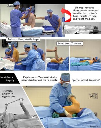
We recommend starting with the patient positioned 180 degrees from the anaesthesia unit in a supine position. Once the patient has been intubated, appropriate vascular access, cardiac monitoring and pulse oximetry equipment is placed so it does not interfere with the surgical site. The donor site and the ipsilateral arm are kept free to prepare the field. The endotracheal tube (ETT) is temporarily disconnected and the patient is placed in a seated position (Figure 1). This exposes the flap preparation boundaries, which include the iliac crest, ipsilateral nipple, near total back and ipsilateral shoulder.
Supine subscapular flap preparation. Photographs depicting the sequential steps for the supine sit preparation (‘prep’) and partial lateral decubitus positions for subscapular flap harvesting. © 2015 Glia Media, printed with permission. ET = endotracheal

Three team members are needed for optimal stabilisation of the patient during the preparation. One team member stabilises the head, neck and ETT, by placing one hand on the back of the head and the other hand over the mouth, grasping the ETT between the fingers. Additional team members support the contralateral back and ipsilateral arm. The back is prepared (scrubbed) to the shoulder, and a sterile drape is placed on the operating table. A shoulder roll and/or headrest may be placed under the sterile drape to improve surgical exposure as needed.
The patient is returned to the supine position while keeping the ipsilateral arm elevated until it is completely prepared, to avoid contaminating the field. The entire ipsilateral arm is then prepared and draped onto an arm board in a supine position. Pressure points on the arm are padded on the arm board prior to draping, and the ipsilateral arm is secured in a supine position with a towel arm sling. The towel sling ensures the arm stays on the arm board securely and in a supine position, to prevent ulnar nerve palsy. This set up and preparation is unique as it also allows for the harvesting of additional radial or ulnar forearm flaps, or the lateral arm flap if needed. The contralateral arm is then padded and tucked. Unlike the lateral decubitus positioning technique, no axillary roll is needed under the contralateral side and no padding is required under the contralateral knee.
The flap donor site is excluded from the head and neck region with drapes. If the posterior aspect of the head and neck is not included in the operative field, then the head will rest on the sterile drape unprepared, and will be excluded once the head and neck are prepared and draped in the usual fashion depending on the procedure.
‘Sit and tilt’ harvesting positioning
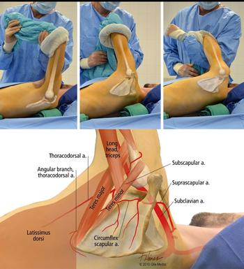
At the time of harvesting, the patient is rolled away from the donor side into a partial lateral decubitus position at the hip and shoulder, to allow placement of two ‘positioning’ stacks of towels (Figure 1). One stack is placed at the midline under the medial border of the scapula, and the second stack is placed at the midline near the hip. This allows the patient to be placed in a partial lateral decubitus position. The placement of the upper stack of towels on the medial border of the scapula is key, as this ‘delivers’ the scapula sufficiently for surgical access (Figure 2).
Scapula position – pedicle anatomy. The patient's arm is manipulated by the assistant to ‘deliver’ the scapula and to position the muscles for optimal identification of the axillary fossa and the triangular space for subscapular flap harvesting. © 2015 Glia Media, printed with permission.

The assistant then holds the ipsilateral arm with the elbow flexed and in line with the shoulder. This positions the muscles for optimal identification of the axillary fossa and triangular space. The assistant may retract or relax the shoulder girdle to facilitate exposure. Alternatively, a Mayo stand or a Spyder arm can be used to facilitate this position (Figure 2).
The subscapular system flap is then harvested in the standard fashion.Reference Paek, Boa, Revol, Servant, Harris and Danino 9 , Reference Albin, Babinski, Maroon and Jannetta 10 The donor site may be closed prior to flap inset, or packed and closed after inset and anastomosis. Positioning towels are removed and can be replaced for closure if necessary. A two-team approach may be utilised if the arm and retracting assistant are sequestered properly from the head and neck.
Results
From July 2014 to December 2016, the described technique was performed for 147 subscapular system flaps (120 latissimus flaps and 27 scapular flaps) from patients with a wide variety of body habitus, ranging from underweight (body mass index (BMI) = 17.7 kg/m2) to morbidly obese (BMI = 45.2 kg/m2). We did not observe limitations in its use, and there were no complications regarding repositioning in our series.
Discussion
Subscapular system flaps are ideal for head and neck reconstruction procedures given their versatility and favourable outcomes at both the donor and the recipient site.Reference Urken 1 , Reference Har-El, Bhaya and Sundaram 12 – Reference Pohlenz, Klatt, Schon, Blessmann, Li and Schmelzle 15 Free flap surgical procedures are complex and time-consuming; hence, each aspect of the procedure must be carefully planned and executed, in order to minimise the potential for negative outcomes. Peri-operative patient positioning is a modifiable risk factor that can be optimised to reduce operative times and the associated risks of prolonged time spent in the operating theatre. Multiple studies have shown that surgeons avoid harvesting subscapular system flaps because of concerns about repositioning.Reference Har-El, Bhaya and Sundaram 12 – Reference Pohlenz, Klatt, Schon, Blessmann, Li and Schmelzle 15
By using the ‘sit and tilt’ preparation technique (a single-preparation technique) head and neck surgical teams can avoid some of the logistical hurdles that would have otherwise precluded the use of a subscapular system flap. Simultaneous harvesting is also possible. Future studies may be performed in order to accurately quantify the difference in time spent in the operating theatre using the ‘sit and tilt’ preparation. This would require matching surgical indications, and identifying specific time spent in the preparatory and closing stages.
The ‘sit and tilt’ technique provides a safe, effective and viable method for harvesting subscapular system free flaps without extensive patient repositioning, and may reduce operative time. It is our hope that by utilising this method, head and neck surgeons will be more willing to take advantage of this extremely versatile anatomical region.