Introduction
The bone-anchored hearing aid (BAHA) is a well established treatment for patients with recurrent ear infections or ear malformation who cannot tolerate conventional hearing aids worn partially or totally in the ear. Bone-anchored hearing aids have also been approved for use in cases of unilateral sensorineural hearing loss, to improve sound localisation and discrimination.
Stabilisation of the implant is achieved by a process of osseointegration, which is a direct structural and functional connection between ordered, living bone and the surface of the load-carrying implant.Reference Bränemark, Bränemark, Zarb and Albrektsson1 Osseointegration is dependent on the implant material's biocompatibility, the implant design and structure, the status of the recipient bone, the surgical approach and the loading conditions.Reference Albrektsson, Bränemark, Hansson and Lindström2
Pure titanium is used for BAHA® implants because of its unique property of forming an oxide layer on the metal surface, which facilitates the process of osseointegration.Reference Albrektsson, Bränemark, Hansson and Lindström2
The failure rate of BAHA osseointegration is 10 per cent.Reference Drinias, Granström and Tjellström3 Previous histological examinations of titanium implants removed due to chronic pain have shown inflammatory cells of varying density, with the presence of a soft tissue zone.Reference Mylanus, Johansson and Cremers4 Such a histological picture may also be due to the presence of bacterial biofilms, but other causes should also be considered.
We report two cases, one of osseointegration failure and the other involving implant removal for other reasons. Both implants were examined by transmission electron microscopy and histopathological analysis.
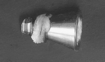
The extruded implants were immersed in histological formalin as soon as practicable. After at least 4 hours' fixation (usually longer), any tissue between or adherent to the fixture threads was carefully removed and placed into a buffer solution (Figure 1). This tissue was post-fixed in osmium tetroxide, dehydrated in a graded series of ethanol and embedded into Araldite resin (Taab Laboratories Equipment Ltd., Aldermaston, UK). After polymerisation of the resin, semi-thin (1-μm) sections were cut from the resin block and stained with toluidine blue for light microscopy. In addition, ultrathin sections (100 nm) were cut from the same resin blocks, using a Reichert OMU4 Ultracut ultramicrotome (Leica Mikrosysteme GmbH, Vienna, Austria), for electron microscopy studies. Ultrathin sections were collected onto 200 mesh copper grids and stained with uranyl acetate and lead citrate. The sections were examined under a Philips CM10 transmission electron microscope (Philips, Eindhoven, Netherlands) and digital images recorded.
Patient one's bone-anchored hearing aid device after it had fallen out.

To enable histopathological analysis, samples were taken from the fixture threads and fixed in formaldehyde (10 per cent buffered), processed, sectioned and finally stained with haematoxylin and eosin.
Cases
Patient one
A 68-year-old man with chronic supprative otitis media and mixed hearing loss of 80 dB had a right-sided BAHA abutment fitted under local anaesthesia in July 2004. He had no co-morbidity. The implant was placed using the recommended technique.Reference Tjellström5 There were no post-operative complications, and the hearing aid was attached in December 2004.
In May 2007, the patient presented with his extruded implant (Figure 1) and a six-month history of pain around the BAHA abutment.
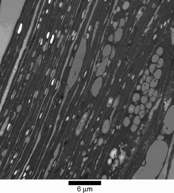
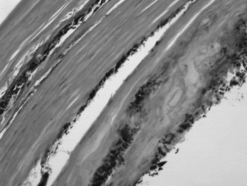
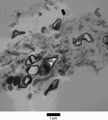
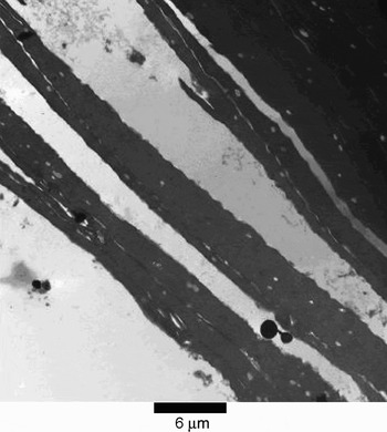
The implant was examined under electron microscopy (Figure 2). Histopathological analysis confirmed the electron microscopy findings of homogenous keratin with bacterial colonies. Occasional nucleated keratinocytes (i.e. parakeratosis) were seen (Figure 3). This tissue was situated between the titanium and the living bone interface. There was evidence of dead bacteria as shown by a remaining Gram-positive wall (Figure 4). Such bacteria did not show any evidence of organisation into a biofilm.
Electron photomicrograph showing keratin from the thread of patient one's titanium implant.

Photomicrograph of tissue taken from patient one's implant, showing homogeneous keratin with bacterial colonies. Occasional nucleated keratinocytes (i.e. parakeratosis) are seen. (H&E; ×200)

Electron photomicrograph of tissue taken from the threads of patient one's titanium implant, showing moribund Gram Positive bacteria.

Patient two
A 72-year-old man with bilateral chronic otitis media was implanted with a right-sided Cordelle™ BAHA (Cochlear Bone Anchored Solutions AB. Mölnlycke Sweden) in August 2005. There were no post-operative complications.
However, over time the patient obtained progressively less benefit from his BAHA; at this stage, his speech discrimination score was 27 per cent with the BAHA. In January 2007, he underwent a right cortical mastoidectomy and myringoplasty to repair the perforation in his good, hearing ear. The BAHA was removed at the same time, as the patient was scheduled to receive a right cochlear implant.
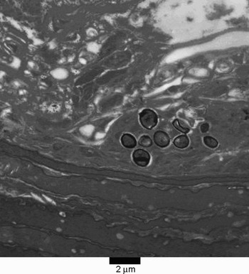
The removed titanium implant was sent for electron microscopy (Figure 5). This showed evidence of keratinocytes between the titanium and the living bone, confirmed on histological examination. There was evidence of bacteria but not of bacterial biofilm formation (Figure 6).
Electron photomicrograph of tissue taken from the threads of patient two's titanium implant, showing keratin.

Electron photomicrograph of tissue taken from the threads of patient two's titanium implant, showing Gram Positive bacteria.

Discussion
Osseointegration has a 10 per cent failure rate over a 10-year period in temporal bone; this increases in the older population.Reference Drinias, Granström and Tjellström3 No cause has been found thus far.
Our findings suggest that keratin may play a role in understanding osseointegration failure.
Analysis of our first patient's implant showed a dense proliferation of keratinocytes, developing over a three-year period and leading to failure of osseointegration. Our second patient's titanium implant was removed surgically, 17 months after implantation, for other reasons. Hence, no failure of osseointegration had occurred; even so, there was still evidence of the presence of keratinocytes, although these were not as abundant as in the first case.
Our analyses showed keratin to be present between the threads of the BAHA fixture, providing a barrier between the living bone and the titanium implant. The cause of such keratinocyte migratation to this interface can only be hypothesised. Osseointegration failure could be due to a chronic wound healing process, such as that seen in cholesteatoma.Reference Huisman, de Heer, Ten Dijke and Grote6 Activation of keratinocytes by an infective or inflammatory process of the skin flap could over time lead to bone resorption and migration of keratinocytes to the bone–titanum interface. In both our cases, there was evidence of dead bacteria but no evidence of bacterial biofilms, suggesting a previous infective period as a possible trigger for keratinocyte migration and proliferation.
• Bone-anchored hearing aids are well established, implanted devices
• This paper reports two cases in which a titanium osseo-integrated device was extruded or removed
• In both cases, keratin was found in association with the implant
• Entrapment of keratin between implant and bone may be a factor in implant extrusion
Keratinocytes have an innate immune function. Staphylococcus aureus can activate keratinocytes, via toll receptors, to release cytokines.Reference Mempel, Voelcker, Kollisch, Plank, Rad and Gerhard7 Transforming growth factor b is a cytokine released by activated keratinocytes and has been identified in cholesteatoma, which is now believed to be a chronic wound healing process.Reference Huisman, de Heer, Ten Dijke and Grote6
Vascular bone is required in order to achieve osseointegration. With increasing age, bone vascularity decreases,Reference Drinias, Granström and Tjellström3 and it has been shown that irradiation of bone can lead to implant instability.Reference Granström8 One could hypothesise that deterioration of bone quality with age could disrupt the bone–titanium interface and activate keratinocytes, which could then migrate into the gap between the bone and the titanium implant. An insidious skin infection could also activate keratinocytes.
If the two processes are combined, one could hypothesise that as the bone–titanium interface is interrupted by decreased vascularity, and as chronic wound infection progresses along the path of least resistance, keratinocytes may migrate between the bone and the titanium implant.
This paper represents the first report in the world literature providing evidence of a possible role of keratinocytes in osseointegration failure. However, one must question whether keratinocytes cause osseointegration failure or whether they are a byproduct of this process. How and why keratinocytes implant at the bone–titanum interface can only be hypothesised. Further investigation of the causes of such keratinocyte proliferation is needed.
In the future, we plan to examine all extruded BAHAs, using the above technique, in order to investigate these questions.