Introduction
Research on the technology now known as flattening filter-free (FFF) beams began in earnest in the mid-2000s. Initial studies comprehensively characterised the properties of FFF beams and produced compelling evidence of the potential benefits of the technology for cancer radiotherapy. Soon after the publication of these data, Varian Medical Systems (Palo Alto, CA) released the TrueBeam STx, a linear accelerator capable of delivering FFF beams. In 2011, Hrbacek et al.Reference Hrbacek, Lang and Klock1 reported on the commissioning of the first clinically used accelerator of this type operated in the FFF mode. Around the same time, George et al.Reference Georg, Knöös and McClean2 reviewed prior publications and the data available at that time. The broad clinical implementation of accelerators capable of delivering FFF beams soon followed. The American Association of Physicist in Medicine and the Institute of Physics and Engineering promptly reported much-needed guidelines for the clinical implementation of FFF beams.Reference Xiao, Kry and Popple3,Reference Budgell, Brown and Cashmore4
FFF beams are well suited for lung stereotactic body radiotherapy (SBRT) because the treatment typically involves small target volumes and large doses per fraction. Early studies of the use of FFF beams for lung SBRT mainly focused on the feasibility of the approach and its potential to reduce the beam-on time (BOT). For example, Vassiliev et al.Reference Vassiliev, Kry and Chang5 reported that differences in doses to the planning target volume (PTV) and to critical structures between FFF plans and plans using the conventional flattening filter (FF) beams were not clinically or statistically significant. However, the median BOT per field was reduced from 25 s with FF beams to 11 s with FFF beams, a reduction sufficient to facilitate the delivery of breath-hold and gated treatments. Boda-Heggemann et al.Reference Boda-Heggemann, Mai and Fleckenstein6 also showed that FFF beams convey a BOT benefit. In a treatment planning study of intensity-modulated radiotherapy, in which a dynamic multi-leaf collimator or volumetric modulated arc therapy (VMAT) was used for fluence modulation, they found that 10-MV FFF beams could be delivered in one breath hold, which reduced the overall fraction delivery time from approximately 30 to 15 minutes. Similarly, Ong et al.Reference Ong, Dahele and Slotman7 reported a reduction in BOT by a factor of approximately 2 when 10-MV FFF beams were used for Rapid Arc treatments. In addition, Prendergast et al.,Reference Prendergast, Fiveash and Popple8 studying a relatively large cohort of 82 lung SBRT patients, of whom 31 were treated with 10-MV FFF beams, reported that the average treatment time with FFF beams was 11·4 minutes, whereas that for conventional treatments with FF beams was 21·1 minutes. The average patient immobilisation time for FFF beams (32·9 minutes) was also shorter than that for FF beams (47·0 minutes). Finally, Gasic et al.,Reference Gasic, Ohlhues and Brodin9 who used FF and FFF beams with energies of 6 and 10 MV to develop four VMAT plans for each of 20 patients with early-stage lung cancer, found that differences in target coverage, conformity and homogeneity were not significant and that doses to organs at risk (OARs) were ‘comparable’ between the FF and FFF treatment plans. Compared with those for the FF-VMAT plans, the average beam delivery times for the FFF-VMAT plans were reduced by 55·3% (6 MV) and 75·0% (10 MV).
More recent studiesReference Lu, Lin and Lin10,Reference Tambe, Fryer and Marsden11 have primarily focused on the dosimetric characterisation of treatment plans. Lu et al.Reference Lu, Lin and Lin10 designed VMAT plans using FFF beams for 12 patients with stage I lung cancer using three fractionation schemes (4 × 12 Gy, 3 × 18 Gy, and 1 × 34 Gy). They found that the 6-MV plans had significantly lower doses to all OARs than the 10-MV plans did. Accordingly, the normal tissue complication probabilities (NTCPs) for the lung and chest wall for the 6-MV 4- and 3-fraction plans were approximately 10% lower than those for the 10-MV plans. The mean BOTs for the 6-MV 4-, 3- and 1-fraction plans were 2·2, 3·3 and 6·3 minutes, respectively, whereas those for the 10-MV plans were 1·5, 2·0 and 3·5 minutes, respectively. Similarly, Tambe et al.,Reference Tambe, Fryer and Marsden11 comparing 6-MV FFF, 10-MV FFF, and 6-MV FF VMAT plans, reported that 6-MV FFF plans achieved significantly better OAR sparing than 6-MV FF or 10-MV FFF plans did, without significant differences in target volume coverage.
Using Monte Carlo simulations of a dose delivered by seven 6-MV coplanar FF or FFF beams, Vassiliev et al.Reference Vassiliev, Kry and Wang12 investigated the impact of tumour size and lung density on the dose distributions in a lung tumour and its vicinity in a virtual thorax phantom at a spatial resolution of 0·5–1 mm. The FFF beams achieved consistent, albeit modest, target dose enhancement. The study also showed that lung density is an important factor that affects dose distribution, especially near the lung–tumour interface.
Pokhrel et al.Reference Pokhrel, Halfman and Sanford13 investigated dosimetric differences between 6-MV FF and FFF VMAT plans for a cohort of 13 patients with early-stage non-small cell lung cancer. The use of 6 MV was based on previous studies,Reference Lu, Lin and Lin10,Reference Tambe, Fryer and Marsden11 showing that best OAR sparing was achieved with 6-MV FFF beams. They found that, for the prescribed dose of 30 Gy in one fraction, FFF beams improved target conformity and lowered doses to OARs. The dose reductions to the lung, heart, oesophagus and ribs were all significant, and the BOT was reduced by a factor of 2·3. Using patient data, they also showed the significant impact of lung density on tumour coverage.
Navarria et al.Reference Navarria, Ascolese and Mancosu14 reported on treatment outcomes for a cohort of 46 patients with inoperable early-stage lung cancer treated with 10-MV FFF VMAT. They compared the dose distributions and treatment outcomes for these patients with those for 86 patients treated with 6-MV FF 3-dimensional conformal radiotherapy. The prescription for all patients was 4 × 12 Gy. In all cases, an ‘adequate’ dose conformity to the target was achieved. No further data on tumour coverage were reported. Consistent with other studies, a significant reduction in doses to the ipsilateral lung was achieved with FFF beams. No data for other OARs were presented. The BOTs for the FFF and FF plans were 1·5 and 8·3 minutes, respectively. At 1 year, the local control rate of the FFF arm (100%) was significantly higher than that of the FF arm (92·5%; p = 0·03). In the FFF arm, 17·4% of patients had grade 1 or 2 pulmonary toxicity, and 4% had grade 3 pulmonary toxicity. The FF arm had higher rates of grade 1, 2 and 3 toxicity at 24%, 42% and 9%, respectively.
The data summarised above demonstrate that lung SBRT with FFF beams is feasible and substantially lowers the BOT. Navarria et al.Reference Navarria, Ascolese and Mancosu14 reported remarkable treatment outcomes with FFF beams, although their study had a short follow-up period. They also reported dosimetric differences between FF and FFF treatment plans only for the lung; therefore, the FFF dose distribution properties responsible for achieving the 100% tumour control rate were unclear. Only Pokhrel et al.Reference Pokhrel, Halfman and Sanford13 performed a detailed dosimetric comparison of FF and FFF treatments. Additional dosimetric comparisons of FF and FFF treatments are needed to determine the extent to which FFF beams can improve the therapeutic ratio of lung SBRT. Our study provides data that complement those reported by Pokhrel et al.Reference Pokhrel, Halfman and Sanford13 We used a different treatment technique, static beams and compared different dosimetric indices, covering all indices for which treatment planning goals are specified in current directives to guide routine planning.
Materials and Methods
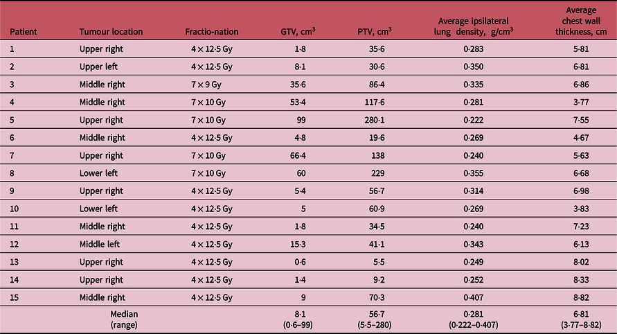
The study was approved by the Institutional Review Board. Fifteen patients who previously received SBRT for early-stage lung cancer at our institution were selected for this study. The cohort size was chosen based on preliminary estimates of tumour control probabilities (TCPs). The patients’ characteristics are summarised in Table 1. The table includes the average ipsilateral lung density, derived using the method described by Pokhrel et al.,Reference Pokhrel, Halfman and Sanford13 as well as the average chest wall thickness, calculated as the average of all wall thicknesses measured along the central axis of each beam. All treatments used conventional 6-MV FF photon beams. We imported the original treatment plans into Eclipse 13·6 (Varian Medical Systems). The analytical anisotropic algorithm dose calculation model was configured using representative beam data for a TrueBeam accelerator operated in 6-MV FFF mode. After replacing conventional FF beams with FFF beams of the same nominal energy (6 MV), we re-optimised all the plans without changing the beam setup. The optimisation strategy was to match the PTV coverage, verify that gross tumour volume (GTV) coverage was not compromised and attempt to lower the dose to OARs, starting with the lung. In most cases, FFF beams improved the target coverage, although this was not an explicit optimisation goal. Other OARs were the oesophagus, heart, spinal cord and chest wall.
Patient characteristics

Abbreviations: GTV, gross tumour volume; PTV, planning target volume.
For each FF or FFF treatment plan, we calculated multiple dose–volume histogram indices characterising target coverage and doses to the OARs. We also calculated the homogeneity index (HI), defined as HI = (D2% – D98%)/D50%,15 and the conformity index (CI), which is the ratio of volume receiving at least the prescription dose, V(R x ), to the PTV.
Tumor control probability
To calculate the TCP, we followed the method described by Chetty et al.Reference Chetty, Devpura and Liu16 The following equations for the TCP were derived by Webb and NahumReference Webb and Nahum17 and Guckenberger et al.Reference Guckenberger, Richter, Wilbert, Flentje and Partrige18 for a single fraction. We modified them to account for the effect of fractionation. Let α and α be parameters of the linear-quadratic model of cell survival, M the total number of voxels in the target volume,
 $N_i^{\left( 0 \right)}$
the initial number of clonogenic cells in the ith voxel, Di the total dose delivered to the ith voxel and Fx the number of fractions. For an individual patient with a given α, the TCP is
 $${\rm{TCP}}\left( \alpha \right) = \mathop \prod \limits_{i = 1}^M \exp \left\{ { - N_i^{\left( 0 \right)} \cdot \exp \left[ { - \alpha {D_i} - \beta {F_x}{{({D_i}/{F_x})}^2}} \right]} \right\}.$$
For a population, it is assumed that α is normally distributed with parameters
 ${({\alpha _{av}},{\sigma _\alpha })}$
). Therefore, the average TCP for the population is
 $${\rm{TCP}} = {1 \over {\sqrt {2\pi \sigma _\alpha ^2} }}\mathop \int \limits_{ - \infty }^{ + \infty } {\rm{TCP}}\left( \alpha \right) \cdot \exp \left[ { - {{{{\left( {\alpha - {\alpha _{av}}} \right)}^2}} \over {2\sigma _\alpha ^2}}} \right]d\alpha. $$
The model parameters are the same as those given by Chetty et al.Reference Chetty, Devpura and Liu16: αav= 0·292 Gy-1, σα=0·037 Gy-1 and β= αav/10 Gy, and the initial number density of clonogenic cells is 107 cm-3. The number of cells in a voxel
$N_i^{\left( 0 \right)}$
is the product of the number density and the voxel volume. The voxel volume and dose to the voxel Di can be derived from the differential dose–volume histogram.
Normal tissue complication probability
To calculate the NTCP for the lung, we followed the method developed by Selvaraj et al.Reference Selvaraj, Lebesque and Hope19 who fit several models to radiation pneumonitis data from patients who received SBRT. The authors found that the Lyman–Kutcher–Burman model with a logistic local dose–effect relation for perfusion loss derived from single-photon emission computed tomography imaging data provided ‘modest improvement’ over other models. In this model, the NTCP is calculated using the following equations:
 $${\rm{NTCP}} = {1 \over {\sqrt {2\pi } }}\int_{ - \infty }^t {\exp } \left( { - {{{x^2}} \over 2}} \right)dx,$$
 $$t = {{{\rm{EUD}} - {\rm{T}}{{\rm{D}}_{50}}} \over {m \cdot {\rm{T}}{{\rm{D}}_{50}}}},$$
 $${\rm{EUD}} = {{\rm{D}}_{50}}{\left\{ {{{\left[ {\mathop \sum \limits_{i = 1}^N {1 \over {1 + {{({{\rm{D}}_{50}}/{{\rm{D}}_i})}^k}}} \cdot {{{V_i}} \over V}} \right]}^{ - 1}} - 1} \right\}^{ - 1/k}}.$$
In these equations, EUD is the equivalent uniform dose, TD50 is the dose to an organ resulting in a 50% complication rate, m is the slope of the NTCP curve, D50 is the local dose causing 50% damage, k characterises the steepness of the local dose–effect curve, D i is the dose to the ith voxel, V i is the voxel volume and V is the total volume of the organ. The best-fit values of the model parameters were as follows: TD50 = 24·9 Gy, m = 0·38; D50 = 28·7 Gy and k = 2·2.
Uncomplicated tumor control probability
The uncomplicated TCP (UTCP) is the probability of achieving tumour control without complications. It is defined as UTCP = TCP × (1 – NTCP).
Statistical analysis
Continuous variables are summarised by the median and range across subjects. All reported p-values comparing the FF and FFF beams were obtained from the Wilcoxon signed-rank test, using the paired values for FF and FFF for each subject. To assess the association of an anatomic feature with differences between FF and FFF beams, we performed univariate linear regression with the parameter ratio for FFF to FF as the response variable; p-values < 0·05 are considered to be significant. The analysis was performed in MATLAB R2017b and R version 3·5·0.
Results and Discussion
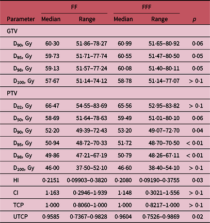
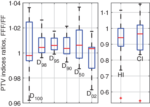
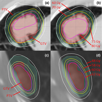
Target coverage data are summarised in Table 2 and Figures 1 and 2. In all cases, the FFF beams achieved PTV coverage that was equivalent or improved compared with that achieved with the FF beams. The improvement was statistically significant for the D98, D95, D90, HI and UTCP. The FFF beams also improved the cohort’s median D98 by 1·9%. In most cases, the TCP was close to 1. In the case with the lowest TCP, the FFF plan improved the TCP by 2·0%. Figure 1 shows box plots for the dosimetric indices characterising the PTV dose distributions. FFF beams improved all median values (red lines), except that of D100, which was essentially the same as that achieved with FF beams (the median FFF/FF ratio was 0·998). FFF beams similarly improved GTV coverage. The improvements in the D95 and D98 were significant. The cohort’s median D98 was improved by 1·6%. Figure 2 shows a comparison of FF and FFF dose distributions near the target volume for patients 4 and 6, who had relatively large and small tumours, respectively. For patient 4, the 75- and 80-Gy isodoses in the FFF plan covered a much larger portion of the GTV than they did in the FF plan (Figures 2A and 2B). For patient 6, the 60- and 62-Gy FFF isodoses covered the superior part of the GTV better than the FF isodoses did (Figures 2C and 2D).
Target coverage data
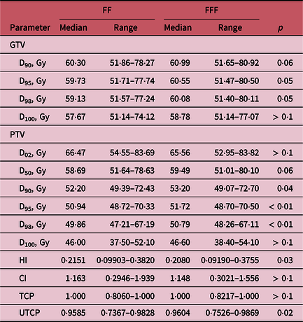
Abbreviations: FF, flattening filter; FFF, flattening filter-free; GTV, gross tumour volume; PTV, planning target volume; HI, homogeneity index; CI, conformity index; TCP, tumour control probability; UTCP, uncomplicated tumour control probability.
Box plots of dosimetric indices characterising PTV coverage. Red lines are the medians, blue boxes are interquartile ranges, black dotted lines are ranges and red crosses are outliers.

Examples of dose distributions near the GTV in two patients. Panels A and B show the distributions for patient 4, who had a relatively large tumour; C and D show the distributions for patient 6, who had a small tumour. Panels A and C are FF plans; B and D are FFF plans.

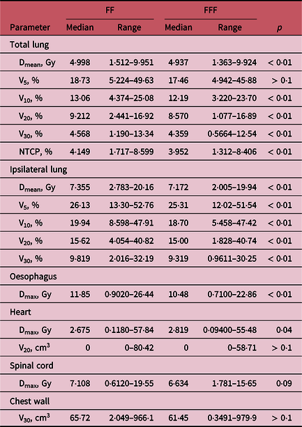
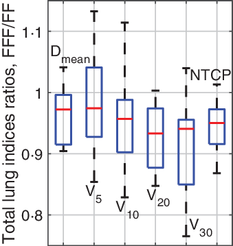
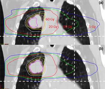
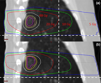
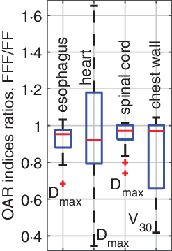
Doses to OARs are summarised in Table 3 and Figures 3–6. In most cases, FFF beams lowered doses to OARs. Reductions in the mean lung dose, V10, V20 and V30, were all statistically significant. Lower doses resulted in significantly lower NTCPs. The cohort’s median NTCP for FFF plans was 5·0% lower than that for FF plans. Box plots of the above indices for the total lung are shown in Figure 3 and clearly demonstrate the substantial lung sparing achieved with FFF beams. As for the ipsilateral lung, FFF plans significantly improved the mean lung dose, V5, V10, V20 and V30. The cohort’s median mean lung dose and V20 in the FFF plans were 2·6% and 4·1% lower, respectively, than those in the FF plans. Figures 4 and 5 show the distributions of lung doses received by the lung in the FF and FFF plans for patients 4 and 6, respectively. In the FF plan in Figure 4, as compared with the FFF plan, the 10-Gy isodose extends farther into the contralateral lung, and the 10- and 20-Gy isodoses extend inferiorly farther into the mediastinum and ipsilateral lung. In the FF plan in Figure 5, as compared with the FFF plan, the 20-Gy isodose extends farther anteriorly and inferiorly into the normal lung. Figure 6 shows box plots of the maximum doses to other OARs (the oesophagus, heart and spinal cord) and the V30 (cm3) for the chest wall. FFF plans lowered the maximum doses to OARs for most patients. The improvements were statistically significant for the heart and oesophagus. For a few patients, the maximum dose to the heart with FFF plans was 20–60% higher than that with FF plans. For all these plans, the doses to the heart were much lower than the tolerance level; thus, we did not pursue further dose reduction.
Doses to organs at risk

Abbreviations: FF, flattening filter; FFF, flattening filter-free.
Box plots of parameters characterising dose distributions in the total lung. Red lines are the medians, blue boxes are interquartile ranges and black dotted lines are ranges.

An example of dose distributions in the lung for patient 4. Panels A and B show FF and FFF plans, respectively (coronal plane view). The white dashed lines provide a reference for comparing the isodoses.

An example of dose distributions in the lung for patient 6. Panels A and B show FF and FFF plans, respectively (sagittal plane view). The white dashed lines provide reference for comparing the isodoses.

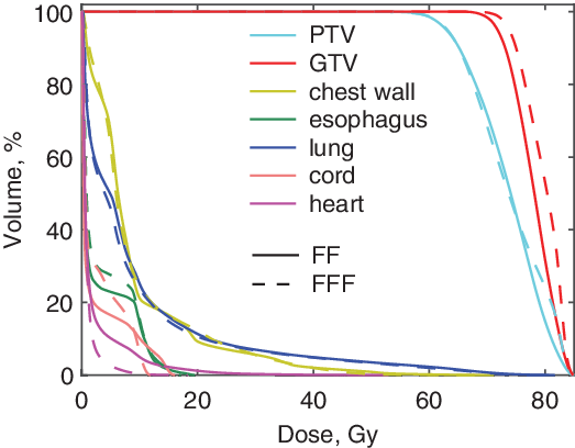
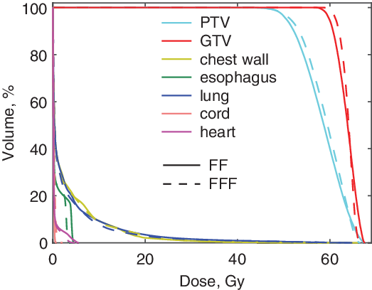
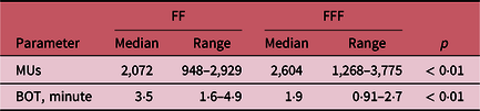
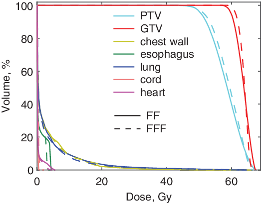
Additional representations of dose distributions, convenient for comparing FF and FFF plans, are shown in Figures 7 and 8. These figures show the dose–volume histograms for patients 4 and 6. Achieving lower doses to the OARs was more difficult for the larger tumour (patient 4). In this case, we substantially lowered the maximum doses to all the OARs. For the spinal cord and oesophagus, this led to larger volumes receiving low doses. In both patients, the FFF plans improved GTV coverage, especially for volumes above 90%, which translated into modestly increased doses to the parts of the GTV that received the lowest doses in the original FF plans. This GTV boost was not an explicit planning objective; rather, our main objective was to achieve equivalent PTV coverage between FF and FFF plans. This approach is well illustrated in Figure 7, which shows that achieving very similar PTV dose distributions in both plans led to higher GTV doses in the FFF plan. The differences in GTV coverage reflect the differences in the physics of FF and FFF beams: FFF beams have higher doses in the build-up region,Reference Vassiliev, Kry and Wang12 and near the beam’s central axis. The number of monitor units (MUs) per plan was on average 26% higher in FFF plans. We could lower this number, but that was not necessary, because the MU rate for the FFF beams (1400 MU/minute) was much higher than that for the FF beams (600 MU/minute), which resulted in a much lower BOT (Table 4). Also, higher MU numbers in the FFF plans were unlikely to result in higher out-of-field doses because in the FFF configuration, the main source of scattering in the beam-line, the flattening filter, is absent.
Box plots of parameters characterising doses received by organs at risk. Red lines are the medians, blue boxes are interquartile ranges, black dotted lines are ranges and red crosses are outliers.

Dose–volume histogram for patient 4.

Dose–volume histogram for patient 6.
Treatment delivery parameters

Abbreviations: FF, flattening filter; FFF, flattening filter-free; MUs, monitor units, BOT, beam-on time.
We analysed the data for potential associations between anatomic characteristics of the cohort and quantities describing the differences between FF and FFF treatment plans. We performed univariate linear regression analysis. The predictor variables were PTV volume, ipsilateral lung density and chest wall thickness. The latter was calculated as the average wall thickness measured along the beam’s central axis for all beams in a plan. The response variables were the FFF/FF ratios of the following parameters: D98, HI, CI and TCP; mean dose, V20 and NTCP for the total lung; maximum doses to the oesophagus, heart and the spinal cord; V30 (cm3) for the chest wall; and total MUs and BOT per treatment fraction. Overall, we tested 3 × 13 predictor–response pairs, none of which showed strong evidence of association. Given our sample size, this negative finding may be due to limited power. A larger cohort is needed to establish associations conclusively.
Conclusions
By replacing FF beams with FFF beams of the same nominal energy in lung SBRT treatment plans, we were able to lower doses to OARs without compromising tumour coverage. Reduced doses to the ipsilateral lung were particularly consistent for all dose levels from 5 to 30 Gy. At the same time, target coverage was not compromised; rather, it actually improved in most cases. Hence, our findings provide evidence that the therapeutic ratio in lung SBRT can be improved by replacing FF beams with FFF beams. Nevertheless, in clinical practice, the choice of FFF over FF beams for individual patients should not be automatic; rather, it should be based on a comparison of FF and FFF treatment plans.
Acknowledgements
J.A. Munch of MD Anderson’s Department of Scientific Publications provided editorial assistance.
Financial Support
This study was supported by the National Institutes of Health through a grant R03 CA211143. In addition, this work used MD Anderson’s Biostatistics Resource Group, which is supported by the National Institutes of Health through grant P30 CA016672.
Conflicts of Interest
The authors have no conflicts of interest to declare.