Introduction
Kawasaki disease is an acute vasculitis involving the coronary arteries in children, especially those less than five years old. Coronary artery aneurysms occurred in about 25% of acute Kawasaki Disease patients without optimal treatment. Furthermore, coronary artery stenosis is induced in about 1–2% of acute Kawasaki disease patients in the late period. Reference Kato, Sugimura and Akagi1 On the other hand, Ichinose et al. reported that systemic artery aneurysms were found in 19 (3.1%) among 616 patients after Kawasaki disease. Reference Ichinose, Akagi, Inoue and Kato2 Moreover, systemic artery aneurysms developed in 10% of patients with giant coronary aneurysms caused by Kawasaki disease. Reference Tsuda, Hamaoka and Suzuki3 Axillary artery aneurysms and internal iliac artery aneurysms are known to be involved. Reference Sasaguri and Kato4,Reference Hoshino, Tsuda and Yamada5 However, abdominal aortic aneurysms and renal artery aneurysms are extremely rare. In Ichinose’s report, there was one abdominal aortic aneurysm and five renal artery aneurysms. Reference Ichinose, Akagi, Inoue and Kato2 Yamada et al. reported that the incidence of abdominal aortic aneurysms was 8 (0.9%) out of the 875 patients between 1971 and 1993, and two of the eight patients died. Reference Yamada6 Either the characteristics of abdominal aortic aneurysms or the clinical course of the renovascular hypertension due to Kawasaki disease have not been reported except in some previous case reports before. Their diagnosis can often go unnoticed. We report two adult patients who had rare systemic artery lesions with a history of Kawasaki disease, with a review of the literature.
A patient with a distal abdominal aneurysm
A 38-year-old male with Kawasaki disease at the age of 2 months underwent coronary artery bypass grafting to the right coronary artery and left anterior descending artery because of coronary artery stenoses. In the screening by thoracic-abdominal CT before the operation, calcification of the distal abdominal aorta was detected, and an abdominal aortic aneurysm with stenosis was diagnosed (Figure 1, middle, right). His blood pressure was 110/70 mmHg. He had been asymptomatic, and there were no differences in his extremities’ blood pressures. The collateral arteries were well developed.
Distal abdominal aortic aneurysms at 2 years and 38 years after the onset of Kawasaki disease. (Left) Aortogram at 2 years and 9 months. A slight dilatation of the distal abdominal aortic aneurysm was found, and the bilateral common iliac arteries were slightly dilated. (Middle, right) computed tomography angiograms at 38 years. A stenosis with calcification was detected in the distal abdominal aorta. At the bifurcation of the common iliac arteries, dilatations with calcification were found.

He had a history of Kawasaki disease at the age of 2 months. From the 6th day, intravenous immunoglobulin of 100mg/kg/day for 5 days was administered to treat acute Kawasaki disease. The fever persisted for 16 days. On the 17th day, aneurysms of both coronary arteries were diagnosed by two-dimensional echocardiography. Aspirin, dipyridamole, and warfarin were given. Two years later, he was referred to our hospital. He underwent cardiac catheterisation, and his selective coronary angiograms at 2 years and 9 months old showed segmental stenosis of the right coronary artery and left anterior descending artery. Looking back at his initial aortogram, there were dilatations of the distal abdominal aorta and the left common iliac artery (Figure 1, left). However, we had overlooked them as important abnormal findings. He had a perfusion defect in the anteroseptal wall of the left ventricle on the dipyridamole-loaded radioisotope 201Tl myocardial perfusion imaging, and aspirin had been given in the outpatient clinic. His perfusion defect improved with the development of collateral arteries during his growth. However, he complained of chest pain at the age of 36 years. His coronary angiograms at 37 years old did not remarkably change compared with the previous coronary angiograms. In the myocardial 13N-ammonia positron emission tomography findings, the myocardial flow reserve ratios of the right coronary artery, left anterior descending artery, and left circumflex artery were 2.12, 1.95, and 3.02, respectively.
A patient with renovascular hypertension
A 26-year-old female visited our hospital because of a headache. Her blood pressure was 154/90 mmHg, height 152 cm, and weight 52.0 kg, respectively. Her total cholesterol, low-density lipoprotein cholesterol, and triglycerides were 230 mg/dL (normal 128–219), 163 mg/dL (<140), and 73 mg/dL (30–149), respectively. Spironolactone, in addition to aspirin, was given. Her plasma renin and aldosterone levels were 6.4 ng/mL (0.2–3.9) and 131 pg/mL (<173), respectively. Her renal function was within normal limits. In the renal echocardiogram and Doppler, a stenosis at the orifice of the right renal artery was detected, and the peak flow velocity was 3.6 m/s (Figure 2, left). There was a peak time delay during the excretion phase of the 99mTc MAG renogram, but there was no laterality. A magnetic resonance angiogram of the abdominal aorta showed severe stenosis of the orifice of the right renal artery (Figure 2, right). Percutaneous transluminal renal angioplasty was recommended. However, she hesitated to undergo the procedure. Her hypertension has been treated with calcium antagonists and angiotensin-converting enzyme inhibitors. The collateral arteries were detected.
Renal artery stenosis at the age of 26 years. (Left) Renal ultrasound examination revealed a severe stenosis at the orifice of the right renal artery. The peak velocity was 3.6 m/s. (Right) A magnetic resonance angiogram showed a stenosis of the orifice of the right renal artery (yellow arrow). The collateral arteries were found (orange arrow).

She visited our hospital at the age of 6 years because she had a history of Kawasaki disease at the age of 6 months. From the 8th day, intravenous immunoglobulin of 200mg/kg/day for 5 days was administered to treat acute Kawasaki disease. Her fever persisted for 14 days. On the 29th day, giant aneurysms of both coronary arteries were diagnosed by two-dimensional echocardiography. Aspirin was prescribed. Two months after acute Kawasaki disease, she underwent cardiac catheterisation, and selective coronary angiograms revealed giant aneurysms of both coronary arteries, and her aortogram was judged to be normal at the previous hospital. The blood pressure of the ascending aorta was 115/68 mmHg. Coronary angiograms at the age of 20 months showed a severe localised stenosis of the left anterior descending artery. She underwent coronary artery bypass grafting with the left internal thoracic artery at the age of 23 months. She again had acute Kawasaki disease at the age of 3 years, and intravenous immunoglobulin of 400mg/kg/day for 5 days was administered. There were no changes in the coronary artery lesions after the second episode of Kawasaki disease, and the graft was patent. The blood pressure of the ascending aorta was 106/65 mmHg. She was followed up every year by an electrocardiogram and two-dimensional echocardiography in our hospital. Her blood pressure at the outpatient clinic was as follows: 20 years 131/84 mmHg, 22 years 132/71 mmHg, 23 years 116/75 mmHg, 24 years 128/79 mmHg, and 25 years 133/91 mmHg. The CT angiogram at the age of 26 years revealed mild dilatation of the right coronary artery with calcification and patency of the left internal thoracic artery graft. The left ventricular ejection fraction by two-dimensional echocardiography was 66%. We suspected that the stenosis of the right renal artery wall was thickening due to acute Kawasaki disease.
Discussion
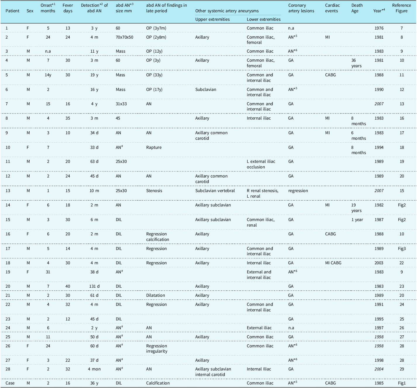
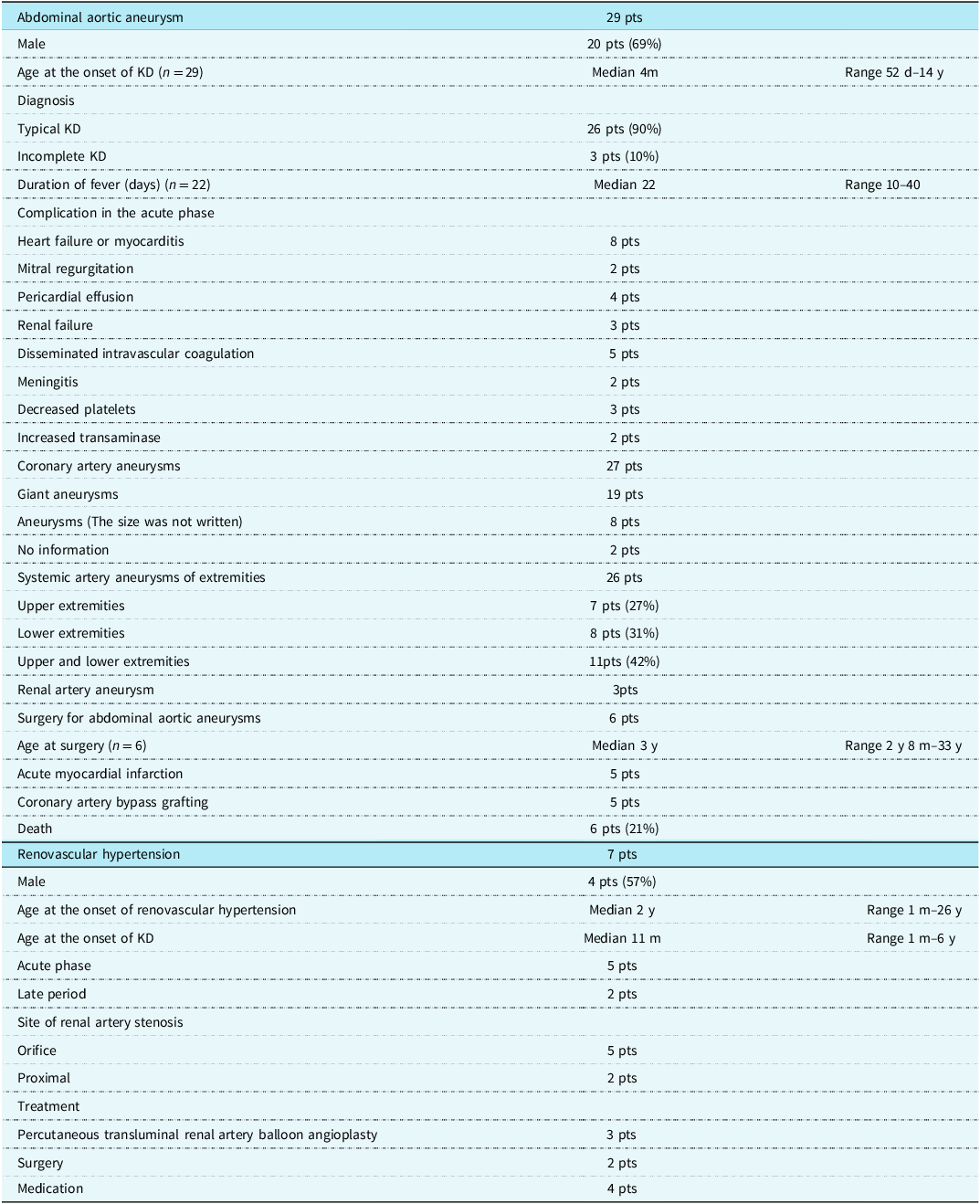
Twenty-nine patients with abdominal aortic aneurysms caused by Kawasaki disease since 1974 were reviewed; however, the information was minimal in some abstracts. There were 23 patients from the references from 13 articles, 8 abstracts, and 6 patients in our institution (Tables 1 and 2). There were 20 males and 9 females. The age at the onset of acute Kawasaki disease ranged from 52 days to 14 years, with a median of 4 months. The number of patients with an age of onset of acute Kawasaki disease of less than or equal to 6 months was 19 patients (66%), and the number of patients with an age of onset of acute Kawasaki disease of less than or equal to 24 months was 26 patients (90%). In patients with systemic artery aneurysms caused by Kawasaki disease, the age of most patients with abdominal aortic aneurysms is younger than that of patients without abdominal aortic aneurysms. Reference Hoshino, Tsuda and Yamada5 In small infants, within 3 months, the vessel of the elastic artery may be weak and likely to be involved due to severe acute vasculitis. Furthermore, twenty-three patients (79%) had had acute Kawasaki disease in the 1980s. Most patients did not undergo massive intravenous immunoglobulin therapy for severe inflammation in the acute phase.
Characteristics and outcome of patients with abdominal aortic aneurysms caused by Kawasaki disease

OP = operation; AN = aneurysm; DIL = dilatation; MI = myocardial infarction; CABG =coronary artery bypass grafting; n.a = not available; Because it was not written in the report, we cannot write it.
* 1 The onset of acute Kawasaki disease.
* 2 The interval from the onset of Kawasaki disease to the detection of the abdominal aortic aneurysm.
* 3 Mass means abdominal aortic aneurysm which is palpable by hand. In this article, dilatation (DIL) of the abdominal aorta is defined as a diameter of less than 2-fold the abdominal aorta diameter in its aortogram.
AN#,The diameter size wasnot written, and the figure of the aortogram wasnot present in the report. Therefore, the size of the aneurysm was unknown.
* 4 The year of the report. Italic year’ s references were the reports in the late period.
In our patients, the year of the onset of acute Kawasaki disease is written.
* 5 Giant aneurysms were strongly suspected, although a patient had stenotic lesions.
* 6 The size of coronary artery aneurysms were not written.
A large palpable abdominal mass by hand or abdominal distention was a clue to the diagnosis in six patients. Reference Takeuchi, Nishikawa and Kano7–Reference Ohashi, Yamaguchi and Tachibana9 The mass was detected by abdominal sonograms and confirmed in aortograms by cardiac catheterisation, and their size ranged from 60 mm to 70 mm. The abdominal aortic aneurysms in the remaining 21 patients were also diagnosed on the aortograms. In others, confirmation was performed by sonogram or autopsy on each patient. The diagnosis of abdominal aortic aneurysms within 1 year after the onset of acute Kawasaki disease was confirmed in twenty-three patients. In the remaining six patients, their abdominal aortic aneurysms were diagnosed in the late period. Reference Tsuda10–Reference Kondo, Tanaka and Shikano12 The interval from acute Kawasaki disease to its diagnosis ranged from 3 years to 33 years, with a median of 11 years (n = 6). In a 15-month-old boy, coronary artery aneurysms had been diagnosed when he had acute Kawasaki disease. However, his abdominal aortic aneurysm was first diagnosed with the first aortogram 4 years after acute Kawasaki disease. Reference Shiono, Isobe, Murakami and Hayashi13 The initial aortogram was performed because of his family history of Marfan syndrome. The diagnosis of abdominal aortic aneurysms is difficult unless aortograms are performed at an optimal time. Some small abdominal aortic aneurysms might have regressed within 1 year after the onset. It might often be overlooked in some patients. Inflammation of the aorta occurred in most patients during acute vasculitis. Hamashima et al. reported acute aortic vasculitits in 82% of autopsies, and described that the peak time of inflammation due to acute Kawasaki disease differed based on the vessel size from a pathological viewpoints. Reference Hamashima14 In small arteries, such as the coronary arteries, the peak of inflammation is around 10 days, that for micro-vessels is within 10 days, and that for large vessels is around 20 days: the larger the vessels, the later the peak of inflammation. Formation of abdominal aortic aneurysms is later than that of coronary artery aneurysms, which can also lead to missing detection of abdominal aortic aneurysms. The tool for the diagnosis of abdominal aortic aneurysms in the 1980s was only aortography by cardiac catheterisation. At present, the detection of abdominal aortic aneurysm by sonogram and magnetic resonance is possible noninvasively. Therefore, screening should be performed in infants with severe acute vasculitis. In the late period, calcification detection by low-dose CT is an appropriate marker of vessel involvement.
Regarding the shape of the aneurysm, there were ball-type and spherical-type aneurysms, depending on the aneurysm’s width and length. Of the thirteen patients for whom the location of their aneurysms had been written, six had an aneurysm below the celiac artery, and seven had an aneurysm that was below the renal artery. An abdominal aortic aneurysm complicating dilation of the common iliac arteries was found in 11 patients. Most abdominal aortic aneurysms involve the distal abdominal aorta around the bifurcation at the common iliac arteries, and that is the characteristic of abdominal aortic aneurysms caused by Kawasaki disease. The outcome of abdominal aortic aneurysms also depends on the size. Six patients underwent surgery for large aortic abdominal aneurysms. The aneurysms were resected, and Y grafts with prosthetic vessels were replaced. The procedure was successful in all six patients. The age at surgery ranged from 2 years and 8 months to 33 years, with a median of 3 years (n = 6). The interval from the onset of acute Kawasaki disease to the surgery was from 4 months to 19 years, with a median of 3 years. In 15 patients, there were findings in the late period, more than 1 year after the onset of Kawasaki disease. Residual abdominal aortic aneurysms were found in nine patients. Regression of the abdominal aortic aneurysms was found in five patients, and either calcification of the regressed aneurysm or irregularity of the abdominal aorta was detected in some patients (Figures 1 and 3). Furthermore, although severe abdominal aortic stenosis occurred, collateral arteries had developed in one patient. Reference Miura15 The patient with abdominal aortic stenosis complicating renal artery stenosis had had hypertension. An autopsy was performed in three patients, Reference Nonaka, Okuyama, Yoshimura and Takagi16–Reference Mori M.Kurozumi and Akagi18 and the pathological findings were recorded in three of the six patients with surgery. There were thrombi and calcifications in the abdominal aortic wall. Reference Yamada6 In the microscopic findings, there were severe calcifications in the intima and disruption or a lack of elastic fibres in the aortic wall. Reference Takeuchi, Nishikawa and Kano7,Reference Font, Mehta, Streusand, O’boyle and Kretzer17
Changes in the serial abdominal aortograms. A 5-month-old boy had bilateral giant aneurysms and multiple systemic aneurysms. (Left) Aortogram at 9 months old. A distal abdominal aortic aneurysm was found. Dilatations of the bilateral common iliac arteries and aneurysms of the bilateral internal iliac arteries were found. (Middle) Aortogram at 1 year and 8 months old. The abdominal aortic aneurysm had regressed. (Right) Aortogram at 22 years old. There was an irregularity in the aortic wall where the abdominal aortic aneurysm had regressed. The bilateral internal iliac aneurysms had regressed.

Claudication of the left leg due to an occlusion of the external iliac artery in the late period occurred in one patient. Reference Fuyama, Hamada and Uehara19 Regarding the systemic artery aneurysms in the upper and lower extremities in 26 patients with aortic abdominal aneurysms. Although dilatation of the ascending aorta was complicated in one patient, it regressed by 1 year after the onset of Kawasaki disease. Reference Ninomiya, Onoda and Hanji20 Regarding the renal artery, renal artery aneurysms were found in three patients, and renovascular hypertension was complicated in one patient with a stenosis at the origin of the renal artery. Reference Miura15 Abdominal aortic vasculitis can involve the orifice of the renal arteries (Figure 4). Inflammatory change was in general remarkable at the branching area of the vessels, particularly marked near the portion that branched directly from aorta. Reference Amano, Hazama and Hamashima21
Abdominal aortic aneurysms complicating renal artery aneurysms. (Left) An aortogram in an 11-month-old boy 6 months after the onset of Kawasaki disease. Abdominal aortic aneurysms and dilatations of the bilateral renal artery aneurysms were found. Dilatations of the right common iliac aneurysm were also found. (Right) Aortogram in an 8-month-old girl 2 months after the onset of Kawasaki disease. Abdominal aortic aneurysms were found, and the orifice of the bilateral renal arteries was dilated.

Information on the acute phase of Kawasaki disease, cardiac sequelae, and the outcomes in patients with abdominal aortic aneurysms is limited (Table 2). Typical Kawasaki disease was observed in 26 patients, and incomplete Kawasaki disease in three patients. The persisting fever ranged from 10 to 40 days, with a median of 22 days (n = 22). Either heart failure or myocarditis occurred in eight patients, and severe mitral regurgitation was observed in two of the eight patients. Renal failure and pericardial effusions were found in three patients and four patients, respectively. Disseminated intravascular coagulation occurred in five patients. Those findings indicate severe general vasculitis due to acute Kawasaki disease. Nineteen of the 27 patients complicated with coronary artery aneurysms had giant aneurysms. In the remaining eight patients, the size of the coronary artery aneurysm was not noted, and in two patients, no information on coronary artery aneurysms was available. It was suspected that almost all patients had had giant aneurysms. Five patients had acute myocardial infarctions, and five patients underwent coronary artery bypass grafting. Reference Tsuda10,Reference Tsuda, Minami and Kobayashi22 The incidence of cardiac events was high because most of the patients had had giant aneurysms. Six patients (21%) died. There were four deaths among the children and two deaths among the adults. A death due to a rupture of an abdominal aortic aneurysm in a 7-month-old boy occurred 34 days after the onset of acute Kawasaki disease. Reference Mori M.Kurozumi and Akagi18 Two patients died of acute myocardial infarction 3 months and 7 months after the onset of Kawasaki disease, respectively. Reference Nonaka, Okuyama, Yoshimura and Takagi16,Reference Font, Mehta, Streusand, O’boyle and Kretzer17 An autopsy was performed on both patients. Another 1-year-old boy died after taking a bath (Figure 4, left). A 36-year-old male patient with a 2-vessel coronary artery occlusion died suddenly 33 years after the replacement of an abdominal aortic aneurysm. He had dropped out of our outpatient clinic. Reference Tsuda10 Another female patient with a history of myocardial infarction and hypoxic brain damage in childhood also died suddenly at the age of 19 years (Figure 4, right). The cause of the above three deaths remains unknown. The incidence of deaths was also high, and it was speculated that cardiac sequelae led to deaths in most cases. Reference Tsuda, Arakaki and Shimizu30 Most of the patients, except for those with an abdominal aortic mass, have been asymptomatic for abdominal aortic aneurysm from childhood to young adulthood. A rupture of abdominal aortic aneurysms is rare.
Results of references reviewed on abdominal aortic aneurysms and renovascular hypertension

KD = Kawasaki disease.
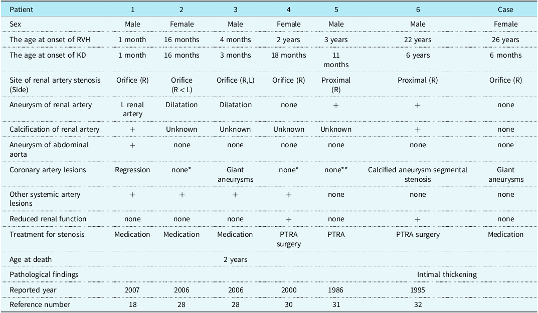
Although renovascular hypertension with a history of Kawasaki disease in children and young adults was also reported, its prevalence was scarce. Plasma renin activity was elevated in all six patients in whom the value was documented. Their occurrence was reported in both the acute phase and the late period. Reference Miura15,Reference Tsuda, Arakaki and Shimizu30–Reference Nagao, Yamamto and Urabe33 (Tables 2 and 3). In the references, up to now, there have been four acute patients and three late-onset renovascular hypertension patients after acute Kawasaki disease in this population. Pathologically, intimal thickening was detected in the renal artery stenosis. Reference Falcini, Calabri, Simonini, Vitale, De Simone and De Martino31,Reference Morishita, Mawatari and Yoshimi34 It was also reported that renal artery stenosis after acute severe Kawasaki disease vasculitis, which induced renovascular hypertension in children. One patient developed abdominal aortic stenosis after a regressed abdominal aortic aneurysm. Reference Fuyama, Hamada and Uehara19 Primarily, there has previously been only one report of adult-onset renovascular hypertension before. Reference Morishita, Mawatari and Yoshimi34 At the onset of adulthood, the patients complained of headaches due to severe hypertension. This long latent period supports the idea that the late appearance of problems may be related to the development of intimal thickening of the renal artery with ageing after acute vasculitis. The renal artery stenosis in the above patient at our institution was also speculated to be the cause of renovascular hypertension. Usually, systemic artery stenosis caused by Kawasaki disease occurs where systemic artery aneurysms existed during acute Kawasaki disease vasculitis. Five patients had a stenosis at the orifice of the renal artery including our patient. Two patients had a stenosis at the middle of the renal artery, which was speculated to occur after regression of an aneurysm. The orifice of the renal artery might also be related to severe acute aortic vasculitis or the regression of abdominal aortic aneurysms. Those situations are difficult to evaluate by an initial aortogram alone. In the treatment, one had a percutaneous transluminal renal artery balloon angioplasty. Reference Nagao, Yamamto and Urabe33 Two patients initially underwent percutaneous transluminal renal artery balloon angioplasty for severe renal artery stenosis; however, they had surgery because of restenosis. Reference Foster, Bernard and Drummond32,Reference Morishita, Mawatari and Yoshimi34 Four patients had medication. One of the three patients had been treated for hypertension for about 20 years with medication after his acute illness. Reference Miura15 Surgery is needed to relieve severe renal stenosis in patients with reduced renal function. Reference Watanabe35
Characteristics and treatment in patients with renovascular hypertension

RVH = renovascular hypertension; KD = Kawasaki disease; PTRA = Percutaneous transluminal renal artery balloon angioplasty.
* Coronary assessment was performed within 1 month after Kawasaki disease, and abnormalities were not detected.
** Coronary angiogram showed normal arteries 6 months after Kawasaki disease.
Renovascular hypertension caused by Kawasaki disease is very rare, and it may also be related to abnormalities of the abdominal aorta from the viewpoint of the vessel site. Although 3 of the above 29 patients with abdominal aortic aneurysms had renal artery aneurysms or stenosis, renovascular hypertension was only observed in one of the three patients (Table 1). Three (43%) of the seven patients with renovascular hypertension caused by Kawasaki disease had no coronary artery lesions. Renovascular hypertension caused by Kawasaki disease is not always complicated by coronary artery aneurysms, which may differ from other systemic artery aneurysms complicated with severe coronary artery lesions.
Renovascular hypertension is an important but infrequent cause of hypertension in children and young adults. Murashita et al. reported a 4-year-old boy who underwent surgery for fibromuscular dysplasia due to an unknown infection. His left renal artery stenosis with hypertension was found incidentally during evaluation for Kawasaki disease. Reference Murashita, Kanemitsu and Fukunaga36 The cause of renal artery stenosis in this patient remains unknown. It was considered that acute Kawasaki disease cannot be denied as the cause of the disease. Odagiri et al. reported a 3-year-old boy who had an abdominal aortic aneurysm with renovascular hypertension. Reference Odagiri, Yoshida and Kawashima37 It was documented as a non-specific inflammatory disease of unknown origin that was not Kawasaki disease or Takayasu disease. As a cause of its effects on children, there are conditions such as fibromuscular dysplasia, Takayasu arteritis, polyarteritis nodosa, etc. Reference Bayazit, Yalcinkaya and Cakar38 Renovascular fibromuscular dysplasia tends to affect women between 15 and 50 years of age. Most patients are asymptomatic for many years, and it is discovered accidentally. Stenosis with renovascular fibromuscular dysplasia is often found in the middle of the renal artery, rather than at the orifice of the renal artery. Takayasu arteritis is a chronic vasculitis with aortitis. Kawasaki disease is one of the most common childhood vasculitis, leading to inflammation and sometimes dilatation of medium-sized vessels. We realised that Kawasaki disease can be one of the causes of renovascular hypertension in children and young adults, although its incidence is infrequent.
The above results indicate that the patients who have severe acute vasculitis within 6 months are a high-risk group. Although any medium-sized vessel in the body can be involved, investigation and follow-up of systemic artery aneurysms in affected children and adults is not always performed, in contrast to that of detecting coronary artery lesions. Most of this population has yet to reach 50 years of age. The occurrence of systemic artery lesions in patients with a history of Kawasaki disease should be considered carefully from now on, especially in patients who have coronary artery aneurysms caused by acute Kawasaki disease at an extremely young age, less than 6 months. Screening and re-evaluating systemic artery lesions by magnetic resonance or low-radiation CT angiography in such adult patients may be helpful. Reference Jone, Tremoulet and Choueiter39
Conclusion
Two adult patients with a severe acute vasculitis under the age of 6 months had abdominal aortic stenosis and renovascular hypertension, respectively. Systemic artery lesions caused by Kawasaki disease have been overlooked because the incidence is very low. Calcification detection by low-dose CT in the late period is an appropriate marker of vessel involvement.
Acknowledgements
We thank Dr. Akiyuki Kotoku and Dr. Yoshiaki Morita in the department of radiology for photographing computed tomography angiogram and magnetic resonance angiogram. We thank Mr. John Martin for his kind English language consultation.
Financial support
This research received no external funding.
Competing interests
The authors declare no conflict of interest.
Ethical standard
All procedures performed in studies involving human participants were in accordance with the ethical standards of the institutional committee with the 1964 Helsinki declaration and its later amendments or comparable ethical standards.