Hypoplastic left heart syndrome can be readily diagnosed by echocardiography. The following sections will highlight the key aspects of diagnostic imaging by echocardiography in foetuses and newborns suspected of having hypoplastic left heart syndrome. In addition, technical aspects of echocardiographic evaluations are emphasised for each stage of surgical reconstruction.
The International Society for Nomenclature of Paediatric and Congenital Heart Disease (www.ipcc.net) offers the following definition of hypoplastic left heart syndrome:Reference Tchervenkov, Jacobs and Weinberg1
Hypoplastic left heart syndrome is synonymous with the term hypoplasia of the left heart and is defined as a spectrum of cardiac malformations with normally aligned great arteries without a common atrioventricular junction, characterized by underdevelopment of the left heart with significant hypoplasia of the left ventricle including atresia, stenosis, or hypoplasia of the aortic or mitral valve, or both valves, and hypoplasia of the ascending aorta and aortic arch.
Prenatal assessment
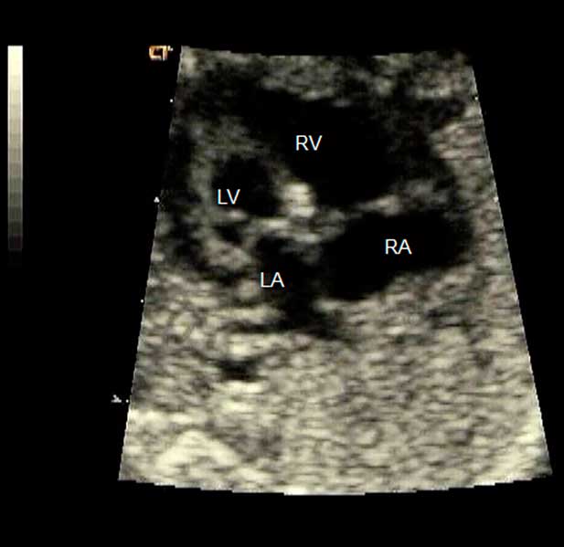
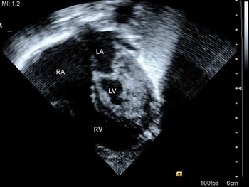
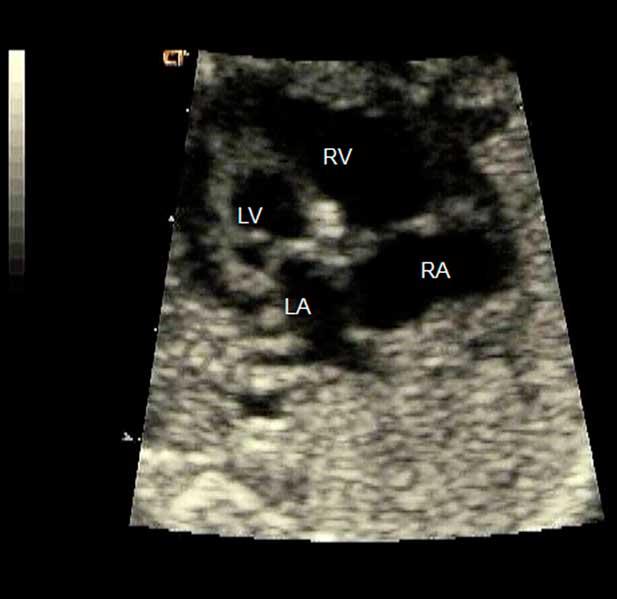
Hypoplastic left heart syndrome is one of the more common prenatally diagnosed congenital cardiac lesions due to the very abnormal appearance of the heart in the four-chamber view on routine screening with obstetrical ultrasound (Fig 1). Hypoplastic left heart syndrome is a spectrum of anomalies that presents by foetal echocardiogram as early as the first trimester or as late as the third trimester. This variation in presentation is because hypoplastic left heart syndrome can have an evolving pathology during gestation. These patients need to be followed up serially to best prognosticate outcomes and plan for postnatal intervention.
Four-chamber view of a foetal heart with hypoplastic left heart syndrome. There is an obvious discrepancy in size between the left- and right-sided structures (LA = left atrium; LV = left ventricle; RA = right atrium; RV = right ventricle).
The spectrum of hypoplastic left heart syndrome includes varying degrees of obstruction of the aortic and mitral valve, including:
• aortic atresia with mitral atresia,
• aortic atresia with mitral stenosis,
• aortic stenosis with mitral stenosis, and
• aortic stenosis with mitral atresia.
When there is both aortic and mitral atresia, the resultant left ventricle is slit-like or completely absent. In contrast, aortic atresia with mitral stenosis or aortic stenosis with mitral stenosis results in a diminutive left ventricle that is very round and “tense appearing”, with the apex of the heart formed by the right ventricle. The endocardial fibroelastosis and poor left ventricular function are secondary to the poor egress of blood flow out of the ventricle. Less commonly, in some patients with hypoplastic left heart syndrome, the left ventricle is of normal size or even dilated. This presentation situation is typically seen with a higher degree of mitral valve patency. Even in these cases, the left ventricle will be hypocontractile and often have endocardial fibroelastosis as well. The left ventricle will often become smaller in size as gestation progresses. With colour Doppler assessment in the four-chamber view, there will be virtually no filling of the diminutive left ventricle. If the mitral valve is patent, it is important to exclude the presence of coronary sinusoids in the left ventricular wall. This evaluation will require placing the colour box over the ventricular septum and the left ventricular free wall and using a low Nyquist limit to look for evidence of sinusoidal flow. This evaluation must be followed up serially, as sinusoids may not present until later in gestation.
The four-chamber view is also the ideal view for assessing the right ventricle and tricuspid valve. It is important to observe the morphology of the tricuspid valve and to rule out any tricuspid insufficiency. Significant tricuspid regurgitation is a poor prognostic sign both for foetal health and for surgical outcomes after birth. It is important to keep in mind the degree of tricuspid regurgitation when assessing right ventricular function. Following up the right ventricular function using the Tei Index allows a quantitative measure that corrects for right ventricular morphology and atrioventricular valvar insufficiency. A Tei Index should be followed up serially and any decrease in function, as predicted by an increase in the Tei Index, should raise concerns about the constriction of arterial ductus and/or foetal acidosis.
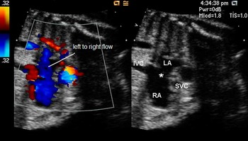
Both the four-chamber and bicaval views allow a two-dimensional look at the patency of the oval foramen as it opens into the right atrium (Fig 2). Patients with a restrictive oval foramen or an intact atrial septum have a worsened prognosis and may not respond to medical intervention in the postnatal period. This poor prognosis is secondary to high pressures in the left atrium that change the pulmonary vasculature. These changes cannot be acutely reversed, and include abnormal pulmonary musculature and atrialisation of the pulmonary veins. The patency of the oval foramen cannot be reliably assessed by two-dimensional visualisation alone. Colour Doppler should show laminar left-to-right shunting. The Doppler pattern should be non-restrictive, biphasic, low velocity – that is, between 20 and 40 centimetres per second – and return to the baseline. Another, and arguably more reliable, means of assessing the patency of the oval foramen is by Doppler and colour interrogation of the pulmonary veins. When the oval foramen is patent in a foetus with hypoplastic left heart syndrome, the reversal of flow in the pulmonary veins with atrial systole will be minimal. However, when the oval foramen is restrictive, the A wave is increased and peaked, with significant reversal of flow in the pulmonary veins during atrial systole. An intact atrial septum shows more of a “to-and-fro” flow pattern. A ratio of the velocity-time integral of the pulmonary venous forward flow over the velocity-time integral of the pulmonary venous reversal of flow that is less than five has been found to be predictive of a need for postnatal balloon atrial septostomy.Reference Michelfelder, Gomez, Border, Gottliebson and Franklin2 Some advocate the use of maternal hyperoxygenation to look for vasoreactive response of the foetal pulmonary vasculature in cases when the oval foramen is suspected to be restrictive. A non-reactive test, defined as less than 10% decrease in the pulsatility index in the branch pulmonary arteries after 10 minutes of 60% inspired oxygen given to the mother, was predictive of a need for immediate intervention at birth.Reference Szwast, Tian, McCann, Donaghue and Rychik3 The assessment of the pulmonary veins must be followed up serially to ensure that the oval foramen is not closing over time. If the oval foramen is becoming more restrictive, an appropriate plan for delivery must be established because immediate balloon atrial septostomy or surgical intervention may be necessary. Certainly, a discussion with the family about the change in prognosis is appropriate.
Bicaval or sagittal view of a foetal heart with hypoplastic left heart syndrome. Colour Doppler showing left-to-right shunting across the atrial septum (*), which appears non-restrictive based on laminar pattern of flow (LA = left atrium; RA = right atrium; SVC = superior caval vein; IVC = inferior caval vein).

The aortic annulus and aortic arch can be challenging to assess. The aortic annulus can occasionally be seen by tilting the transducer anteriorly from the four-chamber view, or in the parasternal views, and will typically measure less than 3 millimetres in diameter. A sagittal view will show the aortic arch and the head and neck vessels, with retrograde flow seen in the ascending aorta and transverse aortic arch. A discrete coarctation of the aorta may also be visualised. The “three-vessel view” at the base of the heart may not show the transverse aortic arch well. However, colour Doppler will show flow in the opposite direction of the adjacent pulmonary artery/arterial duct. This finding confirms retrograde flow in the transverse aorta from the arterial duct. The Doppler profile in the arterial duct in hypoplastic left heart syndrome is altered by the increase in flow across the duct in this lesion. The Pulsatility Index of the arterial duct should remain at the upper limits of normal or just greater than normal (3.0), because the systolic flow is greater than normal and the diastolic flow should remain unchanged in these patients.
In the foetus with hypoplastic left heart syndrome, the brain receives blood via retrograde filling of the head and neck vessels from the arterial duct. In order to encourage enough flow of blood, the foetus with hypoplastic left heart syndrome will decrease the cerebral vascular impedance in a phenomenon called “cephalisation”. The Doppler profile in the middle cerebral artery shows an increase in diastolic flow as compared with systolic flow, resulting in a lower Pulsatility Index than in the foetus with normal cardiac anatomy. The mechanism and significance of this finding remains unclear, although it is well known that these children are at high risk for adverse neurological sequelae.Reference Berg, Gembruch, Gembruch and Geipel4
Foetal echocardiography plays a vital role in the diagnosis and prognosis of hypoplastic left heart syndrome. Careful assessment of the physiology of the foetus as it progresses through gestation helps to predict outcomes and need for immediate postnatal intervention. These assessments are also critical in those cases in which prenatal intervention is attempted, including balloon dilation of the aortic valve and atrial septostomy. These foetal procedures are currently not routine practice and therefore not included in this review. However, the echocardiographic steps outlined here will help identify those patients who may be candidates for these procedures in the future.
Echocardiographic assessment before Stage 1 palliation – Norwood procedure
A comprehensive echocardiographic evaluation should be undertaken when hypoplastic left heart syndrome is suspected in an infant. A standardised echocardiographic analysis should include all appropriate imaging views and should be guided by a segmental approach to cardiac anatomy. The overall goal is to provide accurate diagnosis and complete haemodynamic information to aid with clinical management and surgical planning.
The objectives of the first echocardiogram in the newborn period are to
• evaluate the anatomic and physiologic components of the hypoplastic left heart: left atrium, mitral valve, left ventricle, aorta – valve, ascending aorta, arch, isthmus;
• evaluate the anatomy and the physiology of the interatrial communication – if present;
• evaluate the anatomy and the physiology of the arterial duct;
• evaluate the anatomy and physiology of the right ventricle and the tricuspid valve;
• evaluate the anatomy and physiology of the coronary flow;
• establish the presence or absence of other associated abnormalities.
Subcostal views
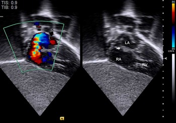
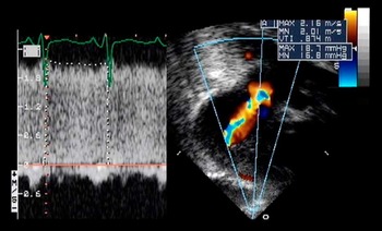
Cardiac position and segmental connections can be easily established at the beginning of the evaluation with a good sweep in the subcostal long-axis view. At the level of the diaphragm, the hepatic veins come together with the inferior caval vein to drain into the right atrium. As the transducer is tilted towards the cardiac base, the coronary sinus, right atrium, and right ventricle come into view. A dilated coronary sinus should alert the echocardiographer to the possibilities of a persistent left superior caval vein or anomalous pulmonary venous connection to the coronary sinus. The right atrium is typically dilated and hypertrophied because of the volume load from obligate prenatal left-to-right shunting through the patent oval foramen. The right ventricle is also seen dilated as compared with the diminutive or “slit-like” left ventricle. Once this view is obtained, careful assessment of the atrial septum, pulmonary venous connections, size and function of the ventricular chambers, and the atrioventricular valves should be performed. Specific anatomy of the atrial septum including size and location of atrial communications should be established. Frequently, there is a leftward deviation of the superior attachment of the primary atrial septum – septum primum (Fig 3). The severity of atrial restriction can be assessed by using colour or pulsed-wave Doppler. The mean gradient is obtained by tracing Doppler flow of the interatrial shunting over two to three cardiac cycles (Fig 4).
Typical deviation of the atrial septum in hypoplastic left heart syndrome. Subcostal view showing leftward and superior deviation of the atrial septum (arrow; LA = left atrium; RA = right atrium; RV = right ventricle).

Restrictive atrial septum in hypoplastic left heart syndrome. The flow jet across the atrial septum is aliased; its velocity remains above baseline throughout the cardiac cycle as shown by continuous wave Doppler interrogation.

Pulmonary venous anatomy and drainage should be carefully assessed, as anomalous pulmonary venous connections and/or drainage occurs in about 5–10% of patients with hypoplastic left heart syndrome. In cases of highly restrictive or intact atrial septum, a decompressing vein may originate from the left atrium and drain to a variable location. Decompressing veins can become stenotic; therefore, a full view of its course of drainage, along with haemodynamic assessment with Doppler interrogation, is important.
Further anterior angulation of the transducer typically demonstrates a dilated right ventricular outflow tract and pulmonary trunk – main pulmonary artery. The tiny ascending aorta can sometimes be seen, but probably can be visualised better in the subcostal short axis.
The subcostal short axis provides confirmation of the cardiac anatomy and assessment of ventricular function. Normal connection of the systemic veins to the right atrium is confirmed with the transducer angled towards the right side of the heart. The extent of interatrial communication can be readily demonstrated. Colour flow mapping and Doppler interrogation of atrial shunting should be performed. As the sweep continues towards the apex of the heart, one should be able to evaluate the right ventricular outflow tract, ascending aorta, and right ventricular function.
Apical views
The four-chamber view provides comparison of the relative sizes of the ventricles (Fig 5). The right ventricle is dilated and hypertrophied. The left ventricle is small, muscle bound, and non-apex forming. The endocardial surface of the left ventricle is often “echo-bright”, which may represent areas of endocardial fibroelastosis; the extension of “echo-brightness” should be documented. The ventricular septum should also be carefully examined for any defects.
Apical four-chamber view in hypoplastic left heart syndrome. The diminutive left ventricle is muscle bound and non-apex forming. This chamber has an echogenic and smooth endocardial surface, suggesting extensive endocardial fibroelastosis (RA = right atrium; LA = left atrium; LV = left ventricle; RV = right ventricle).

The four-chamber view is also excellent to assess the morphology and function of the mitral and tricuspid valves. The annulus of the mitral valve is typically hypoplastic with either atretic or severely stenotic leaflets. The patency of the mitral valve should be determined with the aid of colour flow mapping and Doppler assessment of the inflow into the left ventricle. In cases of stenosis of the mitral valve without evidence of mitral regurgitation, the echocardiographer should be alerted to the likelihood of ventriculo-coronary sinusoids, which can be demonstrated in apical views. The tricuspid valvar morphology and function should be assessed carefully. Displacement of the leaflets, abnormal chordal attachments, and abnormal attachments of the papillary muscles can be best seen in this view. It is important to identify and quantify tricuspid regurgitation, as the presence of significant regurgitation of the tricuspid valve indicates a poor prognosis.
Anterior scanning shows the aortic valve, ascending aorta, and pulmonary trunk – main pulmonary artery. Doppler studies of the aortic outflow can give information about the patency of the aortic valve. The pulmonary valve should also be assessed for any stenosis or regurgitation.
Parasternal views
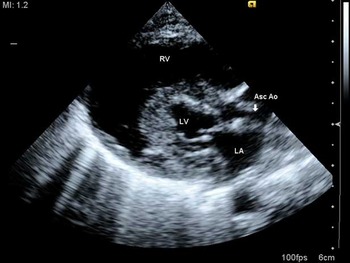
Standard parasternal views are useful for evaluation of ventricular function, and size and morphology of the cardiac valves (Fig 6). The long-axis views show a dilated right ventricle anteriorly and a small left ventricle posteriorly. The ventricular septum is usually intact. The aortic valve is frequently atretic or severely stenotic. Subaortic obstruction may be present. The ascending aorta is frequently small, that is, less than 5 millimetres in diameter. The mitral valve is usually imperforate, but when patent the leaflets appear thickened and doming with shortened chords. A supravalvar mitral ring may be present. Angling the transducer inferiorly allows the echocardiographer to assess the tricuspid valve and its function. Angling superiorly opens the right ventricular outflow tract and dilated pulmonary trunk – main pulmonary artery.
Standard parasternal long-axis view in hypoplastic left heart syndrome. The right ventricle forms the apex of the heart. The mitral valve annulus, left ventricle, and aorta are severely hypoplastic and incapable of supporting systemic output (RV = right ventricle; LV = left ventricle; LA = left atrium; Asc Ao = ascending aorta).

Ventricular sweeping in parasternal short-axis views provides excellent assessment of the mitral subvalvar area and any defect of the ventricular septum. The presence of a single papillary muscle or “parachute mitral valve” can be ascertained. Short-axis views also allow the echocardiographer to determine the morphology of the aortic valve and coronary arteries. The coronary arteries typically have a normal distribution. The right coronary artery is usually prominent in relation to the left coronary artery because of a dominant right ventricle. However, if there are significant coronary-to-left ventricle fistulous connections, then there may not be significant difference in size between the left and right coronary arteries.
Sliding the transducer to the high left parasternal view allows the echocardiographer to visualise the pulmonary artery branches and arterial duct. Tilting the transducer posteriorly, the echocardiographer can assess pulmonary venous drainage. In cases of anomalous pulmonary venous connection, again, it is important to follow the full length of the decompressing vein, as obstruction may occur at any point along its course.
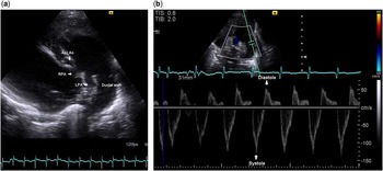
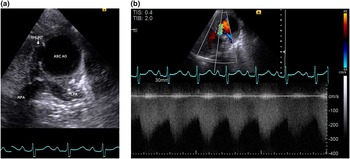
A slight counter-clockwise rotation of the transducer shows the ductal arch, which is usually quite prominent and may be larger than either branch of the pulmonary artery (Fig 7a and b).
(a) Ductal view from a high parasternal window. Pulmonary anatomy and ductal course are demonstrated. A tiny ascending aorta in cross-section can be seen. (b) Bidirectional shunting across the ductal arch with antegrade flow in systole and retrograde flow in diastole is demonstrated by Doppler interrogation (LPA = left pulmonary artery; RPA = right pulmonary artery; Asc Ao = ascending aorta).

Suprasternal notch views
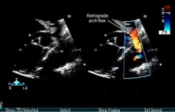
Suprasternal notch views provide full assessment of the anatomy of the aortic arch. Sidedness of the arch should be determined. The aortic arch frequently has a more acute curve than normal. Sizes of the ascending aorta, transverse arch, and isthmus should be assessed. Presence or absence of a posterior shelf and coarctation should be identified and documented. Doppler sampling in the distal segment of the aortic arch usually reveals retrograde flow in systole into the transverse arch and antegrade flow in diastole into the ductal arterial and pulmonary vascular beds (Fig 8).
Suprasternal sagittal view demonstrating a hypoplastic ascending aorta and aortic arch with retrograde flow as shown by colour Doppler.

Echocardiographic assessment after Stage 1 palliation – modified Norwood procedure
The International Society for Nomenclature of Paediatric and Congenital Heart Disease (www.ipcc.net) offers the following definition of the Norwood operation:Reference Tchervenkov, Jacobs and Weinberg1
The Norwood operation is synonymous with the term ‘Norwood (Stage 1)’ and is defined as an aortopulmonary connection and neoaortic arch construction resulting in univentricular physiology and pulmonary blood flow controlled with a calibrated systemic-to-pulmonary artery shunt, or a right ventricle to pulmonary artery conduit, or rarely, a cavopulmonary connection.
The Norwood procedure consists of surgical reconstruction and augmentation of the ascending aorta and aortic arch with aortopulmonary amalgamation, an atrial septectomy, and a systemic-to-pulmonary shunt. The systemic-to-pulmonary shunt can be a modified Blalock–Taussig shunt or a right ventricle-to-pulmonary artery conduit, also known as the Sano shunt.
The anatomy of the atrial septum is easily assessed from the subcostal coronal or sagittal planes. Rapid assessment of the atrial septum is especially important when unexpected cyanosis occurs early after Stage 1 palliation, because one potential aetiology of this cyanosis is the inadequate resection of the atrial septum.
A right Blalock–Taussig shunt can usually be seen from the suprasternal short- or long-axis view with angulation of the transducer towards the right side (Fig 9a). Post-operative complications of the Blalock–Taussig shunt include pulmonary overcirculation and stenosis of the shunt. Doppler flow reversal ratio has been reported to provide useful measure of pulmonary/systemic blood flow ratio in some cases.Reference Rychik, Bush and Spray5 Stenosis of the shunt can be difficult to confirm using echocardiography. Poor acoustic windows and artefacts created by the material of the shunt may impair full visualisation of the shunt and the site of stenosis. Gradients of pressure may be underestimated because the Bernoulli equation is not valid in cases of narrowing of a long segment. Nevertheless, a normal Doppler pattern of an unobstructed shunt should demonstrate a characteristic sawtooth spectral pattern throughout systole and diastole (Fig 9b). Narrowing of the colour jet through the shunt or increased turbulence and velocity noted in the innominate artery should raise concerns of stenosis of the shunt.
Shunt profile from suprasternal views following Stage 1 Norwood procedure. (a) The central shunt is seen from the suprasternal long-axis view with the transducer tilted slightly to the patient's right. (b) A typical sawtooth pattern of the shunt as shown by continuous wave Doppler interrogation (LPA = left pulmonary artery; RPA = right pulmonary artery; Asc Ao = ascending aorta).

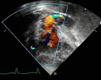
The Sano shunt can be best visualised by subcostal and modified apical views. Modified apical view is obtained by moving the transducer slightly medially from the standard apical view and tilting the transducer to image very anteriorly (Fig 10). In addition, sliding the transducer up towards the mid-sternum will permit excellent viewing of the distal aspect of the conduit and pulmonary arterial branches. Narrowing of the shunt at any point along its course or at its insertion into the pulmonary artery should be identified. Colour and spectral Doppler sampling in the conduit and proximal right and left pulmonary arteries should demonstrate a typical to-and-fro pattern of flow.
Evaluation of the right ventricle to pulmonary artery conduit following Stage 1 Norwood with Sano modification. Image and colour Doppler obtained from modified apical four-chamber view showing its course from the right ventricle to pulmonary artery (RV = right ventricle; LPA = left pulmonary artery; Sano = right ventricle to pulmonary artery conduit).

Evaluation of right ventricular function and tricuspid regurgitation is important and should be performed from multiple imaging planes. Right ventricular dysfunction and significant tricuspid regurgitation can cause acute haemodynamic compromise in the immediate post-operative period.
Echocardiographic evaluation of the neo-aorta and aortic arch is also an important part of the study, with the distal aortic arch deserving special attention. This part of the echocardiographic evaluation is a critical part of serial assessment and is discussed in more detail below. The presence of Doppler acceleration in the distal aortic arch suggests the need for additional evaluation with cardiac catheterisation or magnetic resonance imaging.
Echocardiographic assessment after Stage 2 palliation – bidirectional cavopulmonary anastomosis
The bidirectional cavopulmonary anastomosis – bidirectional Glenn – is most commonly utilised as the second stage of palliation in patients with hypoplastic left heart syndrome; it is an end-to-side anastomosis of the superior caval vein to the ipsilateral pulmonary artery. The cavopulmonary shunt enhances effective pulmonary blood flow but not at the expense of ventricular workload. The overall volume load to the systemic ventricle is decreased. The bidirectional Glenn procedure is typically performed between the ages of 3 and 6 months. Pre-operative echocardiographic evaluation is important to determine patient suitability for the cavopulmonary shunt.
Identification of systemic venous connections is integral in a thorough pre-operative evaluation. The suprasternal view best demonstrates the presence of a left superior caval vein. Bilateral superior caval veins and the presence of a bridging vein are best imaged from the suprasternal short-axis view using colour Doppler imaging. Azygous continuation of the inferior caval vein can best be visualised from the subcostal sagittal view and the subcostal cross-sectional view of the abdomen.
Obstruction at the level of the atrial septum must be identified during the pre-operative evaluation. Flow across the atrial septal defect is best imaged from the subcostal views because the atrial septum is aligned perpendicularly to the sound beam. Unrestrictive flow across the atrial septum is assessed by both colour and spectral Doppler imaging. A mean gradient is obtained by tracing the Doppler flow over two to three cardiac cycles.
The anatomy of the pulmonary arteries must be delineated in the pre-operative evaluation. The size of the pulmonary arteries should be measured and narrowing of the proximal pulmonary arteries should be identified. If obstruction of the pulmonary artery is found pre-operatively, reconstruction of the pulmonary artery can be performed during the cavopulmonary anastomosis.
Assessment of right ventricular function and the degree of tricuspid regurgitation is important. Qualitative assessment of the myocardial shortening of the right ventricle from apical and parasternal views remains the most common means to evaluate the global performance of the right ventricle. Quantitative assessment of the function of the right ventricle is difficult because of the variable geometry of the right ventricle. However, as in foetal echocardiography, the Tei Index allows a quantitative measure of global right ventricular function that corrects for right ventricular morphology.
The ascending aorta and reconstructed aortic arch are assessed for any residual obstruction. Proximal aortic obstruction may occur at the supravalvar region at the suture line, which can be visualised from subcostal, apical, and parasternal windows. The reconstructed arch is assessed using the suprasternal notch windows. An estimate of the right ventricular pressure can be obtained by measuring the peak velocity of continuous-wave Doppler of the tricuspid regurgitant jet. This estimate of the right ventricular pressure can be compared with lower extremity blood pressure as an indirect means of assessing obstruction.
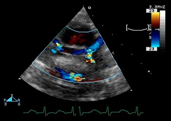
Following surgery, the right bidirectional cavopulmonary anastomosis is best visualised from the suprasternal short-axis view. The entire length of the superior caval vein and anastomosis to the right pulmonary artery can be seen in this view (Fig 11). Colour mapping and Doppler studies should be performed. In the absence of obstruction, flow in the cavopulmonary anastomosis should be laminar and low velocity, with a biphasic pattern of forward flow. With obstruction, the pattern of flow is typically continuous with elevated velocities that do not return to Doppler baseline; in this situation, mean gradient is typically greater than 3 millimetres of mercury when traced over three cardiac cycles.
Bilateral bidirectional cavopulmonary anastomosis – bilateral bidirectional Glenn – from suprasternal long-axis view showing entire course of the pathway. Special attention should be paid to the sites of cavopulmonary anastomosis where narrowing or kinking of the pulmonary artery may occur.

Echocardiographic assessment after Stage 3 palliation – Fontan operation
The last stage of reconstruction for hypoplastic left heart syndrome is the Fontan procedure, which is usually performed between 2 and 4 years of age. The Fontan procedure involves re-routing the blood from the inferior caval vein to the pulmonary arteries most commonly by means of an extra-cardiac conduit or an intra-atrial lateral tunnel. A fenestration, or small opening between the Fontan baffle and the systemic circulation, is sometimes placed during the surgery in order to allow a “pop-off”, which decreases central venous pressure and improves cardiac output at the expense of some degree of desaturation. Some studies have shown that creation of a fenestration can decrease morbidity after the Fontan procedure. The fenestration may decrease the incidence of pleural effusions, as well as possibly decreasing the chance of developing protein-losing enteropathy and other later morbidities associated with the Fontan circulation.
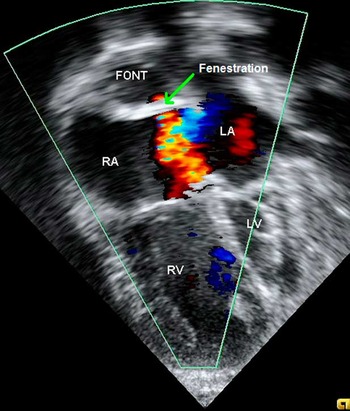
The Fontan pathway is best visualised from the subcostal and apical windows. In the subcostal windows, dilated hepatic and inferior caval veins suggest obstruction or elevated pressure in the Fontan pathway. Any anatomic obstruction in the Fontan pathway – intra-atrial tunnel or extra-cardiac conduit – should be identified with two-dimensional imaging, as well as colour flow mapping, and spectral Doppler interrogation. The colour scale needs to be lowered sufficiently in order to detect disturbances in flow at the low velocities commonly seen in the systemic venous pathway.Reference Snider, Serwer and Ritter6 In the apical view, the Fontan pathway can be seen in cross-section as the posterior structure either within the native right atrium, in the setting of an intra-atrial baffle, or behind the native right atrium, in the setting of an extra-cardiac conduit (Fig 12). If present, a fenestration in the Fontan baffle can be demonstrated by colour Doppler. If a fenestration is present, spectral Doppler interrogation should be performed to determine the transpulmonary gradient, or the pressure gradient from the systemic veins to the pulmonary venous atrium.
Bicaval pulmonary anastomosis – Fontan – with fenestration. Apical four-chamber view showing bicaval pulmonary anastomosis pathway in cross-section localised posterior to the right atrium. Flow across the fenestration (green arrow) can be seen (RA = right atrium; RV = right ventricle; LA = left atrium; LV = left ventricle; FONT = Fontan pathway).

Before and after the Fontan operation, it is important to perform a thorough echocardiographic evaluation of the following features:
• the atrial septum,
• tricuspid valvar regurgitation,
• right ventricular function,
• pulmonary arteries,
• pulmonary veins, and
• aortic arch.
The techniques to evaluate these structures by echocardiography have been previously described in this article in the section “Echocardiographic assessment after Stage 2 palliation – bidirectional cavopulmonary anastomosis”.
Ongoing assessment of the Fontan at routine follow-up
Ongoing surveillance of Fontan physiology at routine follow-up involves assessment of many of the same parameters discussed in the previous section of this article; however, problems of a more chronic nature can also occur following the Fontan operation. One concern following the Fontan operation is dilation of the neo-aortic root. This dilation can lead to disruption of the neo-aortic valvar architecture and subsequently result in valvar regurgitation. This phenomenon can be assessed with serial measurements of the aorta, including measurements of the diameter of
• the annulus of the neo-aortic valve,
• the sinus of Valsalva, and
• the sinotubular junction.
These measurements can be recorded from the parasternal long-axis view and from subcostal windows, as detailed by Cohen et al.7 Another long-term concern after the Fontan operation is the possibility of formation of intracardiac clot or clot within the Fontan pathway.
Echocardiography is critical in the assessment of patients with hypoplastic left heart syndrome. From foetal assessment to ongoing surveillance after the Fontan procedure, echocardiography remains the primary technique for cardiac monitoring in this growing population of children and adults.