Introduction
Ageing is the most important risk factor for the development of visual impairment.Reference Klein, Klein, Linton and De Mets1 The prevalence of visual loss increases substantially after 60 years of age and poor vision is the second most prevalent physical disability in older people.Reference Gohdes, Balamurugan, Larsen and Maylahn2, Reference Dening, Chi, Brayne and Huppert3 Statistics from the Royal National Institute for the Blind (RNIB) suggest that in the UK 20% of people over the age of 75 are living with ‘sight loss’ (RNIB definition: best corrected visual acuity (BCVA) worse than 6/12 in the better seeing eye) and this increases to 50% in those aged 90 and over. Currently, approximately 2 million people in the UK have this degree of visual impairment, predicted to increase to 2.25 million by 2020 and to almost 4 million by 2050.4
Visual impairment in older people is a result of either primary ocular pathology (e.g. cataracts, age-related macular degeneration (AMD) and glaucoma) or systemic disease with associated ocular or visual pathway involvement (e.g. diabetes, hypertension and cerebrovascular disease). Eye problems account for approximately 1.5% of general practice consultationsReference Sheldrick, Wilson, Vernon and Sheldrick5 and, as it has been estimated that over 50% of sight loss could be avoided,4 clinicians involved in the care of the elderly have an important role in the recognition, prevention and management of morbidity related to sight loss in this population.
This review describes the normal ageing changes of the eye and outlines the common ophthalmic diseases affecting older people, recent advances in their diagnosis and treatment, and relevant current research. A systematic review of the literature was carried out using the PubMed database and the search terms ‘ageing’ and the relevant ocular structure, e.g. ‘ageing’ and ‘lens’ combined as follows: ‘ageing AND lens’ in addition to specific searches regarding ‘visual impairment UK’, ‘age related macular degeneration’ and ‘diabetic retinopathy’.
Normal ageing of the human eye
All structures of the eye are affected in some way with age. While age-related changes can reduce the quality of vision, it is important to distinguish age-related physiological changes from pathological changes, particularly those pathological changes that are treatable.
Eyelids
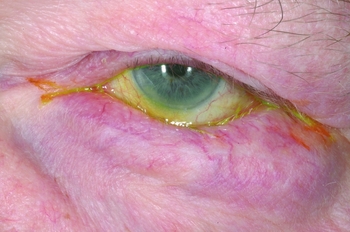
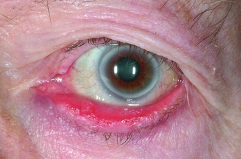
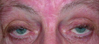
Age results in atrophy of the orbicularis oculi, the muscles that close the eyes. This, in combination with reduced elasticity of the overlying skin and involution and/or dehiscence of lid retractors, can result in entropion (Fig. 1) or ectropion (Fig. 2): outward or inward turning of the lid margin, respectively. The prevalence of ectropion and entropion in the older population has been estimated at approximately 2–4%.Reference Damasceno, Osaki, Dantas and Belfort6, Reference Mitchell, Hinchcliffe, Wang, Rochtchina and Foran7 In the Blue Mountains Study, a marked age-related increase in the prevalence of ectropion was observed: 0.3% of people aged under 60 years, 1.2% of those aged 60–69 years, 6.7% of those aged 70–79 years and 16.7% of those aged 80 years or older.Reference Mitchell, Hinchcliffe, Wang, Rochtchina and Foran7 Likewise, involutional and atrophic changes or dehiscence of the upper lid retractors (levator palprebrae superioris) can result in a droop in the eyelid, known as ptosis.Reference Sridharan, Tallis, Leatherbarrow and Forman8 In some cases this is severe enough to cover the visual axis (Fig. 3). Loss of skin elasticity and involutional defects in the orbital septum (the structure separating orbital contents from the eyelids) can lead to excess upper and/or lower eyelid skin (dermatochalasis) and upper lid fullness (orbital fat herniation), which, like ptosis, can cause hooding and affect the upper visual field. Atrophy of the fat surrounding the globe can lead to the appearance of enophthalmos (eyeball retraction behind lids).Reference Love and Farrior9
Entropion (provided by A Bolton and J Kanksi)

Ectropion (provided by A Bolton and J Kanksi)

Ptosis (provided by A Bolton and J Kanksi)

Tear film
Reduced tear production by the lacrimal gland, decline in mucin production by the conjunctival goblet cells and poor-quality lipid produced by the meibomian glands (sebaceous glands at the lid margin), or any combination of the above, can lead to dry eyes. This is experienced as a dry, gritty sensation or ocular irritation and can be accompanied by intermittent watering in an attempt to lubricate the ocular surface. Tear film break-up time (TBUT), a clinical measurement of the tear stability, decreases with advancing age.Reference Ding and Sullivan10
Cornea and sclera
The corneal endothelial cells line the posterior surface of the cornea and are responsible for maintaining corneal clarity. Their density declines with age, at an average rate of 0.6% per year in normal corneas throughout adult life,Reference Bourne, Nelson and Hodge11 but this does not affect the corneal clarity in the absence of disease. Senile arcus, a grey-white ring inside the edge of the cornea, represents the innocuous deposition of calcium and cholesterol salts. Its presence is strongly associated with age and it is common in those over 60. Corneal sensitivity also declines with age, which may delay healing of epithelial defects, but typically is of little consequence.Reference Niederer, Perumal, Sherwin and McGhee12
Senile scleral plaques are a result of benign calcification and are detected clinically as an area of greyish discolouration, commonly with an elliptical shape, overlying the insertions of the horizontal rectus muscles. They are more commonly seen in older people, with one study reporting a prevalence of 22% in those aged over 80 years.Reference Moseley13
Iris
Ageing changes of the iris include miosis (decreasing pupil size), reduced reactivity to light and slower dilation in the dark.Reference Sridharan, Tallis, Leatherbarrow and Forman8, Reference Birren, Casperson and Botwinick14, Dark adaptation is slower, again due to slower pupillary dilation and miosis.Reference Fotiou, Brozou, Tsiptsios and Fotiou15 The colour of the iris can fade with advancing age, which is analogous to greying of the hair.
Lens
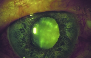
The ageing crystalline lens increases in size,Reference Jonas, Nangia, Gupta, Sinha and Bhate16 decreases in elasticity, becomes denser due to the accumulation of high-density proteins, yellows in colour and can become cataractous (Fig. 4). Reduction in elasticity of the lens and supporting ligaments results in presbyopia; the loss of the ability to read small print makes near work difficult. An initial steep increase in the need for, and strength of, reading correction beginning in the early forties becomes relatively slower but still of significance beyond the mid-fifties – by which time little or no useful accommodation is available.Reference Pointer17 Lens yellowing reduces contrast sensitivity and the associated changes in lens density alter the eye's refractive power, resulting in a ‘myopic shift.’ This acquired myopia (short-sightedness) explains the phenomenon of ‘second sight’ in some presbyopic individuals developing cataract, who become able to read without glasses but experience blurred distance vision.
Cataract (provided by A Bolton and J Kanksi)

Cataract is the commonest cause of blindness worldwide.Reference Pascolini and Mariotti18 In the UK, in 2010, the number of people with partial sight due to cataract was estimated to be around 206,000, and 28,000 with blindness.Reference Minassian and Reidy19 Increasing age is by far the most important risk factor for the development of cataract.Reference Mukesh, Le, Dimitrov and Ahmed20 Surgery is usually performed when the cataract becomes clinically significant, i.e. it is impinging on the daily activities, but other indications include to facilitate examination and disease monitoring when there is a poor view of the fundus, for example in diabetic retinopathy screening, or prevention of secondary complications of cataract, such as lens-related glaucoma.
Decreased quality of vision, glare and blur are common symptoms of cataracts and surgical intervention is generally highly successful. A post-operative visual acuity of 6/12 or better was achieved in 91% of eyes in the Cataract National Dataset audit of 55,567 operations.Reference Jaycock, Johnston, Taylor and Adams21 Modern cataract surgery takes the form of small-incision, sutureless phacoemulsification (to remove the cataractous lens) and insertion of a foldable or injectable intraocular lens (IOL). When choosing which IOL to insert, the refractive aim is most commonly emmetropia (focal point in the distance at infinity) in order to obtain clear vision for distance, i.e. driving or watching television, with no or minimal glasses prescription, but the patient will require glasses for reading, unless a multifocal IOL is used. There are many different IOL options including multifocal aspheric, toric and yellow lenses, which have been developed to optimize outcomes and can be chosen on an individual basis.Reference Agresta, Knorz, Kohnen, Donatti and Jackson22
Vitreous
The vitreous humour is the transparent gelatinous mass located between the lens and retina that makes up the majority of the volume of the globe. Its major component is water, but its structure is characterized by a network of collagen fibrils and hyaluronic acid, which undergoes irreversible changes with ageing. Condensation of the gel, increased mobility of fibrillary structures, and formation of fluid-filled spaces called lacunae occur. As the vitreous humour becomes more liquid, the lacunae coalesce to form larger cavities, eventually followed by separation of the vitreous body from the retina known as a ‘posterior vitreous detachment’ (PVD). The onset of a PVD is usually heralded by flashing lights on eye movement followed by floaters that move in the direction of gaze. Most PVDs are innocuous, but, as they can be complicated by retinal tears or detachment, prompt referral to an ophthalmologist is recommended. The frequency of partial or complete PVD increases with age.Reference Weber-Krause and Eckardt23
Retina and macula
The retina undergoes several structural changes with increasing age, many of which can be detected on clinical examination. These have variable, often insignificant, effects on function.
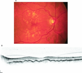
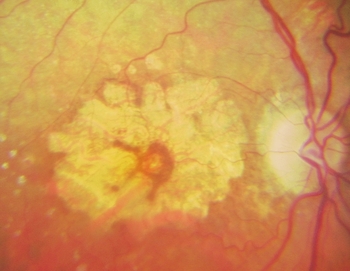
Drusen are yellow-white deposits between Bruch's membrane and the basement membrane of the retinal pigment epithelium (RPE) (Fig. 5A) that result from incomplete digestion or removal of RPE cellular constituents; they are hallmarks of early age-related macular degeneration (AMD) and can be demonstrated more clearly using optical coherence tomography (OCT) (Fig. 5B). OCT retinal imaging captures reflected light from retinal structures to create a cross-sectional image of the retina, which is comparable to histological sections as seen with a light microscope. OCT is used to determine whether active wet age-related macular degeneration is present. Drusen or early AMD have been documented in one in three individuals over the age of 75, and may be present for a long time prior to development of AMD.Reference Klein, Klein, Jensen and Meuer24 It is not inevitable that the presence of drusen will lead to development of wet or dry AMD. Only a proportion of patients will go on to develop advanced AMD. Structural changes in the RPE include hypertrophy, hyperplasia, depigmentation and formation of melanolysosomes, which can lead to gradual atrophy (Fig. 6). Within the RPE there is a progressive accumulation of autofluorescent lipofuscin granules that are highly photoreactive, have the potential for cellular damage via generation of free radicals, and are implicated in retinal pathology in ageing and AMD.Reference Farkas, Sylvester and Archer25, Reference Nag and Wadhwa26
A, age-related maculopathy (ARM) with drusen; B, OCT (optical coherence tomography) of age-related maculopathy with drusen (provided by J Brett and SM Downes)

Geographic atrophy (dry AMD) (provided by A Bolton and SM Downes)

Neural visual pathway
Loss of cells in all regions of the visual pathway from the retinal photoreceptors to the cerebral cortex occurs with age. Visual function may not be as acute as it was in earlier life, despite the presence of an anatomically intact visual pathway.
Age-related macular degeneration
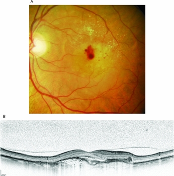
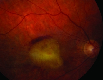
AMD is a major cause of ocular morbidity in high-income countries, accounting for over half of blind and partial sight certifications in the UK.Reference Bunce, Xing and Wormald27 AMD is a complex disease that results from the interaction of genetic and environmental factors. The main problems experienced by patients with AMD include loss of the ability to read, inability to fulfil the Driver and Vehicle Licensing Authority (DVLA) guidelines for driving, and difficulty recognizing faces. It is very unusual to lose peripheral vision, so navigational vision is usually preserved. AMD is classified as early (presence of drusen and/or non-fovea-involving atrophy) or late (exudative AMD or fovea-involving atrophy). The presence of drusen and/or RPE hyperpigmentation is typical of early AMD (Fig. 5A), while geographic (dry) late AMD is characterized by focal areas of RPE hyperpigmentation and atrophy, which coalesce to give rise to central atrophic patches (Fig. 6) and loss of central vision. The exudative or ‘wet’ late type of AMD accounts for the majority of those registered blind with AMD.Reference Economics28 Blood vessels at the level of the choroid proliferate underneath or within the retina (i.e. choroidal neovascularization) and bleed or exude fluid. Exudative AMD can present with sudden onset of central visual loss and/or distortion and, fundoscopically, intraretinal, subretinal and/or sub-RPE fluid and haemorrhage may be present (Fig. 7A). This is shown clearly in the OCT image in Fig. 7B where there is subretinal and intraretinal fluid in the presence of a choroidal neovascular complex (wet AMD). Central vision is lost as a consequence of haemorrhage and fibrosis, leading to a disciform scar (Fig. 8).
A, exudative (wet AMD) with drusen, exudation and haemorrhage (provided by A Bolton and SM Downes); B, exudative (wet) AMD OCT image showing intra- and subretinal fluid and choroidal neovascular complex (provided by J Brett and SM Downes)

Disciform AMD (old scar) (provided by A Bolton and SM Downes)

The most significant risk factors for the development of AMD are increasing ageReference Klein, Klein, Jensen and Meuer24 and genetic predisposition. There is a strong association with DNA sequence variations found within the complement Factor H gene and one single nucleotide polymorphism, Tyr402His, has been shown to be present in 43–50% of patients with AMD.Reference Klein, Zeiss, Chew and Tsai29–Reference Edwards, Ritter III, Abel and Manning31 Individuals with this allele have a five- to sevenfold increased risk of developing AMD, which increases further with smoking and homozygosity.Reference Donoso, Kim, Frost, Callahan and Hageman32 Other risk factors include ultraviolet light exposure,Reference Algvere, Marshall and Seregard33 excess dietary lipids, Caucasian raceReference Clemons, Milton, Klein and Seddon34 and female gender.Reference Rudnicka, Jarrar, Wormald and Cook35
All patients with AMD, regardless of severity (early or late) or type (atrophic or exudative), should be made aware of lifestyle changes that could alter disease progression and potentially improve their long-term prognosis. These include taking a diet rich in green, leafy vegetables (e.g. broccoli, spinach and curly kale) and brightly coloured fruit, as well as eating oily fish twice per week (which contains omega 3 fatty acid),Reference Christen, Schaumberg, Glynn and Buring36 refraining from smoking, and wearing sunglasses (UV protection) in bright, sunny conditions. The Age Related Eye Disease Study (AREDS) demonstrated, in certain forms of AMD (individuals with advanced exudative AMD in one eye and early AMD in the other eye, or with large or intermediate drusen in both eyes), benefit is derived from high-dose vitamin supplements (15 mg beta-carotene, 500 mg vitamin C, 400 IU vitamin E, 80 mg zinc oxide and 2 mg copper);Reference Clemons, Milton, Klein and Seddon34 these findings have been supported by evidence from the Rotterdam study.Reference van Leeuwen, Boekhoorn, Vingerling and Witteman37 However, smokers are advised to avoid the beta-carotene component of the supplement due to an increased risk of lung cancer in smokers taking carotenoids. There are also concerns about the safety of high-dose vitamins and microsupplementation in older people, with particular concerns regarding possible osteoporosis with excess vitamin A. For those who have developed problems with their central vision, advice regarding use of appropriate lighting for visual tasks, large-print books and the provision of low-vision aids such as magnifying glasses, CCTV and other optical devices can usually be provided by low-vision assessment clinics in ophthalmology departments, as well as by local optometrists. The advent of electronic devices allowing on-screen manipulation of contrast, font size and colour can be extremely helpful for patients with decreased central vision. Contact with appropriate sources including social services, visual rehabilitation officers and support groups (e.g. The Macular Disease Society, local associations for the blind, and the Royal National Association for the Blind) can be extremely helpful.
Various treatment modalities have been used for exudative AMD, including argon laser photocoagulation, intravitreal steroid injections, photodynamic therapy (PDT) and surgical techniques, but current treatment almost always involves the use of anti-vascular endothelial growth factor (anti-VEGF) agents such as ranibizumab (Lucentis) or bevacizumab (Avastin) given by intraocular injection. Results of the MARINAReference Rosenfeld, Brown, Heier and Boyer38 study, which compared the use of monthly ranibizumab versus sham injections, strongly favoured anti-VEGF treatment both in terms of stabilization of vision (95 vs 62% at 1 year) and significant improvement in vision (34 vs 5% at 1 year). The ANCHORReference Brown, Kaiser, Michels and Soubrane39 trial showed that ranibizumab has a similar advantage over PDT, the previous ‘gold standard’ treatment. VISIONReference Gragoudas, Adamis, Cunningham and Feinsod40 investigated the efficacy of another anti-VEGF agent, pegaptanib (Macugen), and, though effective, the results were not as good as for ranibizumab. PIERReference Regillo, Brown, Abraham and Yue41 and PrONTOReference Fung, Lalwani, Rosenfeld and Dubovy42 assessed the safety and efficacy of ranibizumab, the results of which support the widely used regimen of three injections at 1-month intervals followed by further injections as required, and there was a low overall rate of complications. Other studies investigating new drugs with a longer action, requiring less frequent injections, are in the pipeline. Comparison studies such as the UK IVAN trial assessing non-inferiority of bevicizumab (unlicensed for this indication) versus ranibizumab as well as continuous and discontinuous dosing, and cost effectiveness, have shown potential for significant savings to the NHS by use of bevicizumab, and non-inferiority at 1 year, but questions regarding the safety profile of bevicizumab need to be further assessed.Reference Chakravarthy, Harding, Rogers, Downes, Lotery, Wordworth and Reeves43 Patients offered anti-VEGF treatment need to be counselled about the risk of endophthalmitis (intraocular infection), which, though rare (approximately 1 case in every 1000 injections), often leads to significant and permanent loss of vision. There is also a potential increased risk of arterial thromboembolic events (e.g. cerebrovascular accident or myocardial infarction) in high-risk patients undergoing anti-VEGF therapy and, as a result, reduced dose treatment is sometimes offered, though this is still under investigation. In the UK, the National Institute for Health and Clinical Excellence (NICE)44 guidelines permit the use of ranibizumab for patients with exudative AMD presenting with a visual acuity of between 6/12 and 6/96 and no permanent structural damage to the fovea. Treatment consists of three intravitreal injections at monthly intervals followed by further injections on an as-required basis. Regular, initially monthly, follow-up with OCT imaging is essential.
There is no specific treatment for the atrophic, or dry, type of AMD. The natural history of geographic atrophy is an increase in atrophy over time, with a corresponding enlargement in a patient's central scotoma or ‘blind spot.’ When geographic atrophy affects the fovea there is a significant reduction in visual acuity.Reference Sunness, Margalit, Srikumaran and Applegate45 Geographic atrophy is a degenerative, rather than neovascular disorder, so effective treatments are likely to be different from those used in exudative AMD. Current research into potential therapies is based on pathogenesis: antioxidants (AREDS2Reference Chew, Clemons, Sangiovanni and Danis46: lutein, zeaxanthin and omega-3), visual cycle inhibitorsReference Schmitz-Valckenberg, Mössner, Fleckenstein, Wiedemann and Holz47 (e.g. fenretinide), chronic low-grade inflammation (corticosteroids: fluocinolone is neuroprotective), complement inhibitorsReference Reynolds, Hartnett, Atkinson and Giclas48, apoptosis (GATE study) and mitochondrial damage (DICER1 and BMP4).Reference Zhu, Deng, Xu and Hinton49
Diabetic retinopathy
Diabetic retinopathy is the commonest cause of visual impairment in young adults and is now an increasing problem among the elderly in the developed world. The prevalence of diabetes has been steadily increasing and is predicted to more than double by 2025.Reference King, Aubert and Herman50 The prevalence of diabetic retinopathy increases with duration of diabetes, with 60% of patients affected after 20 years.Reference Klein, Klein, Moss, Davis and DeMets51 With better diabetic control and increasing longevity, diabetic retinopathy is being seen occurring later in life associated with both types of diabetes.
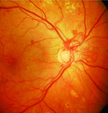
Diabetic retinal disease is typically asymptomatic until relatively advanced and as diabetic retinal treatments are aimed at preventing further visual loss rather than improving vision, early detection and monitoring is essential. Diabetic screening programmes play a crucial role.Reference Fletcher and Chong52 Although some form of diabetic retinopathy will develop in all people with diabetes with increasing age, the rate of progression and severity can be reduced by tight control of diabetes,53, Reference Stratton, Cull, Adler and Matthews54 hypertensionReference Stratton, Cull, Adler and Matthews54 and cholesterol.Reference Stratton, Cull, Adler and Matthews54 Diabetic retinopathy (DR) can be classified into non-proliferative and proliferative stages. Non-proliferative DR is characterized fundoscopically by one or more of microaneursyms, hard exudates, retinal haemorrhages, cotton wool spots, venous beading and intraretinal microvascular abnormalities; proliferative diabetic retinopathy (PDR) requires the presence of new vessels (neovascularization) (Fig. 9). PDR can lead to vitreous haemorrhage, neovascular glaucoma and retinal detachment.
Proliferative diabetic retinopathy (provided by A Bolton and SM Downes)

The mainstay of treatment for PDR is pan-retinal photocoagulation (PRP).Reference Bailey, Sparrow, Grey and Cheng55 The aim of this treatment is to induce regression of neovascularization by eliminating the presence of vasoformative factors (e.g. VEGF) produced by the diabetic ischaemic retina by selectively ablating the ischaemic, less visually important peripheral retina. This is in order to preserve the more useful central vision and prevent neovascular-related vision-threatening complications. Vitrectomy surgery may be necessary in cases of long-standing vitreous haemorrhage and retinal detachment.56
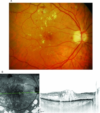
Diabetic maculopathy is the commonest cause of visual impairment in diabetes. Some forms of diabetic maculopathy may benefit from carefully directed macular focal or grid argon laser treatments (Fig. 10A). OCT is very useful in monitoring response to treatment (Fig. 10B). Improvement in vision may be achieved, but the main aim is to stabilize vision. The Early Treatment Diabetic Retinopathy Study (ETDRS) found that macular laser halves the risk of doubling of visual angle over 3 years.57 More recent research suggests that subthreshold micropulse laser is effective and causes less retinal damage.Reference Luttrull and Dorin58 Maculopathy resistant to laser treatment may respond to steroid therapy administered either by intravitreal or sub-Tenons injection,Reference Bonini-Filho, Jorge, Barbosa and Calucci59 but this is associated with significant side-effects including raised intraocular pressure (30–40%) and cataract formation in a significant proportion of those undergoing this treatment.Reference Meyer60
A, diabetic maculopathy (provided by A Bolton and SM Downes); B, diabetic maculopathy. Infrared image on the left side showing pan retinal photocoagulation scars at the arcades and microaneurysms in the macula and posterior pole. OCT image on right, taken through the macula area showing oedema centrally (provided by J Brett and SM Downes).

VEGF, which increases retinal vascular permeability, causes breakdown of the blood–retina barrier and results in retinal oedema, is up-regulated in diabetic retinopathy. Studies have shown ranibizumab (Lucentis) and bevacizumab (Avastin) to be more effective at preserving and improving vision in patients with diabetic macular oedema than placebo or laser treatment alone.Reference Rajendram, Fraser-Bell, Kaines and Michaelides61–Reference Massin, Bandello, Garweg and Hansen65 Approval for the treatment of diabetic macular oedema using ranibizumab was recently given by NICE in the UK.
Retinal vascular disease
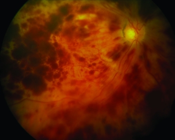
Retinal vein occlusions (Fig. 11) that occur in older people commonly reflect the presence of systemic disease, typically hypertension and hyperlipidemia.Reference Wong, Larsen, Klein and Mitchell66 Hypercoagulable and hyperviscosity syndromes are also risk factors, but are more likely to be found in younger individuals with retinal vein occlusions. Management of these systemic conditions can significantly lower the risk of recurrence and the risk to the fellow eye. Spontaneous visual recovery is possible, but regular ophthalmic review is required to detect and treat potential ocular sequelae of proliferative retinopathy, neovascular glaucoma and macular oedema. Macular laser was the mainstay of treatment for secondary macular oedema in suitable patients, but has been superseded by intravitreal steroid implantsReference Haller, Bandello, Belfort and Blumenkranz67 and anti-VEGF therapy.Reference Campochiaro, Heier, Feiner and Gray68, Reference Brown, Campochiaro, Singh and Li69
Central retinal vein occlusion

Retinal artery obstruction is an important cause of visual loss. The typical presentation is a sudden painless loss of vision, which is usually irreversible. Intermittent loss of vision characterized by symptoms of a ‘curtain coming down over the vision’ is called amaurosis fugax. The site of the obstruction will determine whether the central retinal artery is occluded; if this occurs vision is severely affected and is usually at the level of counting fingers or worse. A branch retinal artery obstruction results in field loss in the supply territory of the vessel. Emboli causing branch retinal artery occlusion (BRAO), or giving rise to amaurosis fugax, commonly originate either from the carotid arteries when carotid stenosis is the usual cause, or from the left atrium due to atrial fibrillation. Such patients are five times more likely to be affected by a cerebrovascular accident, and 30% will have a myocardial infarction within 5 years.Reference Cheng70 Approximately 90% have systemic disease, usually cardiovascular disease; associated risk factors include hypertension, hyperlipidaemia, diabetes, a hypercoagulable state and smoking.Reference Wong, Larsen, Klein and Mitchell66 Occasionally, the cause may be inflammatory as seen in giant cell arteritis and immediate treatment with corticosteroids can be sight and life saving. Management is focused on addressing the systemic risk factors that may improve patient morbidity and mortality, and includes investigation of the origin of the emboli, optimal control of cardiovascular risk factors, and use of anti-platelet agents such as aspirin or clopidogrel, which are usually initiated for secondary stroke prevention.Reference Patrono, García Rodríguez, Landolfi and Baigent71
Glaucoma
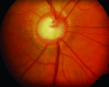
Glaucoma is a leading cause of blindness worldwide.Reference Pascolini and Mariotti18 It affects 1–2% of the white population of the UK over the age of 40 years, increasing to 4–5% in the over-80s. Glaucoma is an optic neuropathy (Fig. 12) characterized by topographical changes in the optic nerve head, with corresponding visual field defects, often with elevated intraocular pressure. It has been estimated that half of all glaucoma in the community remains undiagnosed.Reference Minassian and Reidy19, Reference Mitchell, Smith, Attebo and Healey72
Glaucomatous optic nerve cupping (provided by A Bolton and J Kanksi)

Primary open angle glaucoma (POAG), the most common form of glaucoma, typically presents in later life. It is largely asymptomatic until relatively advanced and is often detected incidentally following a routine eye examination. Risk factors include elevated intraocular pressure (IOP), thin central corneal thickness, the presence of a disc haemorrhage, increasing age, family history, black race, high myopia, hypertension, diabetes and blunt ocular trauma. Due to the asymptomatic nature of the disease, high-risk individuals should be referred for ophthalmic review. First-degree relatives of affected individuals are offered annual screening by optometrists, once they reach 40 years of age. The mainstay of treatment is to lower the IOP, which reduces the risk of progression.73 Earlier initiation of treatment leads to better outcomes.Reference Leske, Heijl, Hussein and Bengtsson74, Reference Lichter, Musch, Gillespie and Guire75 IOP can be lowered by medication (eye drops), laser therapy (e.g. argon laser trabeculoplasty)76 or surgery (trabeculectomy or tube implantation).
Acute angle closure glaucoma, although much less common than POAG, typically presents acutely with a painful red eye, blurred vision with haloes around bright lights, and often nausea and vomiting. This condition demands immediate attention to prevent visual loss. Elevated IOP results from reduced outflow of aqueous due to peripheral iris occluding the trabecular meshwork. This occurs more commonly in hypermetropes who have shorter eyes with narrower iridocorneal angles, which become progressively narrower with increasing age as the crystalline lens increases in size, and the condition is much more common in females.Reference Rüfer, Schröder, Klettner and Frimpong-Boateng77 Treatment is aimed at reducing the IOP and restoring the normal anterior chamber flow of aqueous. Management is initially medical, to lower IOP and restore corneal clarity, before definitive YAG (yttrium aluminum garnet) laser peripheral iridotomy (PI) is performed. The other eye is usually treated with a prophylactic peripheral iridotomy as there is significant risk to the fellow eye.Reference Friedman, Foster, Aung and He78
Approximately 20% of all individuals with open angle glaucoma have an IOP within the normal range. This is known as normal tension glaucoma (NTG). Such individuals have optic nerve heads that seem more sensitive to IOP levels that are within the normal range. It is hypothesized that the vascular supply is compromised or vascular optic nerve head autoregulation is abnormal, rendering individuals with NTG more susceptible to damage at a lower IOP. The association of migraine, Raynaud's phenomenon and cardiovascular disease in patients with normal tension glaucoma supports this hypothesis. The treatment is the same as for POAG; lowering the IOP reduces glaucomatous progression.Reference Schulzer79
Ocular hypertension (OHT) is the condition when the IOP is elevated without evidence of optic nerve damage or visual field loss. A number of individuals with OHT will progress to POAG, and prophylactic lowering of IOP has been shown to reduce this risk.Reference Kass, Heuer, Higginbotham and Johnson80
Increased awareness, advances in imaging (e.g. optic nerve fibre layer analysis) and implementation of NICE guidelines published in 2009 have led to glaucoma being diagnosed at an earlier stage (SR De Silva, Y Riaz, RMJ Purbrick, JF Salmon – unpublished data comparing new patients at Oxford Eye Hospital in 2010 vs 2001).
Neuro-ophthalmological disorders
These include conditions that relate to the optic nerve, visual pathway, pupils, extraocular muscles and eye movements. Neuro-ophthalmic disease typically reflects underlying systemic disease and vascular causes are common. Cerebrovascular accidents give rise to a variety of clinical manifestations, with field defects estimated to affect 20–57% of people who have had a stroke.Reference Pollock, Hazelton, Henderson and Angilley81 Other problems include visual agnosia, visual inattention or cortical blindness, ischaemic sixth and third nerve palsies and non-arteritic anterior ischaemic optic neuropathy. These usually have underlying atherosclerotic risk factors including hypertension, hyperlipidaemia, diabetes and smoking. Individuals with giant cell arteritis (GCA) are at risk of bilateral total visual loss from arteritic anterior ischaemic optic neuropathy and must be treated as an ocular emergency; prompt intervention with urgent steroid therapy (at an adequate dose) can be sight- or even life-saving. Intracranial tumours may produce optic disc swelling secondary to raised intracranial pressure (papilloedema).
Conclusion
Management of eye problems in the elderly involves diagnosis and treatment of preventable or reversible vision loss, identification and modification of associated ocular and systemic risk factors, and practical support with regard to carrying out activities of daily living to ensure independent function. In patients with severe and irreversible loss of vision, significant quality of life improvements can be made through interventions such as certificate of visual impairment (CVI) registration and referral to organizations such as the Royal National Institute for the Blind and the Macular Society.
Eye disease and sight loss are prevalent in the ageing population. As the population becomes increasingly elderly this will have a significant impact on health economics.Reference Minassian and Reidy19 Not only is visual impairment associated with an increased risk of fatal fallsReference Felson, Anderson, Hannan and Milton82 and incidence of depressive illness,Reference Rees, Fenwick, Keeffe, Mellor and Lamoureux83 sight loss has been associated with reduced life expectancy.Reference McCarty, Nanjan and Taylor84, Reference Zheng, Christ, Lam and Arheart85 There is an ongoing and increasing need for public health initiatives to screen for ocular disease, with concurrent implementation of timely treatment together with establishment of robust programmes for the rehabilitation of those who are visually impaired.
Financial support
This research received no specific grant from any funding agency, commercial or not-for-profit sectors.
Conflict of interest
None.