INTRODUCTION AND BACKGROUND
There has been an increasing demand for the radiocarbon (14C) dating of bone material and, in particular, articulated bone groups, as they are demonstrably not residual and can be used with confidence in Bayesian models. However, 14C dating of bone and antler continues to be slightly problematic for many laboratories, as the pretreatment can be challenging, with a number of different methodologies in use. The most frequently used method is based on Longin (Reference Longin1971) (and modifications thereof) to extract the organic fraction of the bone. More recently this has been complemented by ultrafiltration of the collagen protein with the aim of removing non-collagen organic molecules of <30 kDa (Bronk Ramsey et al. Reference Bronk Ramsey, Higham, Bowles and Hedges2004).
The chemical pretreatment of bone is designed to isolate a protein/amino acid fraction, free from non-sample carbon that originated from the post-depositional environment, while simultaneously trying to avoid the addition of further non-sample carbon from the laboratory during the isolation process.
It is assumed that the majority of any potential laboratory-derived contaminant carbon is relatively modern (Vogel et al. Reference Vogel, Nelson and Southon1987). However, the very nature of the collagen isolation process involves the addition of water that is subsequently freeze-dried, and which will inevitably contain trace amounts of carbon that could vary in F14C value. The complex, multiple-step process involved in the isolation of the bone collagen may provide an explanation of why the background associated with bone analysis is often greater than the values attained for other sample types such as wood and carbonate (Tisnérat-Laborde et al. Reference Tisnérat-Laborde, Valladas, Kaltnecker and Arnold2003; Wood et al. Reference Wood, Bronk Ramsey and Higham2010). These studies have also suggested that the background is related to collagen yield and it is agreed that the effect of modern carbon is more significant on small samples. Over a number of years, collagen has been extracted from a range of bone weights and a range of collagen yields obtained. However, within the limits of sample sizes processed in the laboratory, there is no observed relationship between higher F14C values and small sample size. It is also recognized that the isolation procedure used to obtain the alpha cellulose fraction from the interglacial wood, non-bone background standard is also complex, albeit different.
The analytical challenges of obtaining “pure” bone collagen can be magnified by the addition of chemicals that may be used in the preservation and showcasing of bone materials for museum and cataloging purposes. These can potentially add additional carbon contamination and it is crucial that any chemical contamination is identified before the initial bone pretreatment stages commence. For example, the chemical polyvinyl acetate [PVA, with the formula (C4H6O2)n] was formerly used as a compound to stabilize fragile bones. Varnish of various types may often be applied prior to display, while Indian ink and modern chemicals such as Tipp-Ex have been used on bone surfaces to aid curators with identification. Furthermore, animal glue (effectively a modified collagen) has been noted on some samples. Such problems are commonly found with bones that have been in museum or archive stores, therefore the pretreatment of this type of sample should be modified in an attempt to remove these forms of contamination, e.g. by the mechanical abrasion of bone surfaces or by a more complex sequential extraction of the contaminant using solvents. In some cases, where the chemical compound has become an intrinsic part of the bone structure, it may not be possible to isolate the original bone collagen from the contaminant carbon (Healy et al. Reference Healy, Marshall, Bayliss, Cook, Bronk Ramsey, van der Plicht and Dunbar2014). In this paper, only bones of infinite age with respect to F14C, that have not undergone any curation and have had no chemical treatments of any kind applied to them, are used.
Developments such as ultrafiltration have been used to improve the quality of the extracted bone collagen by the removal of small molecular weight compounds (<30 kDa) (Bronk Ramsey et al. Reference Bronk Ramsey, Higham, Bowles and Hedges2004; Higham et al. Reference Higham, Jacobi and Bronk Ramsey2006; Brock et al. Reference Brock, Bronk Ramsey and Higham2007). The <30 kDa fraction is assumed to be either degraded collagen or exogenous material. However, as a side issue, it has been noted that excessive heating of the collagen solution may inadvertently cause the breakdown of intact collagen proteins, generating molecules in this lower molecular size range. The potential effect this ultrafiltration step may have on background bone values is also discussed and it is accepted that the ultrafilters may contain residual contaminant carbon from the humectant present in the filters if imperfectly cleaned prior to use (Brock et al. Reference Brock, Higham and Bronk Ramsey2013). Therefore, in order to monitor the amount and potential F14C value of this humectant, the filtrate solution from one cleaned ultra-filter is retained, combusted, graphitized, and measured on the AMS on a regular basis. Introduction of the ultrafiltration method has been trialled at SUERC and the F14C values for routine background measurements using the ultrafiltration method are also included here.
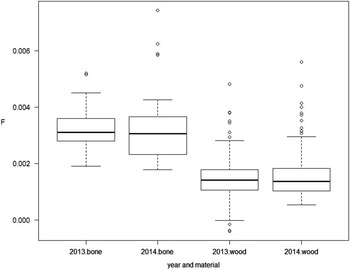
The laboratory employs a one-step “background subtraction” method in the 14C age calculation procedure. For non-bone samples, an interglacial wood, used in the Fifth International Radiocarbon Intercomparison (VIRI Sample K), is employed as the background standard (Scott et al. Reference Scott, Cook, Naysmith, Bryant and O’Donnell2007, Reference Scott, Cook and Naysmith2010a, Reference Scott, Cook and Naysmith2010b). For every batch of samples measured on the AMS, several standards are prepared and measured to produce a mean background value (Xu et al. Reference Xu, Anderson, Bryant, Cook, Dougans, Freeman, Naysmith, Schnabel and Scott2004; Naysmith et al. Reference Naysmith, Cook, Freeman, Scott, Anderson, Xu, Dunbar, Muir, Dougans, Wilcken, Schnabel, Russell, Ascough and Maden2010; Dunbar et al. Reference Dunbar, Cook, Naysmith, Tripney and Xu2016). In order to obtain a representative bone background F14C value, sub-samples of an infinite age mammoth bone (LQH12) [confirmed in a previous study by Cook et al. (Reference Cook, Higham, Naysmith, Brock, Freeman and Bayliss2012)], are routinely prepared and measured along with every batch of bone samples (Dunbar et al. Reference Dunbar, Cook, Naysmith, Tripney and Xu2016). In recent years (2011–2016), approximately 25% of the samples measured in the SUERC Radiocarbon Laboratory have been bone (virtually all of which are of Holocene age and the vast majority less than one half-life in age) and have undergone collagen extraction, while approximately 65% have been non-bone organic samples. The remainder have included a wide range of different sample types including a few percent of carbonates (mollusc shell and foraminifera) and cremated bone. The F14C values of the wood background, compared with those of the mammoth bone background (measured in the same time frame) suggest that the latter is approximately double (Figure 1) (see also Naysmith et al. this volume) as shown by the central line (median) of each box and this is consistent in both years. Figure 1 also shows the spread of values for the two materials in each of the two years. The central box shows the range [inter-quartile range (IQR)] where 50% of the observations lie, and there is a suggestion that the IQR is narrower for the wood than bone samples. The observations identified by the ° symbol are results that are at least 1.5 times the interquartile range (Q3–Q1) from the upper or lower quartile, and it can be seen that there are more such observations for the wood rather than the bone samples, however, this might be expected since proportionately there are approximately 7 times more wood than bone samples in the series.
Comparison of F14C values: Mammoth bone and Heidelberg wood (2013/August 2014). Circled results are those that are at least 1.5 times the interquartile range (Q3–Q1) from the lower or upper quartile.

AIMS AND OBJECTIVES
The main objectives of this paper are to understand what factors contribute to the variations in the bone background and identify why it is higher than the wood background and what precautions can be taken during sample processing to minimize this offset, working towards a stable, consistent value that is more comparable with the F14C values obtained from the wood background standard.
Therefore, the objectives are to:
-
∙ Directly compare both collagen extraction methods (i.e. with and without ultrafiltration) employed in the laboratory and also to investigate the effect of bypassing the collagen solubilization step to greatly simplify the pretreatment scheme. (This can only be done on really high quality, carefully chosen samples and relies on there being no significant contamination.) This will provide some information on whether the complexity of the pretreatment scheme was responsible for the observed higher background values.
-
∙ Set up and verify a DOC method to measure the F14C of (i) the water drawn from the reverse osmosis source routinely used in the bone collagen extraction procedure, (ii) the water from an alternative UV-treated reverse osmosis system, and (iii) mains water that has undergone no treatment. The object here is to determine if the water used in the collagen solubilization stage is a contributing factor to the variable F14C values.
-
∙ Measure the F14C values from an alternative background bone sample (LQH4), used in the Sixth International Radiocarbon Intercomparison (SIRI Sample C), to establish if lower F14C values are achievable from a similar sample derived from Marine Isotope Stage 7.
MATERIALS AND METHODS
As a prerequisite, all pretreatment steps are carried out in a fume hood and all glass vessels and implements are precleaned by washing in a 5% solution of Decon® 90 (surface active decontamination solution), rinsing with 0.1 M HCl and ultrapure water from a Milli-Q® Elix 5 Water Purification System. This quality of water is used throughout all pretreatments for rinsing and preparation of solutions. Following this cleaning, the glassware and implements used in pretreatment and graphitization are heated at 500°C and allowed to cool overnight. All chemical reagents used are analytical grade or better.
Heidelberg Wood Background Sample
An interglacial oak sample, supplied by the dendrochronology laboratory of the University of Hohenheim is used as the SUERC laboratory’s non-bone organic background standard and is used here as a basis for comparison with the bone background values. The alpha cellulose is prepared using a modification of the technique of Hoper et al. (Reference Hoper, McCormac, Hogg, Higham and Head1998) as described in Dunbar et al. (Reference Dunbar, Cook, Naysmith, Tripney and Xu2016). This wood was used in the VIRI study (Sample K), and has a consensus value of 0.0576±0.0062 pMC (F14C=0.0006±0.0001) (Scott et al. Reference Scott, Cook, Naysmith, Bryant and O’Donnell2007, Reference Scott, Cook and Naysmith2010a, Reference Scott, Cook and Naysmith2010b).
Mammoth Bone Background Sample
The mammoth bone LQH12 (Mammuthus cf. trongontherii) was kindly provided by Dr Katharine Scott (St Cross College, Oxford). The sample originates from Latton Quarry in the Upper Thames Valley. The quarry deposits are mainly of medium to coarse limestone gravels, with minor fine-grained facies. These gravels contain faunal remains including a distinctive small form of mammoth (Mammuthus cf. trogontherii) that imply temperate conditions. A U-series age estimate of >147.4±20 kyr, demonstrates that the bones will be of infinite age with respect to radiocarbon and suggests that the deposits correlate with Marine Isotope Stage 7 (MIS 7) (Scott and Buckingham Reference Scott and Buckingham2001; Lewis et al. Reference Lewis, Maddy, Buckingham, Coope, Field, Keen, Pike, Roe, Scaife and Scott2006).
Bone Collagen Preparation: SUERC Modified Longin
The laboratory practices a modification of the Longin (Reference Longin1971) procedure for the routine extraction of collagen from bone samples. Individual, uniform-sized subsamples of LQH12 are cut and the bone surfaces cleaned to remove any adhering soil and contaminant material using a Dremel multi-tool. The bone fragments are weighed and the weights recorded. After close inspection to ensure no surface contaminants remain, 100 mL of 1 M HCl are added for 24 hr, after which the bone material should appear “jelly like.” The excess acid is then decanted and 100 mL of ultrapure reverse osmosis water are added and the samples heated at approximately 80ºC for 3 hr. When the material is solubilized, the samples are allowed to cool slightly and filtered using a Buchner funnel and pre-furnaced GF/A filter paper. The collagen filtrate solutions are dried down to <20 mL and transferred to weighed vials. They are allowed to cool before being frozen and transferred to a freeze drier until all the solution is removed and the crystalline collagen powder remains.
Bone Collagen Preparation: Ultrafiltration
For the modified ultrafiltration method, the collagen extraction procedure is the same as above up to the point of reducing the volume to <20 mL. At this point, the collagen solution is transferred to pre-cleaned Vivaspin 20TM 30 kDa MWCO PES filters, centrifuged and transferred to pre-weighed 20 mL vials before freeze drying. The collagen samples are then combusted and converted to graphite for AMS measurement as described by Dunbar et al. (Reference Dunbar, Cook, Naysmith, Tripney and Xu2016).
Doublespar Carbonate Background Sample
A geological-age carbonate in the form of Icelandic doublespar, used in the Third International Radiocarbon Intercomparison (TIRI Sample F), is used as the laboratory’s inorganic background standard. Approximately 20% of the doublespar surface is removed by appropriate addition of 1 M HCl. This eliminates potential surface and edge contaminant CaCO3. The sample is then rinsed with ultra-pure reverse osmosis water and dried. For CO2 generation, 0.1 g samples are weighed into hydrolysis units (large, single fragments are selected rather than fine material) where a further 20% of the doublespar is reacted with the appropriate volume of 1 M HCl, under vacuum, and the evolved CO2 discarded. The remaining material is hydrolysed and the CO2 collected and converted to graphite for AMS measurement as described by Dunbar et al. (Reference Dunbar, Cook, Naysmith, Tripney and Xu2016).
Carbon in Laboratory Water (DOC)
A system to evaporate water samples under vacuum, based upon that devised by Burr et al. (Reference Burr, Thomas, Reines, Jeffery, Courtney, Jull and Lange2001), was assembled. This method involved the placement of 2 L of water into a pre-combusted (at 500ºC) quartz vessel connected to a diaphragm pump, via a Dewar flask cooled to –50ºC with ethanol/water (68/32 v/v). The water sample was evaporated until almost dry and the residue pipetted into a pre-weighed quartz insert, dried, combusted, and graphitized (Burr et al. Reference Burr, Thomas, Reines, Jeffery, Courtney, Jull and Lange2001).
Two sources of reverse osmosis water were used for the comparison of water purity. Replicate samples of the reverse osmosis water routinely used in all analyses in the SUERC Radiocarbon Laboratory were taken. The second source, for comparison, was UV-treated reverse osmosis water used in a clean laboratory within SUERC and assumed to be of better quality in terms of carbon concentration, having undergone the additional UV treatment step to destroy organic material. An additional water sample was also taken from the mains source which feeds both water purification systems, to determine the quality before treatment.
RESULTS
Background F14C Values for Heidelberg Wood and Mammoth Bone
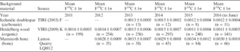
The average background F14C values for all material types measured between January 2011 and June 2016, using the available AMS instruments, are presented Table 1. As the data for each year show, the F14C values for the mammoth bone (LQH12) are approximately double those of the Heidelberg wood and Icelandic doublespar. This difference in the F14C values between the material types is most noticeable in 2015 and 2016, with mammoth bone values of 0.0034±0.0012 and 0.0030±0.0010, compared with Heidelberg wood values of 0.0011±0.0004 and 0.0011±0.0005, respectively (F14C values quoted in this paper are all±1σ). The mammoth bone F14C data presented in the table include both the modified Longin and modified ultrafiltration methods practiced at SUERC.
F14C values for all background types from January 2011 to June 2016 (all measurements). Errors are all±1σ.

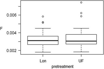
There has been much discussion on the effectiveness and problems associated with ultrafiltration (Brock et al. Reference Brock, Higham and Bronk Ramsey2013); therefore both collagen extraction procedures must be considered as a potential contributing factor to the increased F14C values. However, the running mean values of both the SUERC modified Longin and the SUERC modified ultrafiltration collagen extraction methods (F14C=0.0032±0.0009 and F14C=0.0034±0.0013, respectively) are effectively identical within the uncertainty (based on 2013 and 2014 data) (Figure 2), confirming that there is no difference between the two collagen extraction methods practiced at SUERC and therefore the contribution to the higher variation in bone collagen values exists across the two data sets. Indeed, this supports the results of a previous comparison between the Oxford and SUERC laboratories where Oxford employed their ultrafiltration method and SUERC used only the modified Longin method (without ultrafiltration) (Cook et al. Reference Cook, Higham, Naysmith, Brock, Freeman and Bayliss2012).
Box plot showing the distribution of F14C values from mammoth bone LQH12, prepared by either SUERC modified Longin method or SUERC modified ultrafiltration.

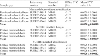
The comparison of the results of these two methods and those for demineralized bone (i.e. bone that has undergone demineralization, but bypassed collagen solubilization (which involves the addition of reverse osmosis water and freeze-drying), are presented in Table 2. These preliminary measurements demonstrate slightly lower F14C values but the difference is not statistically significant (F-test, p-value of 0.1: we have used the convention that a p-value <0.05 demonstrates a statistically significant result).
F14C values for background bone sample, LQH12, with different pretreatment methods.

Measurement of Reverse Osmosis Water (DOC)
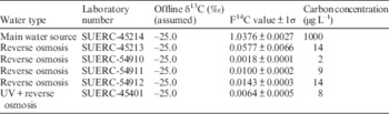
The F14C results for the water analyses are shown in Table 3. They indicate that the mains water has a modern signal, with an F14C value of 1.0376±0.0027, and a significantly higher level of contaminant carbon. Each of the four replicate reverse osmosis water samples from the same source all show significantly reduced carbon contamination and an “old” F14C signal, with three F14C values of 0.0018±0.0001, 0.0100±0.0002, and 0.0143±0.0003 trending towards background values; however, there is a higher anomalous F14C value (0.0577±0.0066). The UV treated water also provided a low F14C value of 0.0064±0.0005. This indicates that the reverse osmosis removes the modern and probably more labile organic material, leaving a very old, intractable organic carbon fraction.
F14C values for the dissolved organic carbon in various water supplies within SUERC.

Bone Sample LQH4
A direct comparison of the routinely used background sample (LQH12) with another mammoth bone (LQH4) (Mammuthus cf.trongontherii, Latton Quarry, Wiltshire, again provided by Dr K Scott) was carried out. This bone was included in the SIRI study [Sample C (Scott et al. Reference Scott, Naysmith and Cook2017)]. A small set of 6 replicate bone collagens extracted from both mammoth bones (LQH4 and LQH12, SUERC modified Longin method) show very similar mean values of F14C=0.0020±0.0001 and F14C=0.0019±0.0003 respectively (Table 4). No statistically significant difference in the means was found (two sample t-test, p-value 0.5).
F14C values for two background mammoth bones samples, LQH4 and LQH12.

Discussion
The F14C data for the background materials used in the SUERC Radiocarbon Laboratory show that a statistical difference exists between the routinely measured values of the Heidelberg wood and doublespar carbonate compared with the mammoth bone samples, the latter being approximately a factor of two higher (Figure 1). The LQH12 mammoth bone data show that the running mean values for both the SUERC modified Longin and the SUERC modified ultrafiltration collagen extraction methods are comparable (Figure 2). In addition, bypassing the solubilization stage produced no significant difference from the Longin and ultrafiltration methods (Table 2). Therefore, we conclude that the complexity of the collagen isolation process is unlikely to be responsible for the additional background contribution to the bone F14C measurements.
The F14C values in the reverse osmosis water samples were variable and low. Also, a maximum of 14 µg L–1 carbon was measured in the water samples, while an average collagen weight of >100 mg was prepared per sample. Therefore, the carbon remaining in the water after reverse osmosis treatment (and UV) will have no significant impact on the bone background values. The variation in F14C observed between the water samples may be a consequence of several influences including the actual mains water supplying the laboratory, and the production and storage of the reverse osmosis water. This could include the maintenance of the filter cartridges, the usage of the system, and the duration that the purified water was stored before use. Maintenance is carried out on a routine basis and water is now routinely prepared on demand for bone analyses.
Data were compared between the routinely used bone background sample (LQH12) and another bone (LQH4) and no difference was observed in the F14C values, demonstrating that it is unlikely to be related to the routinely used sample. One obvious point to note is that all of the replicate bone analyses of LQH4 and LQH12, undertaken independently for this study (Table 4), gave consistent, low F14C values that were less than the averages for each of the last 5 years and indeed approached the values for the wood background. This could indicate the possibility of slight differences in the procedures undertaken by different staff members, perhaps associated with the demineralization process, whereby incomplete demineralization could result in contaminants being held by the phosphate of the hydroxyapatite.
CONCLUSIONS
It can be concluded from the results that the higher background values observed for bone are not related to the complexity of the pretreatment process. Similarly, comparison of two different bone samples deriving from MIS 7 showed no difference in background between them, implying that the actual bone was not the issue. Finally, an investigation into the water quality used in the collagen dissolution process has shown that either reverse osmosis or reverse osmosis/UV treatment is sufficient to reduce the organic matter content to a level that could not influence the background. In addition, these treatments seem to remove all but the very old organic component such that F14C=0.0577±0.0066 was the highest value measured. The focus is now on how well the samples are originally demineralized (could any remaining phosphate in the hydroxyapatite form be able to retain contaminant carbon?). The higher background is not necessarily a problem if it is stable, however, the variability in results is greater than for our wood background and this is reflected in the errors on bone samples we analyze (which tend to be of 1 half-life or less in age) being higher by around 1–6 years on our routine analyses. While the error on the bone background measurements accounts for any differences, reducing this error will help to minimize the error on unknown age samples. We are currently taking steps to standardize the pretreatment regime beyond that of our current procedure.
Acknowledgments
Mammoth bone samples were kindly provided by Dr Katharine Scott (St Cross College, Oxford). A special thanks to Helen Kinch, Iain Murdoch, Kerrie-Anne Lang, and Linzi Straub for sample preparation.