Noonan syndrome is an autosomal dominant disorder characterised by distinctive facial features, short stature, chest deformity, and CHD.Reference Romano, Allanson and Dahlgren 1 Noonan syndrome is caused by mutations of genes that take part in the Ras–mitogen-activated protein kinase signalling pathway, for example, PTPN11, SOS1, and RAF1. A large cohort study revealed that patients with RASopathies have a good cumulative survival along with a higher risk for cardiac events in infants and young adults.Reference Calcagni, Limongelli and D’Ambrosio 2 More than 80% of individuals with Noonan syndrome have cardiovascular involvement that most frequently includes CHD and hypertrophic cardiomyopathy,Reference Romano, Allanson and Dahlgren 1 , Reference Calcagni, Limongelli and D’Ambrosio 2 but only few cases of adolescent or adult Noonan syndrome with coronary artery abnormalities have been reported.Reference Romano, Allanson and Dahlgren 1 , Reference Tartaglia, Gelb and Zenker 3
Case report
A 3-year-old male patient was admitted to our hospital with history of fever for 5 days, and conjunctival hyperaemia. Laboratory findings on arrival included the following: white blood cell count, 15.9×10⁹/L (13.7×10⁹ polymorphonuclear leukocytes); blood platelet count, 550×109/L; aspartate aminotransferase, 55 IU/L; serum sodium, 134 mmol/L; and C-reactive protein, 118.8 mg/L. The fever still continued despite sulbactam/ampicillin administration. Erythema and oedema of hands and feet appeared 7 days after fever appeared. An echocardiogram showed perivascular brightness of coronary arteries. Thus, the diagnosis of Kawasaki disease was established based on clinical findings.Reference Kobayashi, Inoue and Takeuchi 4 Although treatment with aspirin 50 mg/kg/day and intravenous immunoglobulin 2 g/kg started on the 7th day of illness, the fever persisted; thus, additional intravenous immunoglobulin with prednisolone 2 mg/kg/day and urinastatin 30,000 U/kg/day were administrated on day 9. Body temperature went down, and C-reactive protein level decreased on the next day. On day 12, although C-reactive protein level normalised, abdominal distension and systemic oedema appeared with thrombocytosis (1008×109/L). A chest X-ray showed pleural effusion and abdominal echo showed ascites. An echocardiogram was performed every few days from admission, and indicated right coronary artery dilation on day 14, with internal diameter, 4.5 mm; coronary artery z-score, 6.13 SD. An electrocardiogram was normal (Supplementary Fig 1A). Because, on day 16, echocardiogram showed a giant aneurysmal dilatation of right coronary artery up to 8.3 mm (11.18 SD) (Supplementary Fig 1B), infliximab 5 mg/kg was administered concomitantly with ticlopidine, warfarin, and enalapril; then, oedema and thrombocytosis improved gradually.
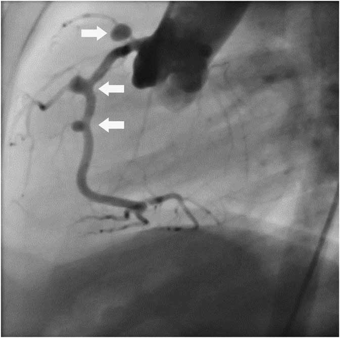
After 3 months, invasive coronary angiography revealed atypical features of coronary artery dilation (Fig 1) and multiple aneurysms/dilatations, 3–4 mm of internal diameter, just distal to branches of right coronary artery; however, no dilatation of the right main coronary artery was found. At 1-year follow-up with echocardiogram revealed gradual regression of right coronary artery aneurysm down to 4.4 mm (5.43 SD).
Atypical coronary artery aneurysms. Coronary arteriograms showing multiple aneurysms/dilatations just distal to branches without the right main coronary artery, 3 months after the onset of Kawasaki disease. Arrows indicate coronary artery aneurysms/dilatations, 3–4 mm of internal diameter (3.3–5.2 SD) in the right coronary artery just distal to branches including the conus branch, right ventricular branch, and acute marginal branch.

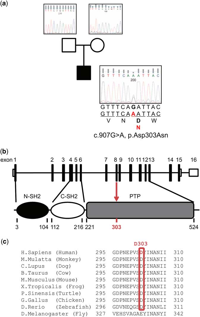
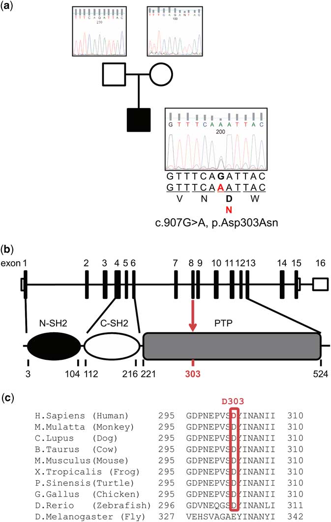
The patient was followed because of clinical diagnosis of Noonan syndrome based on several characteristic symptoms, including shot stature (−2.7 SD), chest deformity, multiple café-au-lait macules, and suggestive facial features (Supplementary Fig 1C–E) without CHD. The clinical suspicion of Noonan syndrome was confirmed by molecular analysis of the PTPN11 gene, which revealed a novel de novo heterozygous missense mutation, c. 907 G>A (p.Asp303Asn) (Fig 2a).
A novel PTPN11 mutation, Asp303Asn. ( a ) Pedigree diagram of the pedigree with the genotype of the PTPN11 mutation, which indicates de novo mutation. ( b ) Location of the mutations on a schematic of the PTPN11 gene map. ( c ) Conservation of Asp303 in vertebrate PTPN11. Homology analysis was conducted using HomoloGene (http://www.ncbi.nlm.nih.gov/homologene/20090), which automatically constructs putative homology groups using complete gene sets from a wide range of species.
Discussion
The PTPN11 gene encodes SHP2, which is a member of ubiquitously expressed protein tyrosine phosphatase with positive regulatory roles in signalling pathways, including Ras–mitogen-activated protein kinase signalling, is the major disease-causing gene in Noonan syndrome.Reference Romano, Allanson and Dahlgren 1 PTPN11 mutations that cause Noonan syndrome are almost always involved in the N-SH2/PTP interdomain binding network, and up-regulate SHP2 function.Reference Tartaglia, Gelb and Zenker 3 We found a novel missense mutation within the PTP domain (Fig 2b). The amino acid residue D303 is conserved among vertebrates (Fig. 2c), and in silico analysis with SIFT® and Polyphen2® predicted that D303N might indicate “damaging”, prediction scores were 0.001 and 0.533, respectively. These findings and the characteristic phenotype indicate that D303N might be pathogenetic mutation that causes Noonan syndrome.
The patient had typical symptoms of Kawasaki disease and a relatively low risk score (<5Reference Kobayashi, Inoue and Takeuchi 4 ). However, the clinical course of Kawasaki disease appeared to be extraordinarily acute deterioration despite additional treatment, which could have resulted in giant coronary artery aneurysms, even though that Kawasaki disease was extremely rare. Indeed, the patient developed severe thrombocytosis and vascular hyperpermeability, which caused severe oedema, ascites, and pleural effusion after pyretolysis and normalisation of C-reactive protein level. These findings might indicate persistent inflammation localised at blood vessels caused by hyperactivation of tumour necrosis factor-α and vascular endothelial growth factor, which were reported to have a key role in coronary artery aneurysms development during acute Kawasaki disease.Reference Dimitriades, Brown and Gedalia 5 , Reference Breunis, Davila and Shimizu 6 Thus, we hypothesised that hyperactivation of SHP2 due to the PTPN11 gain-of-function mutation might affect the development of giant coronary artery aneurysms by excessive inflammation and activation of signalling cascades, because SHP2 could contribute to activation of the signalling pathway of tumour necrosis factor-α and vascular endothelial growth factor.Reference Bode, Schweigart and Kehrmann 7 , Reference Taimeh, Loughran, Birks and Bolli 8
Coronary artery aneurysm is a rare complication only in adolescent or adult Noonan syndrome.Reference Romano, Allanson and Dahlgren 1 , Reference Calcagni, Baban, De Luca, Leonardi, Pongiglione and Digilio 9 The aetiology of coronary artery aneurysms remains unclear; some postulated explanations are that they are caused by a jet flow from bicuspid aortic valve, concomitant hypertrophic cardiomyopathy, a connective tissue disorder, and a direct result of the specific PTPN11 gene mutation.Reference Mauro, Flors, Hoyer, Norton and Hagspiel 10 The patient had no congenital anomaly in coronary arteries or hypertrophic cardiomyopathy that were confirmed by echocardiography before treatment for Kawasaki disease. Calcagni et al noted that the presence of coronary artery aneurysms in the absence of hypertrophic cardiomyopathy raises the possibility that this vascular phenotype is the result of an RAS pathway defect.Reference Calcagni, Baban, De Luca, Leonardi, Pongiglione and Digilio 9 Because SHP2 is involved in the control of cell growth, differentiation, and apoptosis,Reference Tartaglia, Gelb and Zenker 3 a connective tissue defect due to PTPN11 mutations could explain the fragility of vascular walls, particularly of the coronary arteries and aorta.Reference Mauro, Flors, Hoyer, Norton and Hagspiel 10 Indeed, the patient revealed atypical presentation of giant coronary artery aneurysms and multiple coronary sequelae just distal to the branches, subsequent to developing Kawasaki disease. Taken together, we hypothesised that this PTPN11 mutation might affect hyperinflammation caused by Kawasaki disease and vascular fragility in the coronary arteries, which resulted in giant and atypical coronary artery aneurysms in our patient.
Careful follow-up of cardiovascular screening was recommended for all RASopathies, independent of the underlying CHD/hypertrophic cardiomyopathy.Reference Calcagni, Baban, De Luca, Leonardi, Pongiglione and Digilio 9 Our study might indicate that there is a high risk of coronary artery aneurysms due to Kawasaki disease in Noonan syndrome patients, and indicates that earlier additional intervention with therapeutic monoclonal antibodies or cyclosporine is required to reduce cytokine production.
Supplementary material
To view supplementary material for this article, please visit https://doi.org/10.1017/S1047951118001877
Acknowledgements
The authors thank Dr Asami Shimbo, Dr Masato Nishioka, Dr Masayuki Shimohira (Kawaguchi Municipal Medical Center), Dr Eriko Komiya, Dr Makito Sakurai, Dr Yohei Yamaguchi, and Dr Yoshichika Maeda (Tokyo Medical and Dental University) for their clinical support. We are grateful to Dr Kenichi Kashimada, Dr Masatoshi Takagi, and Prof. Tomohiro Morio (Tokyo Medical and Dental University) for their insightful comments.
Financial Support
This research received no specific grant from any funding agency, or from commercial or not-for-profit sectors.
Conflicts of Interest
None.
Ethical Standards
The authors assert that all procedures contributing to this work comply with the ethical standards of the relevant national guidelines on human experimentation (Ethical Guidelines for Medical and Health Research Involving Human Subjects and Human Genome/Gene Analysis Research in Japan) and with the Helsinki Declaration of 1975, as revised in 2008, and has been approved by the institutional committees (the Institutional Review Board of Tokyo Medical and Dental University; article#: 2015-39).
Informed Consent
Written informed consent was obtained from the patient’s parents. In addition, informed consent was obtained from all individual participants for whom identifying information is included in this article.