INTRODUCTION
Heliciculture (snail farming) is becoming an emerging zootechnic industry in Europe in response to the increasing demand of snails for pharmaceutical, cosmetics, and especially alimentary purposes. During the last decades, the required development of reliable helicicultural methods has generated diverse farm designs, which have been based on extensive, intensive or mixed systems (Daguzan, Reference Daguzan1989; Gomot and Deray, Reference Gomot and Deray1990). In extensive systems snails are reared in outdoor fields, while in intensive systems rearing is carried out entirely inside greenhouses or buildings under controlled conditions of humidity, temperature and photoperiod. Finally, in mixed systems, developed in France and widely extended in Spain and especially in Galicia (NW Spain), reproduction and first growing (nursery) are carried out indoors, while fattening is completed outdoors. About 40% of snails consumed in Europe belong to the species Helix aspersa, which includes the subspecies H. a. aspersa, commonly known as petit-gris, and Helix a. maxima, known as gros-gris by its larger size and which seems to exhibit a high potential for growth and fertility (Chevallier, Reference Chevallier1990; De Grisse, Reference De Grisse1991). Gros-gris consumption is relevant in France, but both gastronomy and heliciculture in Spain are still focused principally on petit-gris snails. In spite of this, some farms have already started to rear the subspecies H. a. maxima in our country.
Profitability of mixed farms in Galicia (NW Spain) is being seriously threatened by severe mortalities occurring during the intensive breeding phase, during which overcrowding, and probably reproductive stress, may facilitate the transmission and reproduction of certain pathogens. In addition, snails used for breeding may come from different geographical areas favouring the introduction of diverse pathogenic organisms in farm environments. Previous studies have reported the presence of diverse parasites infecting H. aspersa snails under farming conditions (Morand, Reference Morand1985; Morand and Daguzan, Reference Morand and Daguzan1986; Fontanillas and Pérez, Reference Fontanillas and Pérez1987; Cabaret et al. Reference Cabaret, Morand, Aubert and Yvore1988; Morand, Reference Morand1988a; Morand and Bonnet, Reference Morand and Bonnet1989; Segade et al. Reference Segade, Kher, Lynn and Iglesias2009, Reference Segade, Crespo, Garcia, Garcia-Estevez, Arias and Iglesias2011), but only a few of these studies have used an epidemiological approach (Morand and Daguzan, Reference Morand and Daguzan1986; Cabaret et al. Reference Cabaret, Morand, Aubert and Yvore1988; Morand, Reference Morand1988a; Morand and Bonnet, Reference Morand and Bonnet1989). These kinds of investigation are essential for the control of pathogenic species in any animal production activity, but especially in heliciculture, where chemotherapy strategies have never been practically assessed (Cabaret et al. Reference Cabaret, Morand, Aubert and Yvore1988).
In the present work, we have studied the parasitic infections affecting the snails H. a. aspersa and H. a. maxima in 3 heliciculture farms of Galicia (NW Spain) and their dynamics through the different phases of rearing (hibernation, mating, egg-laying and fattening). In addition, we have also investigated the possible sources of infection and/or reservoirs of certain parasite species in these farms.
MATERIALS AND METHODS
Study design and gastropod sampling
In order to investigate the parasite fauna infecting the 2 H. aspersa subspecies commonly cultured in Galicia (NW Spain), a cross-sectional study was conducted in 3 heliciculture farms between 2005 and 2007. At the time of the study, both intensive reproduction and extensive fattening were being conducted in the mixed system-based farms 1 and 2, while farm 3 was dedicated exclusively to fatten young snails supplied by farm 1. A total of 607 mature H. aspersa snails (427 H. a. aspersa and 180 H. a. maxima) belonging to 11 groups (F1 to F11; see Tables 1 and 2) were examined for the presence of parasites. Except for group F7, which was made up by snails maintained in a fattening greenhouse at farm 3, all groups were collected from different reproduction units at farms 1 and 2 just at the beginning of the 4-month mating period used in both farms. Each reproduction unit consisted of a wooden or metallic framework with the lateral and bottom sides covered by plastic netting. Several uniformly spaced vertical plastic sheets, which served as surface supporting crawling and attachment of snails, were hanging from the upper frame. Snails were fed a commercial snail powder diet containing crude protein (17%), crude fat (1%), cellulose (2·8%), ashes (39%), and vitamins A, D3 and E and Cu as additives. According to the farmers, the population density per unit at the beginning of mating was about 160–180 snails/m2, for petit-gris, and 80–90 snails/m2, for gros-gris. Breeder snails in the groups F1, F2, F6, and F8-F11 had been entirely reared in the farms 1 and 2, while snails in groups F3-F5 had been supplied by other minor associated farms which were dedicated exclusively to fatten the juveniles produced and supplied by farm 1. Since breeder snails from wild stocks are periodically introduced in the farms for reproductive purposes, 3 additional groups of mature H. a. aspersa snails were collected from different fields and gardens in the South of Galicia (group W1: 25 snails from Cambados; group W2: 37 snails from Vigo; group W3: 41 snails from Poio) and included in the cross-sectional study for comparative purposes.
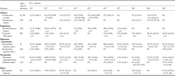
Infection parameters and main sites of infection for the 8 species of parasites which were commonly detected in the farmed (groups F1-F7) and wild (groups W1-W3) Helix a. aspersa snails examined in the cross-sectional study

a Farm 1.
b Farm 2.
c Farm 3.
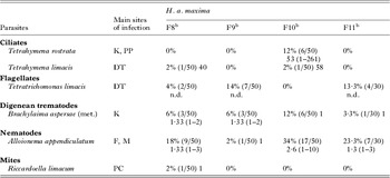
Infection parameters and main sites of infection for the 6 species that were found parasitizing the farmed Helix a. maxima snails (groups F8-11) examined in the cross-sectional study

b Farm 2.
A longitudinal prospective study was also conducted in farms 1 and 2 to investigate the dynamics of the different parasitic infections through the first 3 months of intensive mating which was carried out under indoor intensive conditions (high densities and controlled temperature, light and humidity). Thus, a monthly sampling was carried out during this period in the groups (reproduction units) F1, F2, F6, and F8-F11 examined in the cross-sectional study. In fact, the prevalences and intensities/densities obtained for each parasite and group in the cross-sectional study were considered as initial data (month 0) in the longitudinal study. With very few exceptions, 50–80 snails from each unit were examined monthly for the presence of parasites. Farm 2 had stricter hygiene and handling practices than farm 1, with daily removal of dead/dying snails from the reproduction units. No mortality data were revealed by the farmers.
In order to investigate in which moment of the rearing process the first infections are acquired, 6 egg clutches (90–135 eggs per clutch), 464 newly-hatched, and 147 juvenile snails descending from progenitors belonging to groups F1 and F2 were also analysed for parasites. All eggs examined were from progenitors which were infected at least with T. rostrata since vertical transmission of this ciliate species was previously demonstrated in slugs (Brooks, Reference Brooks1968). Egg clutches were obtained from plastic pots which were disposed at each side of the bottom of the reproduction units. The pots were filled with a commercial non-sterilized soil, which was used as egg-laying substrate. Neonate snails were collected from hatching containers which contained non-sterilized sand as substrate. Both egg-laying and hatching substrates were also examined for the presence of free-living or infective stages of parasites. Finally, the juveniles (2–3 months old; mean weight about 830 mg) were harvested from a grass plot enclosed in a greenhouse at farm 1, where first growing of snails was being undertaken. During this phase, snails were artificially fed the commercial powder diet. At the moment of sampling, numerous slugs belonging to the species Deroceras reticulatum were observed co-habiting the grass plot with juvenile snails and feeding on their diet. These invading slugs (n = 25) were also examined for the presence of parasites to investigate their potential role as reservoirs of these organisms. In addition, we also analysed 47 slugs of the species Deroceras panormitanum, collected from a first growing greenhouse at farm 2, and 38 Cepaea nemoralis snails, harvested from open parks at farm 3, where fattening of H. aspersa snails was being conducted.
Finally, to study the effect of a 3-month artificial hibernation on parasitic infections 86 and 80 petit-gris snails belonging to the same group were analysed before and after the process, respectively. Similar investigations were conducted with a group of gros-gris snails (n = 200; 100 before and 100 after hibernation). All breeder snails included in these studies were submitted to hibernation just after finishing a reproductive cycle.
Parasitological analysis
Snails were transported to the laboratory and maintained at 18 °C under dry environmental conditions promoting inactivity and secretion of the epiphragm until they were analysed for parasites. Each snail was weighed, examined for the presence of mites (Riccardoella limacum) moving on its surface, anaesthetized by intracorporeal injection of a sterile 50 mm magnesium chloride solution, and dissected for parasites. The pallial cavity was exposed by cutting the pulmonary plexus at the level of rectal duct and the mites inhabiting the cavity were counted under the stereomicroscope. Afterwards, the pulmonary plexus, the kidney together with the heart, the digestive system, and the genital apparatus were dissected out and placed in separate Petri dishes. The foot and mantle collar were also excised, each divided into 4–6 fragments, and transferred to a standard Baermann apparatus to detect tissular nematodes (e.g. Alloionema appendiculatum) following a procedure based on that used by Morand and Daguzan (Reference Morand and Daguzan1986). The apparatus consisted of a small funnel with a 1·5 mm plastic mesh on top, on which snail tissues were deposited, and a silicone tubing clamped with a Hoffman clamp connected to its end. The funnel was filled with distilled water to a level to make contact with the snail tissues and the nematodes were collected in a grid-marked Petri dish after 24 h at room temperature and counted under the stereomicroscope.
After dissection, wet mounts made from pulmonary plexus and rectal scrapings and from kidney, bursa copulatrix and digestive gland touch preparations were examined for the presence of parasites using light microscopy. Except for the flagellate species, the total number of parasites observed under the 22 × 22 mm coverslip was counted. In addition, the remaining tissues of the kidney and digestive gland as well as the genital tract were mechanically teased or opened with the aid of forceps and dissecting needles in Petri dishes filled with distilled water and observed under the stereomicroscope to count the total number of helminths. Prevalence and mean intensity/abundance for each species were calculated according to the method of Bush et al. (Reference Bush, Lafferty, Lotz and Shostak1997). In the case of ciliates, mean densities were calculated by using the number of protozoa counted under the cover-slipped area of renal wet mounts.
Each egg clutch was thoroughly washed with distilled water and the eggs were then separated, transferred to a Petri dish with water, incubated for 2 weeks at 18 °C, and observed daily at the inverted microscope for parasites. Pre-washing of egg-clutches was carried out to avoid the potential contamination of eggs with free-living soil nematodes and free-living stages corresponding to the parasitic nematode Alloionema appendiculatum (Morand and Bonnet, Reference Morand and Bonnet1989).
Newborn snails hatched from pre-washed eggs were examined by using 2 different procedures. Thus, 259 snails were disaggregated on slides with the aid of 2 fine needles, cover-slipped and observed for parasites under a light microscope. Each of the remaining 205 individuals examined was washed with distilled water to remove any surface-adhered free-living organisms, transferred to a well of a 24-well culture plate containing 500 μl of distilled water, squashed with forceps, incubated for 4 weeks at 18 °C, and monitored daily under the inverted microscope. This procedure was especially designed to facilitate diagnosis of the nematode Alloionema appendiculatum which easily proliferates on the carcasses of infected snails.
Various samples of the soil and sand substrates used for egg-laying and hatching were examined for the presence of nematodes morphologically compatible with the free-living forms of A. appendiculatum by using the Baermann funnel technique (Coleman et al. Reference Coleman, Crossley and Hendrix2004). The sediment at the bottom of the funnel was collected every 24 h during 72 h and examined under the stereomicroscope. The nematodes observed were isolated and processed for identification as is described below for A. appendiculatum.
Parasite identification
The flagellates Tetratrichomonas limacis and Cryptobia helicogenae were identified by staining the massively infected rectal contents/scrapings or spermatheca touch preparations with Giemsa or with a protein-silver (protargol) impregnation method, respectively. Identification of Tetrahymena spp. ciliates was based on morphological and/or behavioural (cyst formation and autogamy) characteristics as described by Segade et al. (Reference Segade, Kher, Lynn and Iglesias2009). Life-cycle experimental studies and/or morphological techniques (wet mounts, Semichon's acetocarmine staining, and/or silver impregnation for cercarial chaetotaxy) were used to identify the digenean sporocysts and metacercariae as previously described (Segade et al. Reference Segade, Crespo, Garcia, Garcia-Estevez, Arias and Iglesias2011). Nematodes and mites were fixed in 70% ethanol, cleared in lactophenol, and observed under light microscopy.
Histological studies
To study the microhabitat of the different parasites, organs were fixed for 24–48 h in 10% buffered formalin and embedded in paraffin following standard procedures. Sections (4–5 μm) were obtained using a microtome Microm HM-340E (Microm, Walldorf, Germany) and stained with haematoxylin-eosin (H&E).
Statistics
Data from hibernation studies were analysed with Fisher's exact test with Yate's continuity correction, in the case of prevalences, and non-parametric Mann-Whitney test, in the case of mean abundances (GraphPad Instat® 3·06, GraphPad Software, Inc., La Jolla, CA, USA).
RESULTS
Cross-sectional study
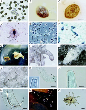
Eight parasite species were commonly found parasitizing H. a. aspersa in heliculture farms (Table 1; Figs 1 and 2). The tissular nematode A. appendiculatum and the renal digenean B. aspersae (metacercariae) were the most prevalent species in farmed snails, being detected in all the groups examined with overall prevalences of 52·9% and 49·2%, respectively. The intestinal flagellate T. limacis and the renal ciliate T. rostrata were found in 6 of the 7 groups with overall prevalences of 15·9% and 3%, respectively, while the genital-dwelling flagellate C. helicogenae and the mite R. limacum were observed infecting 17·1 and 7·3% of farmed snails in 5 groups. Finally, the presence of B. aspersae sporocysts was confirmed in 2·3% of farmed snails belonging to 4 groups, while both the intestinal ciliate T. limacis and the genital-dwelling nematode N. bakeri appeared only in 2 groups with an overall prevalence of 0·5%.
Typical appearance of the main parasites detected in Helix aspersa snails. (A) Numerous T. rostrata ciliates swimming in a wet mount from a pulmonary plexus scraping. Note the presence of 2 dividing ciliates (arrows). Scale bar = 100 μm. (B) Silver impregnated T. rostrata ciliate showing the numerous somatic kineties as well as the cytostomic area (c). Scale bar = 30 μm. (C) Silver impregnated T. limacis ciliate. Scale bar = 20 μm. (D) Giemsa-stained T. limacis flagellate in which the 4 anterior (af) and the free posterior (pf) flagella, the undulating membrane (um), the nucleus (n) and the axostyle (a) can be easily visualized. Scale bar = 5 μm. (E) Numerous pleomorphic C. helicogenae flagellates (arrows) in a bursa copulatrix touch preparation as observed with phase-contrast microscopy. Scale bar = 40 μm. (F) Protargol-impregnated C. helicogenae flagellates showing the anterior (af) and adherent flagella (adf), the nucleus (n) and the species-specific aciculum (ac). Scale bar = 20 μm. (G) Macroscopic appearance of a large whitish sporocyst mass (arrow) of B. aspersae occupying a great part of the digestive gland of a H. a. aspersa snail. (H) Detail of the microcercous cercariae contained in the branched sporocyst of B. aspersae. Scale bar = 100 μm. (I) Mature non-encysted metacercariae of B. aspersae in a wet renal touch preparation. Scale bar = 200 μm. (J) Wet mount from a pulmonary plexus scraping showing a parasitic larval stage of A. appendiculatum (arrows) surrounded by host infiltration tissue. Scale bar = 200 μm. (K) Posterior and cephalic region (inset; phase-contrast microscopy) of an adult male of A. appendiculatum showing the characteristic spicules (s) and gubernaculum (g) as well as the numerous denticles (arrow) on the dorsal metarhabdion of the buccal cavity. Scale bar = 100 μm. (L) Cephalic region of an adult of N. bakeri. Scale bar = 100 μm. (M) Posterior region of a male of N. bakeri showing the characteristic spicules (s) and gubernaculum (g). Scale bar = 50 μm. (N) Numerous R. limacum mites moving in the pallial cavity of a snail (whitish forms are larval and nymphal stages while yellowish forms are adults). (O) Microscopic appearance of an adult R. limacum. Scale bar = 100 μm.

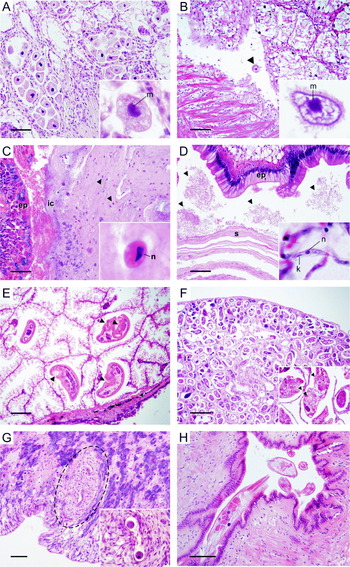
Histological appearance of the different endoparasites infecting Helix aspersa in their respective microhabitats. (A) Numerous T. rostrata trophozoites occupying the lumen of some folds of renal tissue. Note that infected folds lack epithelium which has been ingested by the histophagous ciliates. The presence of the small round micronucleus (m) adjacent to the macronucleus permits to differentiate the ciliate from host cells (inset). (B) A T. limacis ciliate in the lumen of a digestive gland duct. The micronucleus (m) and macronucleus can also help to identify this ciliate (inset). Note that this species presents large vacuoles in the cytoplasm while T. rostrata exhibits a faintly eosinophilic fine granular content. (C) Numerous T. limacis flagellates (arrowheads) embedded in the intestinal content (ep: intestinal epithelium). The elongated nucleus (n) in the eosinophilic cytoplasm can be visualized in sections stained with H&E (inset). (D) Section of a bursa copulatrix heavily infected with C. helicogenae flagellates (arrow heads) with the protozoa distributed luminally between the internal epithelium (ep) and the concentric layers resulting from the degradation of the spermatophore head (s) after copulation. The small rounded nucleus (n) and the elongated kinetoplast (k) can be visualized in some flagellates (inset). (E) Four metacercariae of B. aspersae in the lumen of several renal folds. The presence of suckers (arrowheads) facilitates their identification in histological sections. (F) Section of a digestive gland heavily infected by a branched cercariogenic sporocyst of B. aspersae, with a detail of longitudinally sectioned cercariae (inset) in which the suckers can be easily distinguished (arrowheads). (G) Section of the foot tissues of a snail infected with a larval stage of the tissular parasite A. appendiculatum. Note that the larva is surrounded by a marked granulomatous-like response (encircled). A nematode transversally sectioned is shown in the inset. (H) Section of the genital apparatus showing the luminal microhabitat of the nematode N. bakeri and the absence of pathological changes in the genital epithelium. Scale bars for A, B, C, D and G = 100 μm; for H = 200 μm; for E and F = 400 μm.

Excepting the intestinal flagellate T. limacis, which was not found in wild hosts, both farmed and wild snails were infected by the same species. However, prevalences and intensities of the different parasites varied considerably depending on the origin (Table 1). In farmed snails, this was particularly evident for the species T. limacis (flagellate), C. helicogenae, B. aspersae, A. appendiculatum and R. limacum. Thus, in the group F7, which was collected at farm 3, the prevalences of C. helicogenae (67%) and A. appendiculatum (2·2%) were very different to those observed in the remaining groups (⩽14% for the flagellate and ⩾34%, for the nematode). In addition, in a few snails belonging to this group 2 non-brachylaimid digeneans were detected with very low prevalences (data not shown). One of them, which was assigned to the family Panopistidae, infected the digestive gland as branched cercariogenic sporocysts and the kidney and pericardial sac as metacercariae. In the other digenean species, only xiphidiocercariae-containing sporocysts were found in the digestive gland.
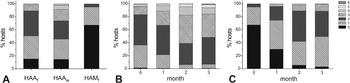
As can be seen in Fig. 3A, the overall proportions of non-infected, single- and mixed-infected hosts were similar in both farmed (15, 35·6 and 49·4%, respectively) and wild petit-gris (14·6, 31 and 54·4%), with most of the multiparasitized snails being infected by 2 to 4 species (only 1 farmed and 2 wild snails were co-infected with 6 and 5 species, respectively).
Percentage of uninfected (0), single-infected (1) and multiparasitized (2 to 6 species) snails observed in the cross-sectional study (A) and during intensive mating of Helix a. aspersa (B) and H. a. maxima (C) (HAAf and HAAw: farmed and wild H. a. aspersa, respectively; HAMf: farmed H. a. maxima).

In general, the number of parasitic species infecting H. a. maxima from heliciculture farms, as well as their respective parasitological parameters, were notably lower than in H. a. aspersa (Table 2). Thus, 67·2% of gros-gris were non-infected, 27·8% presented monospecific infections and only 5% were multiparasitized with 2 species (Fig. 3A). The helminths A. appendiculatum and B. aspersae (only metacercariae were detected) were the most common parasites in this subspecies, being detected in the 4 groups examined but with global prevalences (18·8 and 7·2%, respectively) and mean intensities very much lower than those observed for petit-gris snails. Neither C. helicogenae nor N. bakeri were found in this host. In addition, the presence of R. limacum was incidental with only 1 snail being infected by 1 mite.
Apart from the parasitic species, the free-living nematode Rhabditis axei was observed in scraping preparations from the pulmonary plexus of both H. aspersa subspecies under farming conditions, with prevalences ranging from 3·1 to 35·1%. Incidentally, some free-living soil nematodes and protozoa, as well as some free-living stages of A. appendiculatum, were also found in intestinal contents after being accidentally ingested by snails.
Dynamics of parasitic infections during intensive reproduction and first growing
The evolution of the different parasitic infections during intensive mating in both H. aspersa subspecies are shown in Figs 4 and 5.
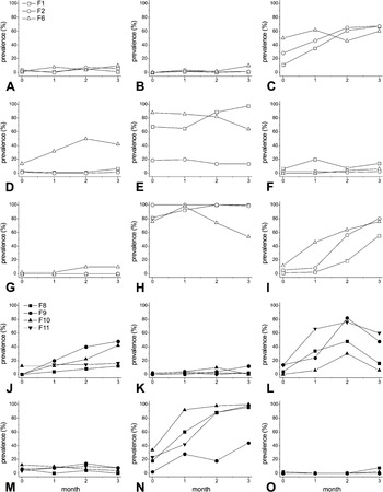
Evolution of prevalence (%) of the main parasites in the 3 groups (F1, F2 and F6) of Helix a. aspersa (A-I) and the 4 groups (F8-F11) of H. a. maxima (J-O) during the 3-month intensive mating period (A and J) T. rostrata; (B and K) T. limacis (ciliate); (C and L) T. limacis (flagellate); (D) C. helicogenae; (E and M) B. aspersae metacercariae; (F) B. aspersae sporocysts; (G) N. bakeri; (H and N) A. appendiculatum; (I and O) R. limacum).

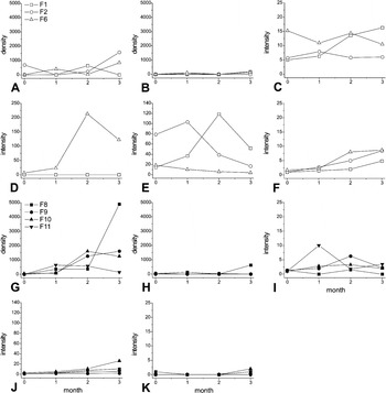
Evolution of the mean intensity/density of the main parasites in the 3 groups (F1, F2 and F6) of Helix a. aspersa (A-F) and the 4 groups (F8-F11) of H. a. maxima (G-K) during the 3-month intensive mating period. (A and G) T. rostrata; (B and H) T. limacis (ciliate); (C and I) B. aspersae metacercariae; (D) N. bakeri; (E and J) A. appendiculatum; (F and K) R. limacum).

No clear rising trends were observed for both ciliate species during the 3-month mating period in the 3 groups of H. a. aspersa studied, with maximum prevalences never exceeding 10% (Fig. 4A and B). On the contrary, the prevalence of the intestinal flagellate T. limacis increased considerably during the first 2 months and slightly thereafter in 2 of the 3 groups of snails under study (Fig. 4C), which exhibited relatively low (11·2%) and intermediate initial values (28·4%). In the third group (F6), which showed the highest starting prevalence (50%), the peak of infection was reached just after the first month. In spite of the fact that the initial values were very different, maximum prevalences observed in the 3 groups were similar ranging between 60 and 67·5%. In the case of C. helicogenae, a clear 2-month upward trend was observed only in the group F6, with the prevalence increasing from 14 to 50% and slightly declining thereafter (Fig. 4D). Initial prevalences of this flagellate in the other two groups were very low (1·3 and 2·5%). Regarding helminths, the infection with B. aspersae metacercariae was characterized by a stable or slight declining pattern except for the group F1, in which a slight raising trend was observed during the last 2 months (Figs 4E and 5C). In this group, the mean prevalence of the cercariogenic sporocysts of this species was ∼12% (ranging from 6·3 to 20%), which was 4–5 times higher than those observed in the other two groups (⩽2·5%, ranging from 0 to 6%) (Fig. 4F). The nematode N. bakeri was only detected in group F6, with parameters being increased only during the second month (Figs 4G and 5D). Thus, its prevalence and mean intensity augmented from 2% and 23 to 10% and 213, respectively, with up to 400 nematodes being counted in 1 snail. In both groups studied at farm 1 (F1 and F2) the pattern of the tissular nematode A. appendiculatum was similar: prevalences maintained practically stable at 100% after this value was reached, and mean intensities increased to values higher than 100 nematodes per host, sharply declining thereafter (Figs 4H and 5E). In the group F1, in which 81·3% of snails introduced in the reproduction unit were parasitized by about 15 nematodes per host, the peaks of infection were registered after the second month, with up to 500 nematodes being detected per host. In the group F2, the prevalence was already 100% at the beginning of the mating period while the peak of intensity was observed after the first month of follow-up. These patterns contrast strongly with that registered in group F6 from farm 2, in which the prevalence increased from 76% to 98% during the first month, and gradually declined thereafter. Moreover, the mean intensity in this group decreased progressively from an initial value of 18·5 to 4, at the end of the study. Finally, in all groups of petit-gris investigated, the presence of the mite R. limacum increased gradually throughout the whole follow-up period reaching final prevalences between 55·1 and 80% and mean intensities in no case exceeding 9 mites per parasitized host (Figs 4I and 5F).
As can be observed in Fig. 3B, the percentage of H. a. aspersa snails supporting 3 or more parasitic species (up to 6) increased progressively from 16·6% at the beginning of the study to about 61% after the second month. A slight decline in this proportion was observed during the third month of follow-up.
In contrast to what was observed in H. a. aspersa, and in spite of the fact that no T. rostrata-infected hosts were initially detected in the snails belonging to 3 of the 4 groups of H. maxima (F8-F11) included in the longitudinal study, infections with this ciliate exhibited a clear rising trend in this subspecies, with prevalences close to 50% being eventually reached in groups F9 and F10 (Fig. 4J). In general, massive infections with this histophagous protozoan (up to about 15 000 ciliates were estimated per touch preparation) were detected after the second-third month of intensive mating (Fig. 5G). The dynamics of the intestinal flagellate T. limacis were also identical in the 4 groups of gros-gris studied, with a gradual increase during the first 2 months followed by a moderate to marked decrease during the last month (Fig. 4L). Maximum prevalences of this protozoan, which ranged from 30% to 82%, were clearly dependent on the starting values. Infections with the tissular nematode A. appendiculatum exhibited a rising trend in all groups of gros-gris investigated (Fig. 4N). Thus, in all cases excepting group F9 prevalences augmented progressively from starting values of 18–34% to maximum values of 96–100%, at the end of the study. Mean intensities in these groups, however, never exceeded 26 (Fig. 5J), contrasting with the high values observed in the H. a. aspersa groups studied in farm 1. In the group F9, in which the starting prevalence was very low (2%), this parameter only reached 44% after the 3 months of follow-up. Mean intensities in this group were maintained at very low levels (<2) throughout the entire study period. Finally, no clear trends were observed in the case of T. limacis (ciliate), B. aspersae (metacercariae), and R. limacum, since prevalences did not exceed 12, 14 and 8%, respectively (Fig. 4K, M and O). In the 2 latter species, mean intensities were never higher than 10 and 1 parasites, respectively (Fig. 5I and K). Neither brachylaimid sporocysts nor the sexually-transmitted species C. helicogenae and N. bakeri were detected in H. a. maxima across the longitudinal study.
As in the case of H. a. aspersa, a trend to multiparasitation was observed during gros-gris mating (Fig. 3C), with the percentage of snails infected by 2 or more species (up to 4) increasing from 5% to 58% over the first 2 months.
None of the parasites registered in breeder snails belonging to groups F1 and F2 was detected in the eggs and newborn snails descending from these snails. However, in spite of eggs and neonates were extensively washed before examination, numerous ciliates morphologically compatible with the morphospecies Tetrahymena pyriformis (Corliss, Reference Corliss1970) were observed in some wells containing eggs and squashed neonates after some days of incubation in distilled water (data not shown). The ciliates were first visualized swimming in the surrounding water, but as shell degradation proceeded, they entered and multiplied within the eggs.
Unlike eggs and newborn snails, the juveniles descending from groups F1 and F2 were found infected with the ciliates T. limacis (29·4%) and T. rostrata (12·6%), the flagellate T. limacis (11%), and A. appendiculatum (6%) after a 2 to 3-month first growing period. The free-living nematode R. axei was also detected in the Baermann sediment from 4 snails and, only in 1 case, in a teased digestive gland.
Study of potential reservoirs/sources of infection
No free-living or infective stages of A. appendiculatum and other parasites were detected in the egg-laying and hatching substrates, which contained some free-living organisms, including the nematode R. axei. Interestingly, the D. reticulatum slugs co-habiting the grass plot from where the juveniles of H. a. aspersa were collected were infected with the same 4 species found in the young snails (prevalences in slugs were 40, 84, 4 and 20%, respectively). Similarly, the other wild invading gastropods collected at farms 2 and 3 also shared some parasitic species with farmed H. aspersa snails. Thus, the slug D. panormitanum was infected by T. rostrata (22·2%), which also infected the helicid C. nemoralis (2·6%), together with the flagellate T. limacis and A. appendiculatum (in both cases 7·9%). A Cryptobia sp. flagellate (7·9%) in addition to Brachylaima sp. and panopistid metacercariae (57·9% and 23·7%, respectively) and some unidentified brachylaimid sporocysts were also found in the latter host species.
Effects of hibernation on parasitic infections
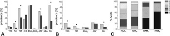
A slight to marked decline was observed in the prevalence of all parasites after hibernation, with the exception of the 2 genital-dwelling species C. helicogenae and N. bakeri, which exhibited a slight to moderate increase, and B. aspersae, which remained practically unaltered (Fig. 6). Only the prevalences of the ciliate T. limacis, the flagellate T. limacis, A. appendiculatum and R. limacum, in H. a. aspersa, and T. rostrata, the flagellate T. limacis, and R. limacum, in H. a. maxima were, however, significantly reduced. In the case of mean abundances, a significant decrease after hibernation was only registered for the mite R. limacum in the case of petit-gris (3·5 vs 0·4; P < 0·0001).
Effect of artificial hibernation on the prevalence of parasites infecting Helix a. aspersa (A) and H. a. maxima (B) and on the frequency of multiparasitic infections observed in both subspecies (C). In graphs A and B, prevalences registered before (light gray) and after (dark gray) hibernation are represented in pairs for each parasite species. Asterisks indicate significant differences between prevalence values obtained before and after hibernation (P < 0·05) (TRO, T. rostrata; TLI, T. limacis (ciliate); TET, T. limacis (flagellate); CHE, C. helicogenae; BRAm, B. aspersa metacercariae; BRAs, B. aspersa sporocysts; AAP, A. appendiculatum; NBA, N. bakeri; RLI, R. limacum). In graph C, HAA1/HAA2 and HAM1/HAM2 mean H. a. aspersa and H. a. maxima before (1) and after (2) hibernation, respectively.

DISCUSSION
Results form the present study revealed that parasite faunas infecting farm-reared and wild H. a. aspersa adult snails were qualitatively similar, with the small flagellate T. limacis being the only species not found in wild hosts. In this case, however, the possibility that very light infections occurring in wild populations have gone unnoticed during examination can not be ruled out. As far as we know, this flagellate has been detected to date only in the slugs Deroceras agreste and Limax flavus (in both cases as Trichomonas limacis) and the snails H. pomatia and Otala lactea (Kozloff, Reference Kozloff1945; Saleuddin, Reference Saleuddin1972). Therefore, it is reported for the first time in H. aspersa and under farming conditions. Interestingly, our results also revealed the absence of the nematode A. aspersae both in wild and farming environments, contrasting with the findings observed in France where this pallial cavity-dwelling monoxenous species has been reported as a frequent parasite in petit-gris snails subjected to intensive reproduction (Morand and Bonnet, Reference Morand and Bonnet1989). Although no negative effects have been associated to date with this nematode, this aspect should be kept in mind before importing allochthonous snails for farming purposes.
With the exception of C. helicogenae, N. bakeri and the sporocysts of B. aspersae, all parasite species detected in H. a. aspersa were also found infecting H. a. maxima. The absence of the two former species, which are sexually-transmitted (Morand, Reference Morand1988a,Reference Morandb; Kozloff, Reference Kozloff2004), may be explained by the apparent natural tendency of both subspecies to behavioural and anatomical reproductive isolation (Gomot-de Vaufleury and Borgo, Reference Gomot-de Vaufleury and Borgo2001), which ultimately could have determined a strict host specificity of both parasites for H. a. aspersa. Finally, the absence of B. aspersae sporocysts is consistent with previous experimental infection studies demonstrating that this subspecies does not support the development of this parasite stage (Segade et al. Reference Segade, Crespo, Garcia, Garcia-Estevez, Arias and Iglesias2011). The absence of sexually transmitted parasites and the low to moderate prevalences exhibited by all infections in the cross-sectional study (in no case exceeding 34%) explain the low multiparasitization rate detected in H. a. maxima. Thus, while only mono- and bi-specific infections were detected in this subspecies, up to 6 parasite species in a single host were observed in farm-reared petit-gris. The lower densities used for fattening of H. a. maxima could contribute to explain these differences by minimizing the transmission of the monoxenous non-sexually transmitted parasites. Apart from these arguments, the possibility that H. a. maxima is more resistant to certain infections than H. a. aspersa should not be entirely ruled out.
According to our observations, and previous studies (Segade et al. Reference Segade, Crespo, Garcia, Garcia-Estevez, Arias and Iglesias2011), most of the parasites detected in this work may be introduced in farms if the physical barriers (e.g. net, fences) used for preventing the entry of predators are not effective enough to block the access of small wild reservoirs (e.g. rodents and gastropods) to the farm facilities. Mice (Mus musculus), for example, which are the final hosts of the tri-heteroxenous digenean B. aspersae (Segade et al. Reference Segade, Crespo, Garcia, Garcia-Estevez, Arias and Iglesias2011), can enter the parks and greenhouses when attracted by snails, and especially by the artificial powdered feed used for fattening. Once in the farm, rodents infected with brachylaimids can pass the miracidium-containing eggs in their feces, which may give rise to cercariogenic sporocysts after being accidentally ingested by H. a. aspersa snails. The cercariae emitted by these first-intermediate hosts will then develop to metacercariae after reaching the kidney of other H. aspersa snails acting as second-intermediate hosts. In addition to rodents, the artificial feed used for fattening also attracts slugs and other wild gastropods which, as it has been demonstrated in this and previous studies (Kozloff, Reference Kozloff1945; Saleuddin, Reference Saleuddin1972; Morand et al. Reference Morand, Wilson, Glen and Barker2004), serve as an important source of certain monoxenous parasites (e.g. T. rostrata, the flagellate T. limacis, A. appendiculatum, etc.). Besides this route, parasites can also be introduced in farm environments when adult snails are collected from uncontrolled wild populations for breeding purposes, a practice commonly used by Galician farmers. In addition to the species cited above, feral petit-gris breeders may be the source of the mite R. limacum as well as the sexually-transmitted parasites C. helicogenae and N. bakeri, all of them being monoxenous and exhibiting a direct mode of transmission (Morand, Reference Morand1988a,Reference Morandb; Kozloff, Reference Kozloff2004; Schüpbach and Baur, Reference Schüpbach and Baur2008a). The presence of the 2 latter species in farm-reared petit-gris, therefore, should be impossible unless infected snails from natural populations are introduced in the reproduction units by farmers, or actively enter the fattening parks. The latter possibility occurred frequently in farm 3, where the prevalence of C. helicogenae was particularly high (67%).
Once in the farm, and especially under the intensive conditions used for mating, overcrowding is the most important factor facilitating the transmission of monoxenous species. This was clearly evidenced by the infection dynamics exhibited by some of these parasites throughout the mating period. Thus, in both H. aspersa subspecies, a general rising trend was observed for T. limacis (flagellate) and A. appendiculatum during the first 2 months, while in the case of C. helicogenae and N. bakeri, which infected petit-gris exclusively, a rise in infection levels was only detected in the group F6 in which initial prevalences of both sexually-transmitted parasites were probably high enough to induce a detectable rising trend. Increasing tendencies, however, were slowed down or reversed during the third month of follow-up resulting, as it was expected, in a decrease in the frequencies of highly multiparasitized snails at the end of the mating period. These findings could be interpreted as a combination of (1) the negative, and probably additive, effects derived from the presence of mixed infections, which could ultimately cause the death of more highly multiparasitized snails, and (2) the progressive reduction in snail densities resulting from the accumulative mortality and sampling removal which may negatively affect the transmission rates of certain parasites. In this direction, it must be underlined that diverse pathological effects have been described in gastropods infected with the histophagous ciliate T. rostrata (Brooks, Reference Brooks1968; Barker, Reference Barker1993; Wilson et al. Reference Wilson, Coyne and Glen1998; Segade et al. Reference Segade, Kher, Lynn and Iglesias2009), the digenean trematode B. aspersae (Segade et al. Reference Segade, Crespo, Garcia, Garcia-Estevez, Arias and Iglesias2011), the nematodes A. appendiculatum and N. bakeri (Morand and Daguzan, Reference Morand and Daguzan1986; Cabaret et al. Reference Cabaret, Morand, Aubert and Yvore1988; Morand, Reference Morand1989; Morand et al. Reference Morand, Wilson, Glen and Barker2004), and the haematophagous mite R. limacum (Fontanillas and Pérez, Reference Fontanillas and Pérez1987; Graham et al. Reference Graham, Runham and Ford1996; Schüpbach and Baur, Reference Schüpbach and Baur2008b). In the particular case of A. appendiculatum, the decrease in intensity observed in groups F1 and F2 after the first and second month, respectively, occurred in both cases after the mean intensity values exceeded 100 larvae, which clearly suggests a strong relationship between parasite burden and host mortality. Death of heavily infected snails could have resulted from the pathological changes induced by this nematode, the hypothetical presence of secondary bacterial infections following larval penetration, and/or the additive effects derived from other concomitant infections. Histological examination of infected pedal (and mantle collar) tissues, however, did not reveal the presence of bacteria in the granulomatous-like lesions surrounding the larval nematodes. Unlike the well-known pathogenic species cited above, the effects of the flagellates T. limacis and C. helicogenae on snail health and growth have not been studied to date. In this sense, recent experimental T. limacis infections carried out in our laboratory (unpublished data), and the histological findings observed in the present study, which demonstrate the absence of any tissue reaction to these luminal parasites, suggest that both protozoa have probably little pathogenic significance.
Unlike T. limacis and A. appendiculatum, the R. limacum infection levels were progressively increased in H. a. aspersa along the mating period (i.e. no apparent slowing-down or decrease was observed after the second month). This mite can pass from one host to another during courtship, mating or other snail activities involving interindividual contact (Schüpbach and Baur, Reference Schüpbach and Baur2008a), although it has recently been demonstrated that transmission via substrate, with mites showing preference to move on fresh snail mucus trails, is also possible (Schüpbach and Baur, Reference Schüpbach and Baur2008a). The latter mode of transmission could explain why increasing trends were maintained during the entire follow-up period in spite of the fact that host densities and copulation rates decreased with time. It must also be underlined at this point that the dynamics of R. limacum observed in H. a. aspersa were surprisingly very different to those registered in gros-gris snails, in which mites were detected only in the groups F8 and F10, and with very low prevalences (up to 8%) and intensities (only 1–2 mites were found in each infected host). Considering the modes of transmission discussed above, these findings could be suggesting that H. a. maxima is not a suitable host for R. limacum, although this hypothesis contrasts with the apparent low host specificity exhibited by this mite (Fain, Reference Fain and Barker2004).
Contrary to what should be expected as a result of the direct life-cycle exhibited by T. rostrata, no increasing trends were observed in the 3 petit-gris groups examined during mating. Prevalences of this ciliate, however, exhibited an upward general tendency in the gros-gris groups under study, with values up to 48% being reached at the end of the mating period. Since the possibility that ciliates infecting petit-gris and gros-gris snails were cryptic species with different infective capacities can be rejected based on previous molecular studies (Segade et al. Reference Segade, Kher, Lynn and Iglesias2009), these dissimilar dynamics could be suggesting differences in the susceptibility of both subspecies to the ciliate. In this sense, recent experimental studies conducted in our laboratory suggest that T. rostrata is unable to infect H. a. aspersa by using oral or trans-urinary routes, and probably enters the snail through small lesions in the tegument or the shell (unpublished data). Future investigations will be required to demonstrate whether T. rostrata may invade the renal organ of gros-gris snails by using oral and/or trans-urinary routes in addition to the trans-tegumental route.
Besides overcrowding, other aspects related to the practices used in the farms studied may also affect the transmission of the parasites detected. Thus, the periodical water spraying used for maintaining humidity may facilitate not only the survival but also the dissemination of certain infective stages. This is the case of the flagellate T. limacis, which is transmitted by the fecal-oral route and apparently lacks the pseudocystic stages observed in other congeneric species infecting vertebrate hosts (Friedhoff et al. Reference Friedhoff, Kuhnigk and Müller1991; Tasca and De Carli, Reference Tasca and De Carli2007). The infective stages of this species, which are continuously passed out in the feces of infected snails, may survive better in the wet environments resulting from spraying. In addition, because most snails in the reproduction units are adhered to the vertical plastic sheets, spraying may also facilitate the downward dispersal of this protozoan species. These arguments are also valid for B. aspersae cercariae emerging from sporocyst-infected snails, and A. appendiculatum third-stage larvae resulting from the free-living cycle of this nematode, which can be completed (1) on decaying gastropod carcasses after death (since the free-living stages of this nematode are bacterivorous up to 24 000 adults and 41 000 larval stages have been estimated to result from 1 infected carcass), and (2) in the soil following the active exit of the fourth-stage parasitic larvae from the pedal tissues of living hosts (Morand and Daguzan, Reference Morand and Daguzan1986; Morand et al. Reference Morand, Wilson, Glen and Barker2004). These particular characteristics make A. appendiculatum an especially abundant parasite in heliciculture environments (Morand, Reference Morand1985; Morand et al. Reference Morand, Wilson, Glen and Barker2004). The high intensities (up to 819 larvae per host) observed at F1 and F2 units belonging to farm 1, where dead snails were not removed daily, and the lower mean intensities (in no case exceeding 26) observed in the 5 units (F6 and F8-F11) studied at farm 2, where removal of moribund and dead snails was conducted daily, stress the importance of carcasses in the transmission of this nematode and the efficiency of removal practices. In the reproduction units studied, where most of snails were adhered to vertical plastic sheets, carcasses were concentrated on the bottom, so the transmission of A. appendiculatum is most probably completed in the horizontal feeding troughs in which, in the absence of rapid removal, the contact between living and decaying snails may occur more easily. Furthermore, the accumulation of water, feed and feces in these troughs can also facilitate the survival and proliferation of the free-living stages of this nematode. In addition to the factors discussed above, the presence of B. aspersae metacercariae in the gros-gris snails from farm 2 and the inability of this subspecies to support the development of the cercariogenic sporocysts of this digenean suggest that infection of H. a. maxima in this farm was associated with the entry of sporocyst-infected petit-gris snails in the parks used for fattening gros-gris. In this particular case, the proximity between fattening pens of both subspecies and the absence of suitable anti-escape measurements undoubtedly facilitated the accidental access of cercariogenic snails to the gros-gris populations. This aspect should also be kept in mind when more than one species (or subspecies) is produced in the same farm.
In spite of the fact that the prevalence of A. appendiculatum in groups F1 and F2 always exceeded 80%, no vertical transmission could be demonstrated for this nematode. Futhermore, no free-living stages belonging to this species were detected in unused egg-laying and hatching substrates. Altogether, these results suggest that (1) contamination of egg-laying substrates and egg-clutches with infective L3 larvae resulting from infected progenitor snails is a key factor in the transmission of this pathogenic helminth to descendants, and (2) washing of egg clutches before incubation prevents the infection of newly-hatched snails with infective larvae derived from progenitors, as it was previously demonstrated by Morand and Bonnet (Reference Morand and Bonnet1989). Unlike A. appendiculatum, R. axei was detected in both substrates examined as well as in the pulmonary cavity and intestinal lumen of both H. aspersa subspecies. This ‘free-living’ nematode, however, is not considered pathogenic (Grewal et al. Reference Grewal, Grewal, Tan and Adams2003) and could act as an endocommensal instead of a true parasite. The absence of T. rostrata in the eggs or neonates examined in this study strongly contrasts with previous findings obtained in experimentally infected slugs, indicating that this ciliate can be vertically transmitted during egg formation (Brooks, Reference Brooks1968). Interestingly, and in spite of the fact that eggs were exhaustively washed with distilled water before examination, only ciliates belonging to the Tetrahymena pyriformis complex were detected in some eggs and squashed newborn snails. Since the possibility that the ciliates observed in the eggs of D. reticulatum were misidentified by Brooks (Reference Brooks1968) can be rejected (the identity was confirmed by using silver impregnation), the differences observed between H. aspersa and D. reticulatum may be related to the presence/absence of this parasite in the albumen gland, which is involved in egg formation (Gómez, Reference Gómez and Barker2001). Thus, while T. rostrata infects this organ in slugs (Brooks, Reference Brooks1968), no ciliates were ever detected in the albumen gland of H. aspersa.
Our results showed that artificial hibernation was accompanied by a reduction in the prevalence of most parasite species and the frequency of highly multiparasitized snails. These findings could be suggesting that certain parasites are especially sensible to the low temperatures used in the process, and/or mortality associated with starvation occurs more frequently in snails supporting mixed infections. Regarding the first possibility, only the mean abundances of R. limacum were significantly affected by the process, which is in accordance with recent studies indicating that larvae, nymphal stages and adults of this species are unable to survive prolonged exposure to 4 °C (Schupbach and Baur, Reference Schüpbach and Baur2010; Haeussler et al. Reference Haeussler, Piza, Schmera and Baur2012). Eggs of this mite, however, can resist hibernation in the host pallial cavity without hatching (Haeussler et al. Reference Haeussler, Piza, Schmera and Baur2012). In agreement with these findings, no reduction was observed in the presence of R. limacum eggs detected in H. aspersa after artificial hibernation (data not shown). Thus, in most snails examined after this process only eggs and mite remnants were detected in the pallial cavity. This resistance capacity is epidemiologically relevant since surviving eggs can serve as a source of new infections when breeder snails are introduced in the reproduction units after resuming activity. Apart from these considerations, it should be expected that, as the result of the complex internal defence responses mounted against different parasites, the highly multiparasitized snails have less energy reserves for supporting the starvation and physiological changes associated with hibernation.
In conclusion, our results confirm the importance of parasitic infections in heliciculture and stress the need to enhance the effectiveness of control strategies. In this sense, farmers should be broadly informed about the risks associated with the presence of heavy parasitic infections and advised on the benefits of preventing the entry of wild potential reservoirs into the farms as well as minimizing the use of wild snails for mating purposes. Nevertheless, if wild breeders are eventually required, special care should be taken in order to select uninfected or lightly infected natural populations. Finally, a rapid removal of dead snails from the reproduction units should also be considered mandatory in order to prevent heavy infections with the pathogenic nematode A. appendiculatum.
FINANCIAL SUPPORT
This work was supported by grant PGIDIT04RAG310015PR (Xunta de Galicia, Spain).