I. INTRODUCTION
Extensive studies are being conducted on the development of chitosan-based biomaterials for various biomedical applications such as wound healing, wound dressing, tissue engineering, and drug delivery. Chitosan-based materials can be easily processed in the form of gels, membranes, sponges, fibrils, beads, scaffolds, or nanoparticles.Reference Muzzarelli1–Reference Ahmed and Ikram3 Zhou et al.Reference Zhou, Yang, Chen, Xu, Lu and Nie4 prepared a composite nanofibrous mat from polyvinyl alcohol and carboxyethyl chitosan nanofibers via electro-spinning and studied the L 929 cell attachment and proliferation. The authors reported that this fibrous mat has the potential to be used as a wound dressing material for skin regeneration. In their later study, silk fibroin nanoparticles were also added and a composite fibrous mat having good biocompatibility and skin regeneration capacity was developed.Reference Zhou, Yang, Liu, Mao, Gu and Xu5 Nguyen et al.Reference Nguyen, Nguyen and Hsieh6 developed a curcumin/chitosan/gelatin composite sponge having improved water uptake ability, wound closure, and antibacterial activity. In another study, chitosan hydrogel was prepared for wound dressing and cell viability tests were conducted using fibroblast cells isolated from rat skin.Reference Ribeiro, Espiga, Silva, Baptista, Henriques, Ferreira, Silva, Borges, Pires, Chaves and Correia7 The authors reported that chitosan hydrogel promoted cell adhesion and proliferation, and chitosan and its degradation by-products were found non-cytotoxic. Another group developed chitosan/polyethylene glycol diacrylate blend films for wound dressing application and evaluated the in vitro cytotoxicity of the films using L929 mouse fibroblasts. The material showed good biocompatibility and non-cytotoxicity.Reference Zhang, Yang and Nie8
For decades, organic and/or inorganic nanomaterials such as polymers, dendrimers, liposomes, micelles, and materials based on carbon, iron oxide, silicon, silver, or gold with different morphologies such as nanotubes, nanowires, nanocapsules, nanofilms, and nanogels have been fabricated and explored as new platforms for diagnostic and therapeutic purposes.Reference Deng, Zhen, Hu, Wu, Xu and Chu9 Among the various inorganic nanoparticles, silica particles have attracted much interest because of their excellent chemical stability, inert nature, thermal stability, low density, low toxicity, and biocompatibility. Silica nanoparticles show good compatibilities with other materials and can also be functionalized easily for chemically binding other active materials.
Over the past decades, a huge volume of research has been conducted on mesoporous silica nanoparticles (MSNs) and hollow silica sphere (HSS) nanoparticles. Because of their large surface area, stability, and biocompatibility, these silica nanoparticles found application in biomedical field, pharmaceutics, and biochemistry.Reference Liberman, Mendez, Trogler and Kummel10,Reference Gui, Wang and Sun11 These particles are used as shield for enzymes or proteins, and as delivery vehicles for drugs because of their porous nature and greater loading efficiency.Reference Liu, Yang, Shen, Gao, He, Liu and Zhu12,Reference Jiao, Li, Zhang, Liu, Di, Zhang, Zhao and Wang13 A number of synthetic techniques exist in the literature for the production of silica nanoparticles.Reference Bao, Shi, Wang, Li and Ma14 Silica nanoparticles having the desired particle size, shape, porosity, and crystallinity can be generated by properly designing the appropriate synthetic route.Reference Chen, Yu, Lv, Zhang, Xing and Yu15,Reference Dong, Wang, Yu, Wang, Zhu, Fu and Ni16
Sol–gel process and template-assisted synthesis are the generally adopted methods for the production of HSSs. The form of nanoparticles can be changed using a shaped template or by adding various dopants as precursors, varying pH, or varying temperature during production. These hollow particles which can carry very bulky payloads or comprise cores may include desirable materials such as metals or various polymers.Reference Liberman, Mendez, Trogler and Kummel10 Monodispersed phenyl groups-containing HSSs were generated by Hah et al.,Reference Hah, Kim, Jeon, Koo and Lee17 Wang et al.,Reference Wang, Liu and Yan18 and Aslan et al.Reference Aslan, Soydan and Bozkurt19 without using any templates. These HSS nanospheres were soluble in organic solvents.
Combining HSS with smart biopolymers is an interesting approach which may be used in biomedical applications such as drug delivery and wound dressing. Fang et al. developed a pH-responsive drug carrier using chitosan-coated MSNs.Reference Chen and Zhu20 Drug-loaded MSN particles were added to the chitosan solution at pH 6 and stirred for 36 h, then centrifuged, washed, and dried. In another set, instead of MSN, methacryloxy propyl trimethoxysilane-coupled MSN was used and drug release was studied from both systems in a narrow pH range. The authors reported that chitosan-wrapped MSNs exhibited targeted drug release. A greater release of drug was observed at pH 6.8, the pH of inflammatory tissues and tumor cells, compared to pH 7.4, the pH of normal cells. It was noted that the material in which chitosan wrapped directly on to MSN showed better pH sensitivity than the material that contained a coupling agent.
For a material to be used in wound healing applications, it should be biocompatible, non-cytotoxic, and non-allergenic and should inhibit growth of microbial organisms. Chitosan possess all the above-mentioned characteristics. Silica materials have the capability to absorb wound exudates. Some researchers have investigated silica–chitosan combinations for hemostatic applications. Sun et al. fabricated a porous chitosan/kaolin composite microsphere through an inverse microemulsion process combined with thermally induced phase separation at −196 °C and found that these porous microspheres are a promising quick hemostat.Reference Sun, Tang, Pan, Wang, Yang and Liu21
In a recent work, a three-component system composed of chitosan, dopamine, and biosilica has been prepared to be used for rapid blood coagulation in hemostatic applications.Reference Wang, Fu, Li, Mu, Zhang, Zhang, Liang, Feng and Chen22 Although some naturally available silica has been investigated for hemostatic applications in combination with chitosan, the wound healing potential of synthetic HSS—chitosan bionanocomposites has not been studied.
In this work, we report the preparation and characterization of chitosan-functionalized HSSs by epoxidizing HSS and chemically binding chitosan onto the HSS to form HSS-epoxy-CHI-X where X represents the percentage (w/w) of chitosan in the bionanocomposite. The epoxidation of HSS via polymerization of glycidyl methacrylate (GMA) monomer has been reported in our previous article, where GMA monomer that contained an epoxy unit was polymerized on the surface of HSS.Reference Aslan, Soydan and Bozkurt19 However, in this study, epichlorohydrin was used to carry out direct epoxidation on the surface of HSS. In the second step, chitosan binding was achieved through a ring-opening reaction between the epoxy-functionalized HSS and chitosan. The developed materials were characterized by FT-IR, XRD, thermogravimetry analysis (TGA), SEM, and TEM. A ss and average pore diameter were measured by the Brunauer–Emmett–Teller (BET) method. The potential of the chitosan-modified HSS material was evaluated for wound closure applications by conducting in vitro scratch assay. It was expected that in addition to the biological activity of chitosan, the HSS present in the system may allow gas exchanges and absorb wound exudates. Therefore, the newly developed material will be a best candidate for wound healing applications.
II. EXPERIMENTAL
A. Materials
Phenyl trimethoxysilane (PTMS) (97%), epichlorohydrin (99%), low-molecular-weight chitosan (mol wt 50,000–190,000 Da, deacetylated 75–85%), and dimethylformamide (DMF) were purchased from Sigma-Aldrich (St. Louis, Missouri). Other chemicals used in the synthesis such as HNO3 and NH4OH were of analytical purity.
B. Methodology
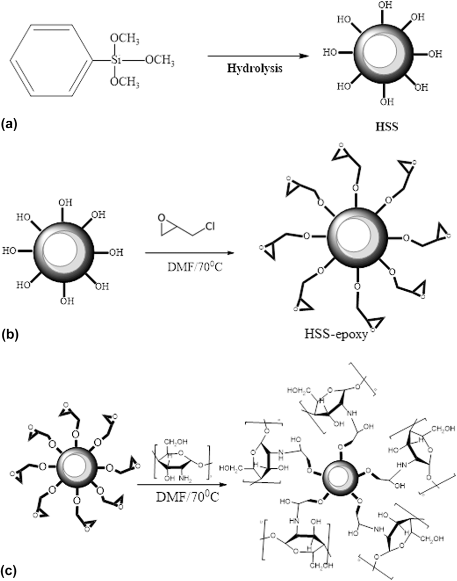
HSS nanoparticles were prepared as reported in our previous work.Reference Aslan, Soydan and Bozkurt19 Briefly, sol–gel method was adopted for developing HSS. PTMS was hydrolysed in an acidic medium (HNO3), and the condensation of the HSS was performed in a basic medium (NH4OH) [Fig. 1(a)]. The precipitated nanoparticles were collected by a membrane filter and washed with water/ethanol mixture and then dried under vacuum.
Schematic representation of the preparation of chitosan-functionalized HSS nanoparticles: (a) preparation of HSS nanoparticle, (b) epoxidation of HSS surface, and (c) binding of chitosan to functionalized HSS.

For carrying out the epoxidation of HSS, a similar procedure described in the literature was used.Reference Alzina, Sbirrazzuoli and Mija23 The HSS was mixed with DMF (0.1 g HSS in 10 mL DMF) and dispersed via ultrasonic bath for 20 min at room temperature. Then, excess epichlorohydrin was added dropwise to the solution at room temperature. Then, the reaction temperature was increased to 70 °C for 1 h. After the completion of reaction, the mixture was filtered and, to remove unreacted epichlorohydrin, was washed several times with ethyl alcohol–water (1:4) solution. Finally, modified HSSs were kept wet for further reaction [Fig. 1(b)].
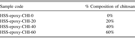
The next step is the binding of chitosan on to the functionalized HSS surface [see Fig. 1(c)] via a ring-opening reaction of epoxy unit on HSS surfaces. HSS-epoxy was dispersed in acetic acid (0.2 g HSS-epoxy, 10 mL for 20%), and chitosan (w/w) in acetic acid (2 g chitosan, 30 mL for 20%) was added to the solution of epoxidized HSS at 70 °C and stirred for 24 h. HSS-epoxy-CHI was precipitated due to cross-linking. The precipitate was washed with water/methanol solution several times, filtered, and dried at 80 °C under vacuum. Different HSS-epoxy-CHI samples were prepared by varying the molar ratio of chitosan, i.e., 20, 40, and 60%. The formulation of the samples used in this study is given in Table I.
Formulation of chitosan-functionalized HSS samples.

C. Characterization
FT-IR spectra were recorded on a Bruker Alpha-P in ATR (Bruker Corporation, Billerica, Massachusetts) in the range of 4000–400 cm−1 (single-reflection diamond crystal). BET measurements were taken using an ASAP-2020 plus, accelerated surface area, and porosimetry (Micromeritics, Norcross, Georgia). Thermal stabilities of the samples were studied using TGA with a PerkinElmer Simultaneous Thermal Analyzer (STA) 6000 (PerkinElmer Inc., Waltham, Massachusetts). The samples (∼10 mg) were heated from room temperature to 700 °C under N2 atmosphere at a heating rate of 10 °C/min. XRD data were attained from a fully automated 9.0 kW anode rotating generator Rigaku Smart Lab X-Ray Diffractometer (Rigaku Corporation, Tokyo, Japan) with two-theta range of 0 to 70°. The surface morphology of the samples was detected using scanning electron microscopy (SEM; JEOL 7001 FESEM, JEOL Ltd., Tokyo, Japan). Samples were sputtered with gold for 150 s before the SEM measurements. For measuring the size of HSS, a transmission electron microscope (TEM; FEI, Morgagni, Czech Republic) was used.
1. In vitro wound healing (scratch) assay
NIH 3T3 fibroblasts cells were seeded in 12-well plates at 1 × 10⁵ cells/well and grown until confluence in complete DMEM supplemented with 10% FBS, 2 mM L-glutamine, 100 U/mL penicillin, 100 µg/mL streptomycin, and 4.5 g/L glucose in a humidified chamber at 37 °C and 5% CO2. To simulate a wound, a straight scratch was made in each well with a P200 pipette tip. The cell debris was removed, and the cells were treated with various formulations of nanoparticles for 24 h. The closure of the scratch was observed under the microscope at certain time intervals, i.e., 0, 14, and 21 h of exposure of scratch to the bionanocomposites. Digital images of cells were captured by a phase contrast microscope (Leica Microsystem, Weztlar, Germany). The closure of the scratch was calculated based on the width difference initially and after 14–21 h of treatment, using the ImageJ software (National Institutes of Health, Bethesda, Maryland) and expressed as a percentage of scratch closure.
III. RESULTS
A. BET surface area and TEM analysis of HSS nanoparticles
BET surface analysis is readily used to precisely measure the specific surface area (A ss) of porous materials. In this study, the BET analysis was performed to measure the A ss and pore size of HSSs. The product showed A ss value of around 4.8 m2/g. The Langmuir surface area was found to be around 8 m2/g. The adsorption average pore diameter (4V/A by BET) was 10.180 nm.
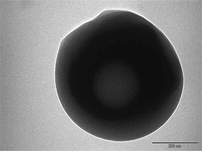
Dimensions of HSS nanoparticles were measured with the help of a transmission electron microscope (TEM). TEM image (Fig. 2) showed that the size of HSS was ∼400–450 nm, and the size of hollow was about 150 nm and thickness of cover was ∼100 nm.
TEM image of HSS nanoparticle.

B. FT-IR analysis
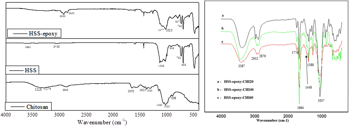
Infrared spectra of chitosan, HSS, epoxy-functionalized HSS samples, and nanocomposites (HSS-epoxy-CHI-20, HSS-epoxy-CHI-40, HSS-epoxy-CHI-60) are shown in Fig. 3.
FT-IR spectra of chitosan, HSS, epoxy-functionalized HSS, and HSS-epoxy-CHI nanocomposites.
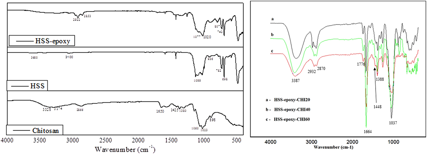
The spectra of HSS showed a strong absorption peak at 1092 cm−1 belonging to Si–O–Si asymmetric stretching. The peaks at 782 and 493 cm−1 could be assigned to symmetric stretching vibration of Si–O, and the peak at 936 cm−1 could be due to bending vibration of Si–OH.Reference Xiang, Ding, Suo, Xu, Gao and Hu24 A weak distinctive Si–OH stretching vibration was displayed at about 3600 cm−1. The peak at 3100 cm−1 was attributed to asymmetrical stretching vibration of C–H groups of aromatic phenyl attached to HSS. These results demonstrated condensation product of PTMS to form HSS.
In the spectra of epoxy functionalized HSS, additional peaks were centered at 2900 cm−1 owing to the presence of methylene groups. Peak at 807 cm−1 indicated the presence of the epoxy group. In the epoxy-functionalized sample, the peak of Si–O–H showed slight change in its position and intensity, which indicates the participation of Si–OH in the epoxidation reaction.
Chitosan characteristic peaks were observed at 1650 cm−1 due to the amide group, 1568 cm−1 corresponding to N–H bending, 3288 cm−1 due to O–H, 2866 cm−1 C–H bending, 1425 cm−1 due to stretching vibrations of the amino group, 1154 cm−1 associated with C–O–C stretching, 1023 and 1060 cm−1 due to C–O-stretching, and 898 cm−1 due to glycosidic linkage.Reference Al-Sagheer and Muslim25
The spectra of HSS-epoxy-CHI nanocomposites showed distinct peaks of chitosan and silica. It can be observed that the peak in the region of 800 cm−1 associated with the epoxy group is absent in the chitosan-bound nanocomposites, which confirms the ring-opening reaction of epoxy. It is expected that amino group of chitosan is interacting with the epoxy group. The shift in the amino group peaks to lower frequency regions observed in the spectra of nanocomposites supports this assumption.
C. X-ray diffraction studies
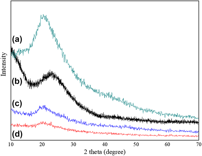
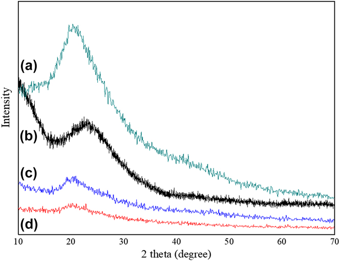
The XRD patterns of the chitosan, HSS, and chitosan-functionalized HSS nanocomposites with a chitosan content of 40% and 60% are shown in Fig. 4. Pure chitosan showed a strong peak at 2θ = 20.5° as reported in the literature.Reference Flores, Lopez, Tabary, Neut, Chai, Betbeder, Herkt, Cazaux, Gaucher, Martel and Blanchemain26 The HSSs were comparatively amorphous in nature, and no highly crystalline peaks were observed, except one broad band around 22°.Reference Chen, Li, Zhou, Li, Wang, Guo and Feng27 For bionanocomposites, the intensity of this peak decreased, which indicates that chitosan-functionalized HSS nanocomposites were more amorphous in nature. However, among chitosan-functionalized HSS samples, the intensity of this broad peak increased with the increase in the percentage of chitosan in the composites.
XRD spectra of (a) pure chitosan, (b) HSS, (c) HSS-epoxy-CHI-60, and (d) HSS-epoxy-CHI-40.
D. Scanning electron microscopic analysis
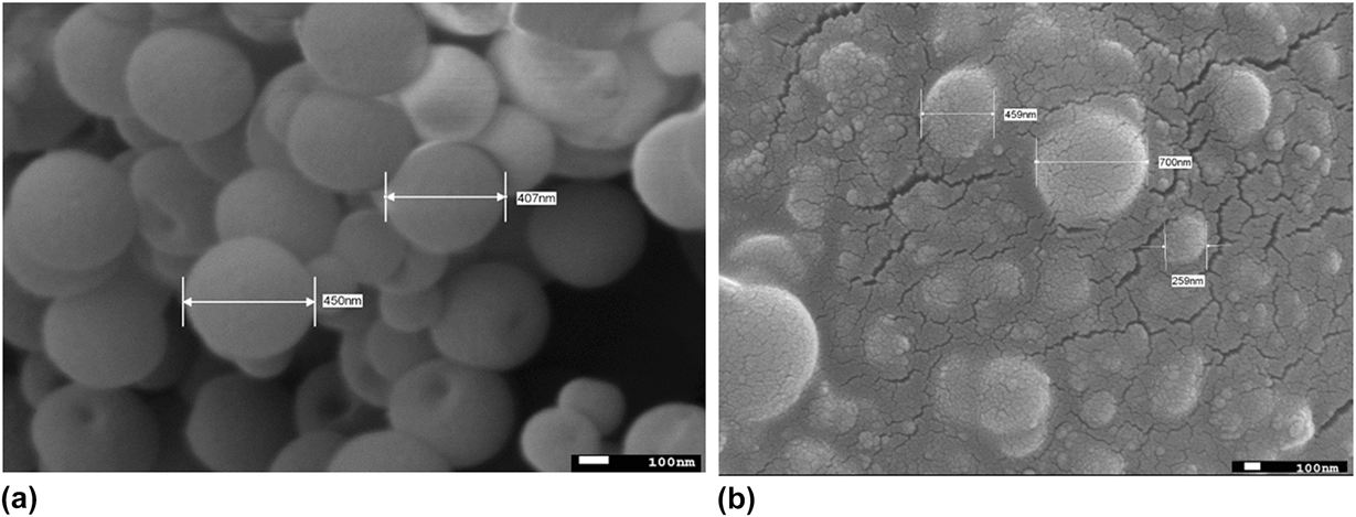
The surface morphology of synthesized HSS and chitosan-functionalized HSS was analyzed using SEM. The SEM image of HSS [Fig. 5(a)] confirms the hollow nature of HSS. As evidenced from Fig. 2, the synthesized HSS showed an average diameter of 400–500 nm. Figure 5(b) shows the SEM image of chitosan-functionalized HSS (40% chitosan). It can be seen that HSS particles were homogeneously dispersed in chitosan and no phase separation was observed between chitosan and HSS after the modification of HSSs with chitosan via the ring-opening reaction of epoxy. As expected, the dimension of chitosan-functionalized HSS nanoparticles increased compared with pure HSS because of the presence of chitosan coating on the surface of HSS.
SEM image of (a) HSS nanoparticle and (b) HSS-epoxy-CHI-40 nanocomposite.

E. Thermal analysis
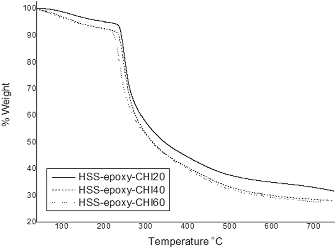
TGA plots of chitosan-functionalized HSS samples are shown in Fig. 6, which show similar pattern, and only a slight variation in thermal stability is observed. Chitosan usually follows a two-step degradation profile: a small weight loss within the temperature range 30–180 °C due to the evaporation of absorbed water and small molecules; and a major weight loss between 250 and 600 °C due to the decomposition of chitosan polymer chains and the degradation of phenyl groups of HSS.Reference Zhao, Yan and Feng28 It is interesting to note that chitosan-functionalized HSS samples exhibited higher thermal stability at lower temperatures when compared to HSS-epoxy sample that contained no chitosan. When the plots of nanocomposites were compared, there were small variations in the initial weight loss of the chitosan-functionalized samples due to the variation in the moisture content in the nanocomposites. Samples with high percentage of chitosan showed greater weight loss. The residue content of the samples was proportionate to the chitosan content in it. Sample prepared with low chitosan content showed higher residue content and samples with high chitosan content showed less residue content due to the complete degradation of the chitosan molecules.
TGA curves of chitosan-functionalized HSS samples.

F. In vitro wound healing (scratch) assay
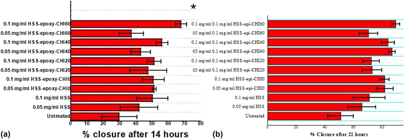
Skin is the largest body organ, which protects our body by acting as a barrier and preventing harmful pathogens from entering our body. However, a wound can occur on the skin either by a physical, chemical, or thermal damage. Wounds and burns are a serious problem in health care. The production process of wound healing is a complex system that involves the regeneration of the damaged tissues and replacement of lost tissues. Wound healing process mainly involves four phases: hemostasis, inflammation, proliferation, and remodelling.Reference Sinno and Prakash29 To investigate the wound healing potential of chitosan-functionalized HSS nanoparticles, each of the newly synthesized formulations in DMSO was tested using in vitro wound healing assay. The wound was simulated by introducing a straight scratch in a confluent culture of NIH 3T3 fibroblasts. The percent closure was observed and calculated 14 and 21 h later. Fourteen hours after introducing a scratch and treating with different nanocomposites, 100 µg/mL of HSS formulation loaded with 60% chitosan had significantly higher percent closure of about 70%, that is, healing, compared to the untreated controls with average percent closure of about 30% [see Fig. 7(a)]. All of the remaining nanocomposites showed slightly higher but not statistically significant increase in percent closure of the initial scratch. Twenty-one hours after introducing a scratch and exposure to different ratios of nanocomposites, the average percent closure of untreated control is increased to about 50%. However, scratch exposed to different amounts of HSS showed average percent closure higher than the controls with most of the nanocomposites having a statistically higher increase in percent closure [see Fig. 7(b)]. This is quite expected due to biological activity of chitosan and the hollow nature of silica which permits exudates absorption and gas exchange. Similar to the 14 h treatment period, the 100 µg/mL of HSS formulation loaded with 60% chitosan had the highest percent closure of about 90%.
Wound healing potential of novel HSS-based formulations in in vitro wound healing assay (a) 14 and (b) 21 h after the scratch and exposure to different formulations (mean ± SEM, n = 4 from a total of four repeated experiments, *: P < 0.05 and **: P < 0.01 compared with the untreated controls).
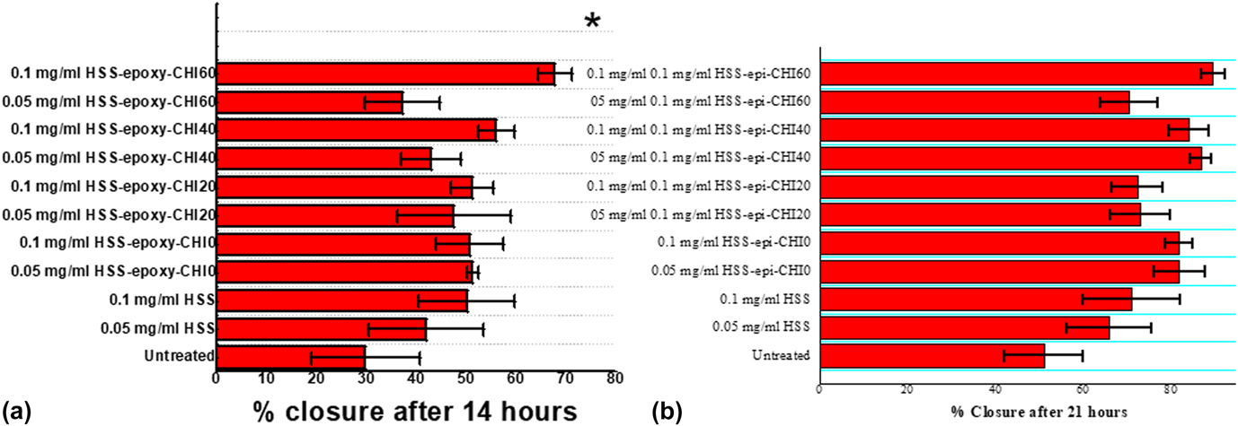
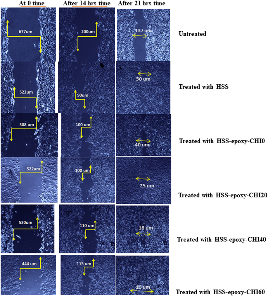
Figure 8 shows the photomicrographs of scratch analysis at various time intervals, i.e., at 0, 14, and 21 h after the scratch and exposure to different chitosan–HSS formulations (concentration—0.1 mg/mL). Compared with the untreated scratch, greater percent closure was observed for the scratch treated with HSS–chitosan formulations. It was observed that as the chitosan content in the formulation increases, percent wound closure increases. Among the different formulations studied, formulation with 60% chitosan content showed greater wound closure. It can be seen that, in all cases, as the exposure time increases, more closure can be achieved. Compared with 14-h-treated samples, more percent closure was observed after 21 h.
Photomicrographs of in vitro scratch test at 0, 14, and 21 h after the scratch and exposure to different chitosan–HSS-based formulations (mean ± SEM, n = 4 from a total of four repeated experiments, *: P < 0.05 and **: P < 0.01 compared with the untreated controls) (100× magnification).
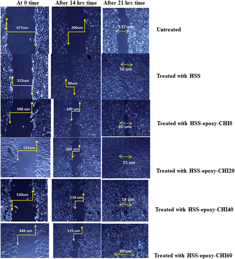
Clearly, in vitro scratch assay was used as a tool to investigate the wound healing potential of the novel composite materials. Percentage closure of the scratch was measured after certain time intervals of exposure to nanocomposites. Greater percentage closure was observed for samples with greater content of chitosan, because of the specific biological features of bionanocomposite. When the time of exposure increased, greater closure was observed. The newly developed material could create a better environment for the wound closure.
IV. CONCLUSIONS
Chitosan-functionalized hollow silica nanoparticles were prepared and characterized by different techniques such as FT-IR, TGA, XRD, SEM, and TEM. BET analysis of pristine HSS was performed, and A ss value was found to be around 4.8 m2/g and the adsorption average pore diameter was found to be 10.18 nm. Binding of chitosan on the HSS nanoparticle was confirmed using FT-IR analysis and SEM analysis. These novel biocompatible nanoparticles were tested for application in wound healing by carrying out in vitro scratch assay using NIH 3T3 fibroblast cells. Out of the analyzed nanocomposites, HSS-epoxy-CHI-60 bionanocomposite exhibited better performance. About 70% wound closure was observed for this sample after 14 h of exposure, and 90% wound closure was observed after 21 h of treatment. The synergic effect of chitosan and HSS leads to better performance of these materials. From the results it can be seen that wound healing ability of the samples is proportionate to the chitosan content in the samples, concentration of nanoparticles formulation, and the exposure time. Scratch exposed to higher concentration of chitosan nanocomposites showed wound closure to a greater extent. It can be summarized that the newly developed functionalized materials have great potential for application in wound care.
Supplementary Material
To view supplementary material for this article, please visit https://doi.org/10.1557/jmr.2018.430.
ACKNOWLEDGMENT
This work was supported by Institute for Research and Medical Consultations (IRMC) - IAU.