“We’re at a point right now where doctors can’t keep up with everything they need to know. We need a machine to help us do that.” – Kevin Hughes, MD Massachusetts General HospitalReference Krisber and Krisberg1
Artificial intelligence implies the application of computers or machines to perform or mimic human thinking without human intervention. Artificial intelligence is transforming almost every aspect of our lives with emerging solutions such as facial and speech recognition, sports analytics and personalised investment recommendations. Historically, the medical field hesitated to embrace artificial intelligence largely due to concerns surrounding patient privacy. This was compounded by a lingering skepticism from when the hype of artificial intelligence failed to meet expectations, now attributed to a lack of available data and computational power. The recent contemporaneous surge of large volumes of available data, advancement of computational power and cloud computing, and the advent of machine learning has returned artificial intelligence to the forefront of medicine.
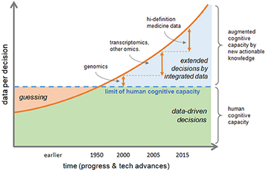
Patients today produce 750 quadrillion bytes of data daily, encompassing 30% of the world’s data production.Reference Erika and Mukherjee2 This surge of accumulated patient-generated data, coined “the healthcare data conundrum”, means that physicians now face 200 times the medical information that a human can process (Fig 1).Reference Erika and Mukherjee2 Currently, medical data remain unstructured in a variety of repositories that do not communicate. There is no seamless and practical method for busy clinicians to integrate and analyse the data into meaningful decision-making clinical tools, and this climate promotes the re-emergence disruptive technologies such as artificial intelligence. A sustained rate in rise of publications centred on advances in artificial intelligence in medicine suggests a changing attitude towards this field. This newfound acceptance of artificial intelligence in medicine mirrors the increased recognition that artificial intelligence offers a reasonable solution to address the escalating volume of patient data burdening our healthcare system. Proponents of artificial intelligence argue that its algorithm-based applications provide a novel advantage to analyse the growing complexity inherent to medical data sets and can detect subtle relationships in the diagnosis, treatment and prediction of many clinical scenarios.
As it stands today, the majority of projects employing artificial intelligence technology lean heavily towards specialties rich in data and imaging such as cardiology, ophthalmology, critical care and oncology. These specialties are able to provide the volume of data necessary for effective algorithmic development. Cardiology specifically is both a perceptual and a cognitive subspecialty that demands complex decision-making, suggesting artificial intelligence implementation in this field has great potential. The prototypical artificial intelligence system would autonomously input patient data into a collaborative database that syncs, interprets and ultimately classifies the patient’s profile to specific disease phenotypes. After classification, the system would compare the patient’s disease phenotype against a large aggregate of shared peer health data and outcomes, current medical literature and ongoing trials. The comparison then offers morbidity and mortality predictions, targeted drug therapies, tailored surgical plans and recommendations for timing of sequential imaging. Comprehensive reviews by Johnson et al Reference Johnson, Torres Soto and Glicksberg3and Krittanawong et al Reference Krittanawong, Zhang, Wang, Aydar and Kitai4 discuss how artificial intelligence can elevate adult cardiology from a research-based endeavour to clinical practice to re-imagine precision cardiovascular medicine. This review paper offers a primer on artificial intelligence and paediatric cardiology by briefly discussing the history of artificial intelligence in medicine, modern and future artificial intelligence technology applications in adult and paediatric cardiology across selected concentrations, and current barriers to implementation of these technologies.
Escalating volumes of data are changing the decision-making process. The accelerated rate of data production, volume and variety now supersedes the limits of human cognitive capacity. From Rossi RL, Grifantini RN. Big data: challenge and opportunity for translational and industrial research in healthcare. Front Digit Humanit; 2020, with permission.

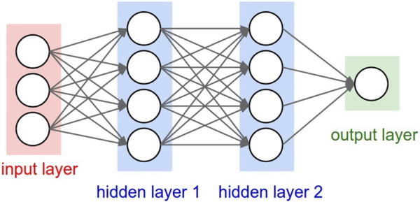
A Depiction of an artificial neural network. Obtained with permission.

Historical significance
History of artificial intelligence
During World War II the pioneer of artificial intelligence, Alan Turing, paved the way for artificial intelligence by designing the precursors of modern computers: electro-mechanical machines named “bombes” to decode German secret transmissions.Reference Benko and Sik Lányi5 In his 1950 publication “Computing Machinery and Intelligence”. Turing aptly predicted that by the year 2000 digital computers would evolve to “thinking machines”.Reference Moor6 The term artificial intelligence was officially coined at the Dartmouth Conference in 1956 by Stanford computer scientist John McCarthy.Reference Moor7 The term referred to the capacity of a machine to perform operations that are analogous to human cognitive capabilities. A year later, Frank Rosenblatt invented the “perceptron”, which marked the first “artificial neuron”.Reference Rosenblatt8 This concept of a three-layer structure with an input, transfer and output layer catalysed the development of the artificial neural networks, a modern deep learning algorithm (Fig 2).
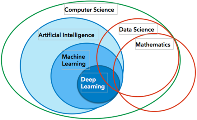
A diagram to orient the reader. Artificial intelligence algorithms are built from big data, and data mining and data science are incorporated. The main branch of artificial intelligence is machine learning and its subtype deep learning. From Chang AC. Artificial Intelligence in Medicine: Principles and Applications. Elsevier; 2020, with permission.

History of artificial intelligence in medicine
Artificial intelligence spread to the medical field in the 1960s with the Dendral experiments,Reference Buchanan and Feigenbaum9 which analysed mass spectra data and the existing knowledge of chemistry to identify possible chemical structures. One of the most notable advances of artificial intelligence in medicine took place at Stanford and Rutgers in the late 1970s through the use of the SUMEX-AIMReference Nakamura10 Computing Resource. This technology enabled researchers to use artificial intelligence methods to solve issues in medicine internationally. Researchers struggled with developing solutions for biomedical problems, eventually creating fuzzy set theory, Bayesian networks and artificial neural networks to address the evolving needs of intelligent computing systems in healthcare. Following the early advances of machine intelligence in the 1970s, two artificial intelligence “winters” occurred due to the suboptimal executions resulting in a depressing outlook on the future of artificial intelligence.Reference Lim11 In 1982, Peter Szolovits’ book, Artificial Intelligence in Medicine, reintroduced artificial intelligence in medicine as a field rich in sophisticated representational and computing techniques.Reference Szolovits12 He argued that artificial intelligence could produce valuable tools for improving healthcare when combined with expert physicians’ insights. In 1987, artificial intelligence regained recognition when the European Society for Artificial Intelligence in Medicine founded a biennial meeting titled Artificial Intelligence in Medicine in Europe.Reference Patel, Shortliffe and Stefanelli13 After a period of latency in the late 20th century, artificial intelligence expanded exponentially in business and government. This surge paralleled the simultaneous growth in computational power and the accumulation of massive amounts of data.
Key components of artificial intelligence
Artificial intelligence
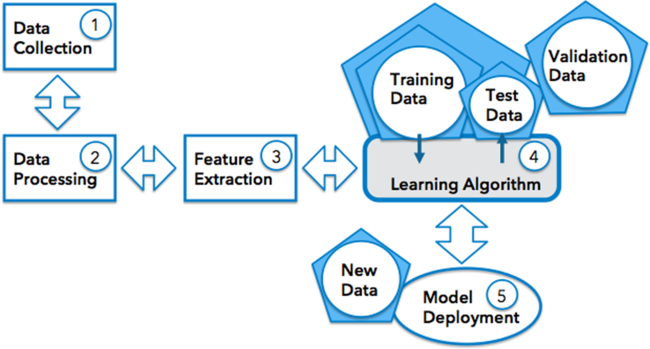
Artificial intelligence-driven analytical algorithms aim to autonomously find hidden insights within large pools of complex sets of medical data without explicitly being programmed. The main arm of artificial intelligence is machine learning, which is further subdivided into deep learning (Fig 3). Algorithmic development occurs by a process nicely outlined in Fig 4. The pipeline includes data collection, data cleaning and processing, feature extraction (selection of imaging data points of interest, medical record information, etc.), task selection (determination of computational approach and tools for model development and training), algorithmic development and final model performance testing. Algorithmic development relies on partitioning the data into a training, validation and test data sets. The training data is the sample of data used to fit models and the validation data is the unseen sample of data used to provide an unbiased evaluation of a model fit on the training dataset, to tune the model hyperparameters (architecture of the neural network) and to compare performances between models to decide which model to use. The final accuracy of the chosen model is determined by feeding the algorithm the test data, new labeled data independent of the training dataset. These six steps in algorithmic development eventually generate automated diagnosis or predictions. Each system has the potential to describe significant relationships in the diagnosis, treatment and prediction of many clinical scenarios. Furthermore, artificial intelligence systems now can perform autonomous time-heavy tasks to improve efficiency in the workplace.
The machine learning workflow includes data collection, data processing, feature selection and algorithmic development. From Chang AC. Artificial Intelligence in Medicine: Principles and Applications. Elsevier; 2020, with permission.

Artificial neural networks
Artificial neural networks are a type of machine learning inspired by the neurons and intricate synaptic interconnections in the brain. These networks have layers of connections with forward and backward data propagation directions. The forward neural network was the first and simplest type of artificial neural networks devised. In this network, the information moves in the forward direction. In contrast, back propagation is a mechanism in which the artificial neural networks can “learn”. Therefore, the difference between the output and the desired output is used to calculate a modification to achieve the desired outcome.
Machine learning
Machine learning, a subfield of artificial intelligence, describes algorithms capable of improving task performance with experience as they learn and adapt from data patterns. Machine learning algorithms can learn from supervised and/or unsupervised training. With supervised learning, the model has labelled data as its dependent variables. Classic methodologies that use supervised learning include support vector machines, neural networks, naive Bayesian classifiers and hidden Markov models. The goal of supervised learning is to learn a function from a provided sample of data and desired outputs that best describes the relationship between input and output of data. Dawes et al used a supervised learning model for the integration of clinical and imaging variables to predict the desired outcome of pulmonary hypertension. This supervised learning model used outcome predictions to discover new regions of right ventricular function on imaging strongly associated with increased survival when compared to conventional parameters.Reference Dawes, de Marvao and Shi14 Unsupervised learning, in contrast, does not have labelled outputs but rather infers the natural structure present within a set of scattered data points with no dependent variables. Classic methodologies include k-means clustering and principal component analysis. The goal with these systems is to learn the inherent structure of the data without using explicitly provided labels. This technology could be instrumental in exploratory analysis to automatically identify structure in the messy data sets innate to medicine, such as the surveillance of clinical variables found in the electronic health records to classify new disease subtypes of congenital heart disease (CHD). A combination of the two techniques could involve unsupervised learning to classify patient responses to cardiovascular drugs into subtypes while supervised learning can monitor for different treatment responses to various interventions.Reference Johnson, Torres Soto and Glicksberg3
Deep learning
Deep learning is a subset of machine learning that attempts to mimic the activity of the layers of neurons in the neocortex through the creation of large arrays of artificial neural networks called convolutional neural networks. Convolutional neural networks are a class of neural networks that consists of one or more “convolutional layers” followed by one or more fully connected layers of neural networks. Convolutional neural networks are particularly good at image recognition and classification. The architecture of a convolutional neural network takes advantage of the 2D structure of an input image or speech signal. The algorithm then learns to recognise patterns in digital representations of sounds, images and/or other data sources. Recent improvements in mathematical formulas and increasing computational power now allow for modelling more layers of virtual neurons with convolutional neural networks (Fig 5). As a result, there have been remarkable advances in speech recognition and natural language processing, computer vision with visual object recognition and detection, and autonomous vehicle driving. A convolutional neural network model for automated tissue classification for Kawasaki disease using optical coherence tomography successfully identified pathological coronary artery changes with 90% accuracy.Reference Abdolmanafi, Duong, Dahdah and Cheriet15 Applications of deep learning will be reviewed in more detail under the imaging section.
An example of a convolutional neural network. From Alsharqi, et al. Echo Res Pract. 2018;5:R115–R125, with permission.

Natural language processing
Natural language processing describes the process of using computer algorithms to identify key elements in everyday language and extract meaning from unstructured spoken or written input. The end goal is to allow the computer to understand spoken as well as written human language.16 Natural language processing uses a specific set of techniques, such as parsing (the analysis of words in the sentence for grammar), followed by a process of rearranging the words to show their proper relationship. One application in medicine is answering unique free-text inquiries that require the synthesis of multiple data sources. Additionally, natural language processing can engage in optical character recognition to transform images, such as scans of care summaries and imaging reports into text files that can then be parsed and analysed. Current areas of interest include conducting speech recognition to allow users to dictate clinical notes or other information that can then be converted to text. A recent study described utilisation of natural language processing for large-scale, automated and accurate extraction of structured, semi-structured and unstructured data from echocardiography reports.Reference Nath, Albaghdadi and Jonnalagadda17
Cognitive computing
Cognitive computing systems continually acquire knowledge from the data fed into them by mining diverse data for pertinent streams of information. Cognitive computing also involves self-learning systems that use data mining, pattern recognition and natural language processing to mimic the way the human brain works. Ideal systems can reason and learn to solve complex problems through the translation of data into knowledge. The International Business Machines Corporation’s Watson is the prototypical example; the platform relies on cognitive computing to interact in natural language to process vast amounts of disparate forms of big data and learn from each interaction.Reference High18 Systems like Watson refine methods for pattern recognition and data processing in real time to anticipate new problems and model possible solutions. Cognitive computing works to create automated information technology with applications including expert systems, natural language programming, neural networks, robotics and virtual reality. Current applications in research include uncovering new insights into relationships among genes, proteins, pathways, phenotypes and diseases, which assist in the creation of individualised treatment plans.Reference Ahmed, Toor, O’Neil and Friedland19
Reinforcement learning
Reinforcement learning attempts to optimise behaviours to achieve maximum rewards with trial and error learning from input data and an outcome. In healthcare, systems can be queried at regular intervals to improve the decision-making process as clinical changes arise, improving future outcomes. A deep reinforcement learning algorithm utilising the publicly accessible Multiparameter Intelligent Monitoring in Intensive Care II database of de-identified patient data was recently applied to heparin dosing.Reference Nemati, Ghassemi and Clifford20 The algorithm learned optimal adult heparin dosing and effectively proposed heparin doses with better expected outcomes than the clinical guidelines through studying sample dosing trials and their associated outcomes.Reference Nemati, Ghassemi and Clifford20 Future applications in paediatric cardiology include determining the optimal dosing of vasopressors, anticoagulation or inotropes to maximise benefits and minimise side effects for post-operative management in the cardiac ICU.
Main applications of artificial intelligence in paediatric and adult cardiology
Precision medicine
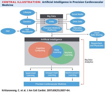
Precision medicine involves a comprehensive approach to disease diagnosis and treatment that incorporates the patient’s genomic makeup, clinical electronic health record data, lifestyle, family history and wearable data together to promote a patient-specific treatment plan. Such a model can transition from a “one-size fits all” approach for management of CHD to patient-specific tailored therapies that acknowledge that subtle variations in CHD lesions merit individualised treatment strategies (Fig 6). The sheer volume of preexisting data and the development of new data sources – such as genome sequencing, advanced imaging studies and electronic transmission of wearable information – will necessitate paediatric cardiologists analyse and incorporate this information into their practice. The inherent strength of artificial intelligence algorithms to handle large data sets makes it a more precise method for classifications of CHD phenotypes, allowing narrowed predictive analytics for novel interventions and improved standardised treatment plans. This ideal system would combine strategies that use artificial intelligence to discover new clinical and imaging markers of deterioration and create a road map for expected disease progression for specific paediatric heart diseases. Such a system could offer useful recommendations on how to optimise the frequency of surveillance echocardiograms, and advanced imaging modalities for the optimal detection of clinical deterioration and timing of interventions.
Appreciation for the precision healthcare model as it pertains to paediatric cardiology relies on a review of key components. The body of this review focuses on artificial intelligence applications in adult and paediatric cardiology across nine main concentrations: big data, cloud computing, decision support, hospital monitoring, biomedical diagnostics, digital medicine and wearable technology, drug discovery, medical imaging and robotics. These main areas were chosen based on current literature and recent developments of artificial intelligence systems.
Big data
Big data refers to infinitely large data sets whose scale, diversity and complexity require more advanced technology than standard processing methods for analysis, interpretation and storage. Information collected from mobile phones, wearable technology, social media, environmental and lifestyle-related factors, socio-demographic, “omic” data (e.g. genomics, metabolomics and proteomics) and electronic health records all contribute to big data. Routine cardiovascular medicine generates a wealth of data that artificial intelligence can apply for novel analytics and interventions. Partnership between big data analytics and artificial intelligence currently is used to discover novel genotypes and phenotypes of heterogeneous syndromes, such as heart failure,Reference Shah, Katz and Selvaraj21 Takotsubo cardiomyopathy,Reference Eitel, Moeller and Munz22 hypertrophic cardiomyopathy,Reference Jacoby, DePasquale and McKenna23 primary pulmonary hypertension,Reference Soubrier, Chung and Machado24 hypertensionReference Soubrier, Chung and Machado24 and coronary artery disease.Reference Raghavendra, Fujita and Gudigar25 Regeneron Genetics Center and Geisinger Health System recently integrated genomic data into electronic health records to discover a genetic variant that results in reduced levels of triglycerides and a lower risk of coronary artery disease.Reference Carey, Fetterolf and Davis26
The movement towards creating large registries and systems to amass data will facilitate the acceleration of research-based artificial intelligence endeavours and increase the power of traditionally based statistical studies. This is particularly important in paediatric cardiology, where randomised control trials are limited by disease rarity and small patient numbers. Lewis et al provided significant insight into outcomes and risks associated with pulmonary artery stenting in children with CHD disease using a multi-institutional database.Reference Moore27 They emphasised the conceivable power of big data in prospective registries. The development of optimal tools therefore relies exclusively on data reservoirs rich in a high quantity and quality healthcare information. Researchers at Massachusetts Institute of Technology spearheaded Multiparameter Intelligent Monitoring in Intensive Care III, an open database for Computational Physiology with de-identified data from over 40,000 critical care patients with newer databases with paediatric patients to encourage more artificial intelligence research development by eliminating privacy and proprietary concerns.Reference Johnson, Pollard and Shen28
Cloud computing
Cloud-based systems are becomingly increasingly attractive to store escalating volumes of healthcare big data and facilitate large-scale cross-institutional collaboration. These large multi-institution collaborations are necessary to amass the volume of data necessary to overcome rarity of paediatric heart diseases. Healthcare data remain in myriad siloed locations, without a common repository and format that enables stakeholders to interact with and produce meaningful artificial intelligence healthcare solutions. In an ideal scenario, there will be a private cloud used to connect healthcare providers, transfer electronic documents and share health information on patients. Hsieh et al argue that the rise in cloud computing will facilitate the intercollegiate application of tele-cardiology services between hospital systems worldwide.Reference Hsieh, Li and Yang29 They predict the global contribution of electrocardiogram and cardiac imaging to big data will lead to the development of an e-consultation program and an e-platform for clinical research and educational purposes. One innovative report described a machine learning and system modelling project to facilitate a five-centre collaborative system that allows rapid structured big data fact-finding and wide dissemination of expertise.Reference Wolf, Lee and Nicolson30 Through the process, the team found that the duration of mechanical ventilation following infant cardiac surgery could impact clinical scenarios and that earlier extubation optimised outcomes. Development of such systems will underscore the need for a focused solution to apply to the volume of data at the point of collection. Disruptive technologies, such as the blockchain protocol, will be essential to facilitate cross-institutional healthcare data sharing while concurrently assuring protection of patient data.
Clinical decision support systems
Clinical decision support systems powered by artificial intelligence automatically filter large quantities of established clinical knowledge specific to each patient’s condition. These systems thereby improve decision-making, clinical workflow and predictive outcome analytics. Machine learning algorithms were deployed to train a large data set of adults with CHD to prognosticate outcomes and facilitate management.Reference Diller, Kempny and Babu-Narayan31,Reference Samad, Wehner and Arbabshirani32 These tools may help combat the big challenges in paediatric cardiology of navigating the diagnosis, prognosis and management in individuals with minor different manifestations of the same disease. Ruiz-Fernández et al developed four artificial intelligence-based neural networks to classify the risk of various paediatric cardiac surgeries.Reference Aczon, Ledbetter, Ho, Gunny, Flynn, Williams and Wetzel33 These neural networks affirmed the feasibility of risk estimation for prediction of post-operative complications or mortality.Reference Aczon, Ledbetter, Ho, Gunny, Flynn, Williams and Wetzel33
Incorporation of artificial intelligence may improve standardised existing models. Ledbetter et al generated a dynamic real-time paediatric ICU mortality prediction risk score using applied machine learning to 10 years of data, which achieved significantly higher accuracy than the standard clinical mortality scoring systems.Reference Aczon, Ledbetter, Ho, Gunny, Flynn, Williams and Wetzel33 Melillo et al leveraged machine learning techniques to heart rate variability detected on Holter monitors from hypertensive patients to predict who would suffer a significant cardiac event with greater accuracy than standard parameters.Reference Melillo, Izzo and Orrico34 Similarly, Weng et al utilised routine clinical data collected from 300,000 patients free from cardiovascular disease to develop a machine learning systems that predicted a first-time cardiovascular event over the next 10 years with improved accuracy from standard cardiovascular risk prediction tools.Reference Weng, Reps, Kai, Garibaldi and Qureshi35 Algorithm-based scoring systems can thus repurpose routine collected patient data and establish preventative models for future patients.
Hospital monitoring
Ultimately, the purpose of hospital monitoring is to generate predictive deviations from the expected recovery course of an illness and to act on that information prior to the onset of an undesired outcome. The DEWS study (deep-learning-based early warning system) revealed that predictive artificial intelligence models using only four vital signs could lead to earlier detection of patients at risk for clinical deterioration in the paediatric ICU.Reference Olive and Owens36 In a multicentre study, ICU researchers successfully developed a personalised algorithm to discriminate between sedation levels based on heart rate variability.Reference Chase, Starfinger, Lam, Agogue and Shaw37 With further refinement, the potential exists for an automated cardiac ICU system that improves monitoring of sedation levels and reduces dose-related complications through algorithmic reading of electrocardiogram data. Sukuvaara et al developed a knowledge-based alarm system for monitoring post-operative cardiac patients that deduced specific pathological conditions from a combination of continuously measured signals.Reference Sukuvaara, Koski, Mäkivirta and Kari38 Intelligent systems such as these hold a future for maintaining patient monitoring when human attention is divided. These systems may also detect subtle signals predictive of future morbidity before they amount to numbers drastic enough for human detection.
The interplay of big data, artificial intelligence and precision cardiovascular medicine. From Krittanawong C, et al. Precision cardiovascular medicine. J AM Coll Cardiol: 20167;69(21); 2657–64. Obtained with permission.

Biomedical diagnostics
Biomedical diagnostics provide information to aid in the screening, detection, diagnosis and monitoring of disease. Several studies show the potential for computer-assisted phonocardiography to correctly analyse and classify heart murmurs for CHD screening.Reference Bhatikar, DeGroff and Mahajan39–Reference Latif, Usman, Rana and Qadir41 Software called Automated Auscultation Diagnosis Devices and Computer-aided Auscultation use multi-layer perceptron neural networks to evaluate discriminative energy frequencies particular to cardiac lesions and distinguish between innocent and pathological murmurs.Reference Zühlke, Myer and Mayosi42 Authors argue that phonocardiography technology can reduce time to diagnosis, triage patients that need immediate care and assist paediatricians in rural communities without access to paediatric cardiologists.
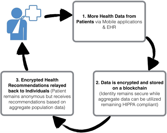
Captured patient biomedical data now stream from electronic health records, genomics, medical imaging, mobile wearable sensors and personal health records. The rise in remote digital health monitoring has shifted medical health awareness, diagnosis and treatment through utilisation of wireless implanted, wearable and external devices. Green et al developed an automated learning classifier to detect hypertrophic cardiomyopathy using photoplethysmography sensors incorporated in commercial smart watches that sense blood volume changes at the skin surface.Reference Green, van Mourik, Wolfus, Heitner, Dur and Semigran43 This may increase the diagnostic detection of asymptomatic patients without a family history to alert providers to screen with an echocardiogram. Patient-worn electrocardiogram monitors continuously store, analyse and wirelessly transmit electrocardiograms with diagnostic yields often higher than that from traditional Holter monitors as well.Reference Rothman, Laughlin and Seltzer44 Natural offshoots could deploy similar technology to Holter monitors routinely ordered in many CHD patients for predicting adverse outcomes especially during the vulnerable inter-stage time period. Analysis of these modalities could translate into downstream information that useful for reducing medical errors and accelerating evidence-based decision-making (Fig 7).
An example of an individual patient’s data can be de-identified, encrypted, and stored on the cloud to amass the volume of aggregate patient data necessary for the development artificial intelligence solutions to relay back personalized clinical inferences.

Digital medicine and wearables
New wearables include smartphone-compatible devices that measure and automatically transfer parameters such as heart rate, blood pressure and weight.Reference Vashist, Schneider and Luong45 Arising companies now offer products that connect to and store data on proprietary artificial intelligence-powered, remotely accessible platforms.Reference Tison, Sanchez and Ballinger47 These platforms pull in vital health data from wearables and analyze those automatically.46 University of California San Francisco published a study that shows that the Apple Watch’s heart rate sensor paired with an artificial intelligence-based algorithm can detect atrial fibrillation.Reference Tison, Sanchez and Ballinger47 Increased sophistication of these technologies may obviate the need for Holter monitors, reducing healthcare costs and the time burden of multiple clinic visits. One study found single-lead electrocardiograms obtained with a smartphone monitor can effectively record supraventricular tachycardia in paediatric patients and predict the mechanism of supraventricular tachycardia as accurately as Holter monitors.Reference Ferdman, Liberman and Silver48 These studies reveal a novel opportunity to detect asymptomatic possibly harmful heart arrhythmias in CHD without an added effort of patients by repurposing popularised, commercially available technology.
Debatably, publicly accessible health monitors now digitalise the patient to construct a virtual medical care system. Real-time feedback from wearables tracking variables such as weight, exercise frequency and blood pressure may motivate paediatric patients to consistently work towards prescribed health timeline goals. Virtual assistants can act as digital healthcare coaches to support patients in the preventative care process. This can allow for remote clinician assessment, increased patient awareness and increased patient ownership of their health. Wearables may be a convenient tool to increase adolescent CHD population compliance and health ownership by allowing them to use popularised technology to track their own progress outside the clinical setting.
Drug discovery
Until recently, the pharmaceutical industry faced years of unprecedented challenges fostering the growing hope for artificial intelligence to accelerate drug discovery and development. Companies using artificial intelligence engines work with structural, functional and phenotypic data in order to predict the biological activity of compounds. Growth in big data and deep neural networks has seeded a string of pharmaceutical firms to partner with artificial intelligence-based drug discovery start-ups. Pharmaceutical companies acknowledge the power of artificial intelligence to optimise drug discovery and development, minimise costs of early drug discovery and anticipate possible side effects at late-stage trials. Some companies have started evaluating all drug candidates against genetic and medical data to validate prospective drugs against genetic targets.Reference Erika and Mukherjee2 The proposed pipeline favours prioritising drugs with a clear genetic target confirmed from their database and retiring drug programmes not sanctioned by their data. With the increasing utilisation of whole-genome sequencing in CHD, there may be a new opportunity for targeted drug development.
Initial studies using artificial intelligence to better understand the structure and function of stem cells in disease progression also show promise to create more targeted drug therapy approaches.Reference Brasil, Pascoal, Francisco, dos Reis Ferreira, Videira and Valadão49 Induced pluripotent stem cell-derived cardiomyocytes now effectively capitulate inherited syndromes, such as hypoplastic left heart syndrome,Reference Jiang, Habibollah and Tilgner50 familial hypertrophic cardiomyopathy,Reference Han, Li and Tchao51 long QT syndromeReference Egashira, Yuasa and Suzuki52 and catecholaminergic polymorphic ventricular tachycardia.Reference Itzhaki, Maizels and Huber53 The successful use of induced pluripotent stem cell-derived cardiomyocytes to model the antiarrhythmic effects of dantrolene in a subpopulation of catecholaminergic polymorphic ventricular tachycardia1patients alludes to the feasibility of stem cell models for individualised drug therapy.Reference Penttinen, Swan and Vanninen54
Other advancements include Stanford scientists’ development of a deep learning algorithm, which can predict the properties of a potential drug compound. Stanford’s deep learning algorithms use very few data points to determine the safest, most effective drug screen for toxicity, side effects and instability to shorten and improve the drug screening process.Reference Kubota55 Artificial intelligence allows for modelling the pharmacokinetics and pharmacodynamics in children that may mitigate barriers to increase safety and drug discovery. Johnson et al developed two machine learning models using microarrays and fibrous cap thickness response to statin therapy to predict the baseline characteristics of patients more likely to achieve an improved response to rosuvastatin.Reference Johnson, Glicksberg and Shameer56
Medical imaging
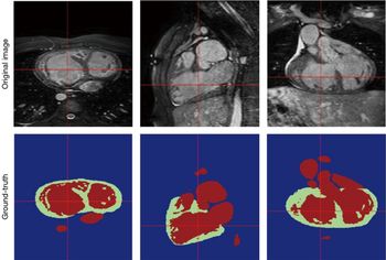
Paediatric cardiology is both a perceptual and cognitive subspecialty with a strong dependence on advanced imaging modalities for clinical decision-making. There is increased use of artificial intelligence in the domain of cardiac imaging ranging from echocardiography to cardiac magnetic resonance imaging for both automated diagnosis and prognosis. Unprecedented amounts of imaging data enable the development of innovative algorithms to automate various stages of the echocardiographic and cardiac magnetic resonance imaging diagnosis workflows. Techniques applied to adult cardiac magnetic resonances now automate chamber segmentation,Reference Lu, Jolly and Georgescu57 view planning for cardiac magnetic resonance acquisitionReference Lu, Jolly and Georgescu57 and cine cardiac magnetic resonance analysis.Reference Wolterink, Leiner, Viergever and Išgum58 Arafati et al recently published a review describing the unmet potential of artificial intelligence to transform adult and paediatric congenital cardiac magnetic resonance for more robust and efficient systems for acquisition, reconstruction and post-processing (Fig 8).Reference Arafati, Hu, Finn, Rickers, Andrew, Cheng and Kheradvar59 Deep learning techniques now enable reconstruction of images from cardiac magnetic resonances for children unable to hold their breath to avoid sedation or critically ill patients where sedation could be harmful.Reference Hauptmann, Arridge, Lucka, Muthurangu and Steeden60 These reconstructed images can achieve quality similar to gold-standard breath-hold cine techniques.Reference Hauptmann, Arridge, Lucka, Muthurangu and Steeden60 Zhang et al described the first fully automated echocardiogram with real-time standard view classification, image chamber segmentation, automated ejection fraction and strain measurements.Reference Zhang, Gajjala and Agrawal61 However, none of the automation software is sophisticated enough to automatically read CHD echocardiograms at this time.
A sample image of segmentation performed on cardiac MRI data from a patient with CHD. The myocardium is green and blood pool is red. From Arafati A, et al. Artificial intelligence in pediatric and adult congenital cardiac MRI: an unmet clinical need. Cardiovasc Diag Ther 2019;9:S310–25. Obtained with permission.

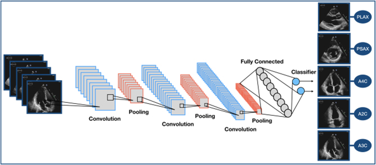
Machine learning also offers diagnostic support through increased efficiency with automated diagnosis of disease states as well as precise ability to detect subtleties vital to diagnostic differentiation. Automation of accurate, reproducible and consistent interpretations reduces the risk of human error and decreases time constraints. Deep-learning algorithms applied to cardiac magnetic resonance proved superior in the detection of pulmonary hypertension to clinicians’ assessment.Reference Diller, Kempny and Babu-Narayan31 Advancements in the intersection of artificial intelligence and echocardiography include algorithms capable of differentiating constrictive from restrictive pericarditisReference Sengupta, Huang and Bansal62 and hypertrophic cardiomyopathy from physiologic cardiomyopathy seen in athletes.Reference Narula, Shameer, Salem Omar, Dudley and Sengupta63 Additional trained algorithms now autonomously detect hypertrophic cardiomyopathy, cardiac amyloidosis, and pulmonary arterial hypertension from echocardiograms.Reference Zhang, Gajjala and Agrawal61 Xu et al recently developed a convolutional neural network model for the segmentation of cardiac structures from the fetal apical four-chamber view.Reference Xu, Liu and Shen64 The authors hope to build off their initial work to create a future screening fetal echocardiography convolutional neural network model capable of autonomous prenatal detection of CHD lesions to reduce missed diagnoses and ultimately improve outcomes.
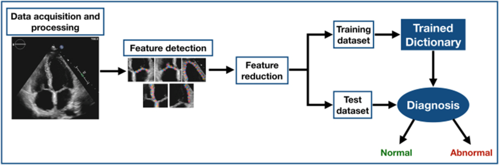
A convolutional network model for artificial intelligence based autonomous view classification, the stepping stone for an eventual machine learning pipeline for automated diagnosis and disease surveillance. From Alsharqi, et al. Echo Res Pract. 2018;5: R115–25, with permission.

Deep learning may be used in the future to detect subtleties that predict the likelihood and time frame for clinical deterioration. Lending support to this strategy, a recent study using automated ejection fraction and strain surveillance could identify breast cancer patients at risk for chemotherapy cardiotoxicity.Reference Zhang, Gajjala and Agrawal61 This study demonstrated the feasibility and effectiveness of a cognitive computing machine learning approach for automated acquisition and interpretations of large pools of echocardiogram strain data.Reference Johnson, Glicksberg and Shameer56 This study showed a proof of concept of how automated data interpretations could save physicians’ time, improve diagnostic accuracy and enable initiation of the appropriate workup and therapy sooner for diseases with challenging diagnostics.Reference Johnson, Glicksberg and Shameer56 Interpretation of imaging data within the clinical context may further precise medicine and clinical decision support systems (Fig 9). Slomka et al argue that the automation process and advanced machine learning will provide patients a conclusion based on massive databases rather than a single doctor’s lifetime of experience while simultaneously cutting down on costs.Reference Dey, Slomka and Leeson65 To this end, Samad et al developed a machine learning pipeline combining imaging parameters and clinical data on tetralogy of Fallot patients. This pipeline was able to identify baseline variables in tetralogy of Fallot patients that were most predictive.Reference Samad, Wehner and Arbabshirani32
Robotic technology
Robotic technology gained increased recognition for its potential to decrease procedural time and cost. Robotic-assisted adult cardiac surgery is well-established as a safe and effective tool in mitral valve repair and replacement, surgical correction of atrial fibrillation, repair of atrial septal defects, intra-cardiac tumour resection and endoscopic coronary artery bypass surgery in adults.Reference Poffo, Toschi and Pope66 Given the complexity of operative and interventional procedures in paediatric cardiology and the minimal size difference between ports and intercostal spaces in paediatric patients, it will likely take decades before a robot will be performing a complete cardiac procedure. Traditionally, robotic technology and imaging systems in paediatric cardiology were limited to thoracoscopic procedures on extra cardiac lesions, ligation of patent ductus arteriosus and division of vascular rings.Reference Suematsu, Mora, Mihaljevic and Del Nido67 However, a newer robotic tool delivery platform enables minimally invasive reconstructive procedures for assistance in the repair of atrial septal defects, ventricular septal defects, complete atrioventricular canal defects, double aortic arch and some tetralogy of Fallot.Reference Bacha and Kalfa68 Vascular robotic systems enhance the performance of cardiovascular procedures by decreasing the occupational hazards encountered by interventionists in the cardiac catheterisation laboratory, while simultaneously providing patients with improved procedural outcomes.Reference Dugas and Schussler69 In the PRECISE trial, interventions on 163 patients using the CorPath system proved to be feasible and safe for relatively modest lesions with a significant reduction in mean radiation exposure time to the patient and operator.Reference Smilowitz, Moses and Sosa70 The REMOTE-PCI study similarly provides a glimpse of what the future of paediatric interventional cardiology may hold. In this study, Madder et al showcased the extent of robotic stenting remotely controlled via interventional cardiologist in a separate room from the patient in the catheterisation lab.Reference Madder, VanOosterhout and Jacoby71 Remote stenting, or “telestenting”, achieved technical success in more than 85% of cases and procedural success in 95% of cases. As in the PRECISE trial, the REMOTE-PCI study showcases the potential to significantly decrease the occupational hazards that have persisted in interventional cardiology, such as orthopedic injuries and chronic irradiation. The study also shows promise to remotely provide more technically skilled interventions to regions of the world where complex interventions are uncommon.
Limitations of artificial intelligence in healthcare
Weak versus strong artificial intelligence and data transparency
Currently, all artificial intelligence projects are categorised as weak because each application performs a limited set of operations. Several artificial intelligence methods support transfer learning, which focuses on storing knowledge gained from solving one problem and applying this knowledge to solve another problem. The quality of prediction models therefore depends on the quality of the data used to create the model. Data derived from the electronic health records potentially affect the results of such models because of inaccuracy or incompleteness. These inaccuracies can stem from failed interoperability between electronic health record systems, little standardisation in entered data and selective measurement.Reference Chang72 The real challenge becomes not gathering the data, but how to “clean it up”. Unlike former rigid systems, however, machine learning can overcome fundamental inconsistencies in “messy” unstructured data sets inherent to medical data. There are reasonable concerns for the effect of large biases in the data sets of machine learning algorithms. Physicians rightfully desire data transparency as the results from the algorithms built from this quality of data intend to influence their clinical decision-making and impact the fragile CHD population. This warrants deliberation before implementation into the patient care model.
Physician buy in
Physician “buy in” remains a necessary and uniquely challenging barrier to widespread adoption of this emerging technology. It will require dispelling the commonly held misconception of artificial intelligence as an antagonistic state of clinician versus machine. As evidenced in this review, artificial intelligence will not supplant physicians. Rather, it could be used by physicians to augment daily decision-making and increase efficiency. By offloading the time constraints of repetitive tasks, physicians can focus on restoring the physician-patient relationship and may have increased job satisfaction, as they find their career again centering on clinical judgement.
Current trajectory and adoption of artificial intelligence in the clinical setting
Many academic centres are investigating the extent of artificial intelligence and machine learning methodology, while a number of private companies are developing and selling products without published validation studies. These disjointed efforts continue to create further silos and barriers to widespread clinic implementation. Unfortunately, even in ideal scenarios when companies complete validated research publications and receive FDA clearance, final adoption in the clinical setting is not guaranteed if the product is not user friendly or easily integrated into the workflow.
Data ownership
Data ownership and regulation is currently the largest roadblock obstructing progression of artificial intelligence in paediatric cardiology. The steady integration of artificial intelligence in paediatric cardiology cannot understate the need for a clear delineation of data ownership. For the first time in the history of medicine, valuable patient data are in the hands of private companies or hospitals that stand to profit from monetising the collective streams of health information gleaned from their consumers or patients. The surge in the consumer wearable device sector has not yet touched on the sticky issue of who deserves to profit from privately collected health information or for how long these private companies should be allowed to retain patient information history. This patient-level and patient-collected information now available to physicians is so new that it is difficult to ascertain what long-term ramifications or practice changing updates will result from interpretation of the data.
At the same time, artificial intelligence delivers the most significant impact free of bias when it is able to draw from voluminous globally sourced de-identified data, a feat not possible if private device companies, hospitals and patients remain in their collective silos. Ideally, patients should be aware of and have a say in how their data are used both for their treatment and for artificial intelligence training and analysis. In their commitment to non-maleficence, paediatric cardiologists may soon find themselves thrust into the role of advocating for standardised regulation of data ownership. Without a clear idea of who stands to profit, it is difficult for profit-incentivised private companies to continue improving and innovating their artificial intelligence designs. In the same vein, it is equally difficult for hospitals to quantify the value of their artificial intelligence when billing for a hospital encounter. Paediatric patients and their families may find themselves reluctant to release their information streams if they are unaware of what data are collected, how their data are used and if they deserve a portion of the profits.
Conclusion
Paediatric cardiology is both a perceptual and cognitive field, rich with robust data sources aligned well with the strengths of the artificial intelligence. Today’s medical education system is devoid of courses in artificial intelligence and currently few paediatric cardiologists are dual-trained. The successful progression of artificial intelligence in paediatric cardiology will likely require collaboration between data scientists and providers to ensure these powerful tools are clinically valuable and ethically aligned. The end goal of this collaboration is to preserve the human-to-human interaction that distinguishes the physician-patient relationship, and medicine as a profession. The core issue remains clarifying how to ethically incorporate artificial intelligence into the healthcare model in a manner that preserves the Hippocratic Oath protecting patient data. If done correctly, the gold standard artificial intelligence model allows physicians to tailor clinical decisions dependent on the patient’s specific health information in the context of large encrypted real-time databases of aggregate patient data to optimise patient outcomes. Physicians will likely experience more job satisfaction with the increased workflow efficiency, freeing additional time to spend at the bedside or to further improve patient outcomes. Patients will likely report improved satisfaction both from more face-to-face time with their physician and from a data-driven management plan with the newfound ability to have larger ownership of their condition through wearable-sourced data. Participating families may feel a component of altruism from sharing de-identified data to contribute to the health and well-being of future patients with CHD. Achievement of this alliance between artificial intelligence, patients, data scientists and physicians could shift evidence-based medicine to intelligence-based medicine.
Financial support
This research received no specific grant from any funding agency, commercial or not-for-profit sectors.
Conflicts of interest
None.
Ethical standards
We have no ethical conflicts of interest to disclose.