Pulmonary arterial hypertension is a rare disease with a very poor prognosis in children.Reference D’Alonzo, Barst and Ayres 1 Prior surgical or interventional series have reported improved functional status and survival after converting severe idiopathic pulmonary arterial hypertension into an Eisenmenger physiology, creating a surgical or percutaneous Potts shunt with direct side-to-side anastomosisReference Blanc, Vouhé and Bonnet 2 , Reference Baruteau, Serraf and Lévy 3 or transcatheter technique.Reference Boudjemline, Patel, Malekzadeh-Milani, Szezpanski, Lévy and Bonnet 4 We report the case of a female child suffering from severe idiopathic pulmonary arterial hypertension with suprasystemic pulmonary pressure, a small atrial septal defect and patent ductus arteriosus.
The baby was diagnosed with idiopathic pulmonary arterial hypertension at the age of 10 months. She underwent transthoracic echocardiography and right-heart catheterisation in another country at the time of diagnosis, not shown at our consultation.
She remained stable on oral medications, such as warfarin, sildenafil 10 mg tid, and furosemide 12.5 mg/day, until 4.5 years of age, when she presented to our department with shortness of breath and dizziness. She was moderately cyanotic, with peripheral oxygen saturation of 86%, without clubbing. Transthoracic echocardiography revealed severe right-chamber dilatation, interatrial, and interventricular septum bowing to the left, elevated pulmonary pressure, a small atrial septal defect (4 mm), and patent ductus arteriosus, both with bidirectional shunting. Cardiac catheterisation confirmed the presence of pulmonary arterial hypertension with near-systemic pulmonary pressure, and angiography showed a small patent ductus arteriosus shunting bidirectionally. Vasoreactivity test with inhaled nitric oxide (20 ppm for 5 minutes) was negative. The patient was treated with triple pulmonary arterial hypertension therapy: sildenafil, 10 mg tid; bosentan, 31.25 mg bid uptitrated to 62.5 mg bid after 4 weeks; and subcutaneous treprostinil, uptitrated from 2 to 24 mcg/kg/minute, and was put in waiting list for lung transplantation. After an initial clinical improvement, 1 year later, the child complained of fatigue, shortness of breath, and faintness.
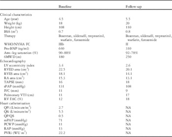
Written informed consent was obtained from the parents before the procedure. The procedure was performed under general anaesthesia. Heart catheterisation showed suprasystemic pulmonary pressure and a tiny patent ductus arteriosus with right-to-left shunting. In particular, indexed pulmonary blood flow was 2.7 L/minute/m2, indexed systemic blood flow 5.5 L/minute/m2, pulmonary-to-systemic flow 0.5, pulmonary artery pressure – systolic, mean, and diastolic – 90/71/59 mmHg, systemic pressure 82/61/48 mmHg, right atrial pressure 13 mmHg, and pulmonary vascular resistance index 22.2 WU m2.
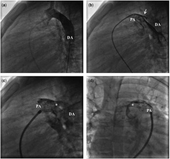
The size for patent ductus arteriosus stenting was empirically chosen based on the patient’s size, aortic diameter at the level of diaphragm (9 mm in our patient), and saturation/haemodynamic changes monitored during the procedure. The ductus arteriosus was carefully approached using a coronary guidewire from the aortic site and stabilised with a pre-mounted 17-mm-long bare metal stent, progressively dilated to 8 mm. The final pulmonary artery to aortic pressure ratio was 1.1 (Fig 1). Arm and leg oxygen saturation at baseline was 86 and 85%, respectively, owing to right-to-left atrial septal defect shunting. At the end of the patent ductus arteriosus stenting procedure, arm and leg oxygen saturation was 92 and 78%, respectively. The procedure was completed successfully without any complication. Postoperative transthoracic echocardiography revealed a widely patent stent with unrestricted flow across the stented patent ductus arteriosus.
Tiny patent ductus arteriosus (white arrow) with exclusive right-to-left shunt, as imaged on aortic and pulmonary angiography. Aortic angiography in lateral view fails to show any left-to-right shunt ( a ). However, a small patent ductus arteriosus (1.2 mm) is imaged on pulmonary angiography ( b ). After stent deployment in the arterial duct, a significant increase in right-to-left shunt can be appreciated, as confirmed by visualisation of the descending aorta ( c and d ). DA=descending aorta; PA=pulmonary artery; PDA=patent ductus arteriosus; *=stent.

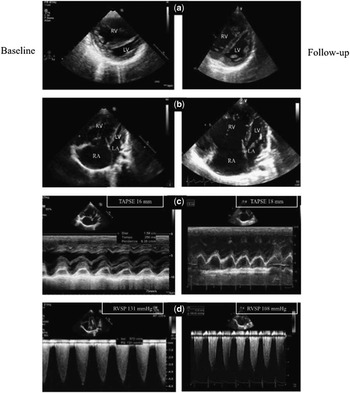
Right ventricular function improved immediately after the procedure and the patient was discharged 3 days postoperatively. She continued the pulmonary arterial hypertension-specific therapy without significant changes after the procedure. At 1-year follow-up (Table 1), a remarkable improvement was observed in clinical status – NYHA functional class from IIIb to II according to the functional classification of the Pediatric Task Force of the Pulmonary Vascular Research InstituteReference Lammers, Adatia and Cerro 5 , functional capacity – 6-minute walk distance from 180 to 250 m, and serology – pro-brain natriuretic peptide concentration from 640 to 310 ng/ml. Echocardiography showed a favourable cardiac remodelling with decreased right-chamber size – right ventricular end-diastolic area in a four-chamber view from 22.5 to 20.1 cm2, right ventricular end-systolic area from 18.1 to 14.1 cm2, and right atrial end-systolic area from 15.3 to 13.1 cm2; increased left-chamber size with reduced interventricular septum flattening – left ventricular eccentricity index from 3.4 to 2.6; and improved right ventricular function – tricuspid annular plane systolic excursion from 16 to 18 mm, that means from −1.8 to 0 z-score related to patient’s age and body surface area,Reference Koestenberger, Ravekes and Everett 6 and right ventricular fractional area change from 12 to 18% (Fig 2). Echocardiography also demonstrated an exclusively right-to-left shunt across the patent ductus arteriosus with a peak velocity of 1.5 m/second (corresponding to 9 mmHg). Haemoglobin increased from 14.4 to 16.1 g/dl because of the right-to-left shunt.
Echocardiographic images showing a favourable cardiac remodelling ( a and b ), reduced right-chamber size and increased left-chamber size, an increase in tricuspid annular plane systolic excursion ( c ) and a reduction in estimated right ventricular systolic pressure and ( d ) from baseline to end of follow-up. LA=left atrium; LV=left ventricle; RA=right atrium; RV=right ventricle; RVSP=right ventricular systolic pressure; TAPSE=tricuspid annular plane systolic excursion.

Patients’ clinical, echocardiographic, and haemodynamic characteristics at baseline, before PDA stenting, and during follow-up.

6MWD=6-minute walk distance; BNP=brain natriuretic peptide; BSA=body surface area; FAC=fractional area change; FC=functional class; LV=left ventricle; mPAP=Mean Pulmonary Artery Pressure; NA=not available; PCWP=pulmonary capillary wedge pressure; PDA=patent ductus arteriosus; PVRi=pulmonary vascular resistance index; QPi=pulmonary flow index; QP/QS=pulmonary-to-systemic flow ratio; QSi=systemic flow index; RA=right atrium; RAP=right atrial pressure; RV=right ventricle; RVED=right ventricular end-diastolic; RVES=right ventricular end-systolic; sPAP=systolic pulmonary arterial pressure; TAPSE=tricuspid annulus plane systolic excursion; VC=inferior vena cava; VTI=velocity time integral; WHO=World Health Organization.
The poor response to medical therapy in children with pulmonary arterial hypertension has prompted the need for alternative approaches to the management or palliative care of this population.Reference Blanc, Vouhé and Bonnet 2 – Reference Boudjemline, Patel, Malekzadeh-Milani, Szezpanski, Lévy and Bonnet 4
Lung or heart–lung transplantation is the conventional procedure for severely ill children with pulmonary arterial hypertension unresponsive to medical treatment.Reference Badesch, Raskob and Elliott 7 The median reported survival after transplantation is 4.5–5.8 years.Reference Keogh, Mayer and Benza 8 – Reference Goldstein, Sweet, Mao, Huddleston and Grady 11 Besides the relatively short survival time, lung or heart–lung transplants are rarely performed because of the lack of available donor organs in this population.
The rationale for creating a right-to-left shunt at the arterial level performing a surgical or percutaneous procedure is to convert idiopathic pulmonary arterial hypertension physiology into a more favourable Eisenmenger physiology. These procedures may result in cyanosis limited to the lower extremities. As a consequence, the upper extremities and the brain are free from the potentially detrimental effects of hypoxia and paradoxical embolism.
Our patient with severe pulmonary arterial hypertension and suprasystemic pulmonary pressure was unsuccessfully managed with maximal medical therapy for pulmonary arterial hypertension – endothelin receptor antagonist, phosphodiesterase-5 inhibitor, and parenteral prostanoid – and finally treated with percutaneous stenting of a small patent ductus arteriosus.
Similarly to Boudjemline et al,Reference Boudjemline, Patel, Malekzadeh-Milani, Szezpanski, Lévy and Bonnet 4 the stent was gradually enlarged by careful sequential balloon inflation to equalise pressures in the pulmonary artery and aorta, to maintain pulmonary blood flow and to limit lower extremity desaturation.
A remarkable improvement in functional status, effort capacity, and echocardiographic parameters was observed immediately after ductus arteriosus stenting and during follow-up, despite a significant desaturation of the lower extremities, with leg oxygen saturation 85–78%. Medical therapy, with bosentan, sildenafil, treprostinil, warfarin, and furosemide, was maintained during follow-up without occurrence of significant complications.
Our good result is consistent with previous case series observations.Reference Boudjemline, Patel, Malekzadeh-Milani, Szezpanski, Lévy and Bonnet 4 The significant clinical and haemodynamic improvement after the procedure reinforces the need to perform a detailed cardiac catheterisation with pulmonary and aortic angiography in all young patients with suprasystemic pulmonary arterial hypertension to look for a patent ductus arteriosus. In addition, it suggests a novel approach for the management of children with severe pulmonary arterial hypertension and suprasystemic pulmonary pressure.
Acknowledgement
None.
Financial Support
This research received no specific grant from any funding agency, commercial, or not-for-profit sectors.
Conflicts of Interest
None.
Ethical Standards
The authors assert that all procedures contributing to this work comply with the ethical standards of the relevant national guidelines on human experimentation and with the Helsinki Declaration of 1975, as revised in 2008, and has been approved by the institutional committees – Monaldi Hospital, Second University of Naples, Italy.