Implications
Piglets of low birth weight have a smaller number of skeletal muscle fibres than their heavier littermates at birth and throughout life, which is associated with impaired growth and a poor carcass and meat quality at market age. An early-postnatal supplementation with l-carnitine to suckling piglets of low birth weight seems only to result in a temporary increase in the number of skeletal muscle fibres, but not to improve overall growth performance as well as carcass and meat quality of the pigs at market age.
Introduction
As a result of prenatal undernutrition, light piglets are less mature at birth than their heavier littermates (Rehfeldt and Kuhn, Reference Rehfeldt and Kuhn2006) and subsequently are disadvantaged during postnatal life. They show a lower growth rate, and finally at slaughter they exhibit poorer carcass and meat quality, as indicated by a smaller meat percentage, a higher degree of fat accumulation, decreased meat tenderness, higher drip loss and a lower pH value (Gondret et al., Reference Gondret, Lefaucheur, Juin, Louveau and Lebret2006; Rehfeldt et al., Reference Rehfeldt, Henning and Fiedler2008a). One possible explanation for the correlation between low birth weight, body composition and meat quality is the low total number of muscle fibres. At birth, low weight piglets exhibit a significantly smaller fibre number than medium and heavy weight piglets, and this ranking is maintained until slaughter (Wigmore and Stickland, Reference Wigmore and Stickland1983; Rehfeldt and Kuhn, Reference Rehfeldt and Kuhn2006). Because of the negative correlation of fibre number with fibre cross-sectional area (FCSA), pigs of low birth weight have highly hypertrophied fibres, which in turn is associated with poor meat quality. In addition, fibre number limits muscular protein accretion, so that nutritional energy is increasingly used to deposit fat (Rehfeldt and Kuhn, Reference Rehfeldt and Kuhn2006).
Although the majority of muscle fibres is formed prenatally in the pig (Wigmore and Stickland, Reference Wigmore and Stickland1983; Rehfeldt et al., Reference Rehfeldt, Fiedler, Dietl and Ender2000), a postnatal increase in total fibre number in the cross-section of m. semitendinosus (ST) and m. psoas major has been observed in domestic and wild pigs (Rehfeldt et al., Reference Rehfeldt, Fiedler and Wegner1987 and Reference Rehfeldt, Tuchscherer, Hartung and Kuhn2008b; Bérard et al., Reference Bérard, Kalbe, Lösel, Tuchscherer and Rehfeldt2011). This increase could be associated with the early-postnatal appearance of a third generation of fibres (Mascarello et al., Reference Mascarello, Stecchini, Rowlerson and Ballocchi1992; Lefaucheur et al., Reference Lefaucheur, Edom, Ecolan and Butler-Browne1995) or elongation of existing short fibres (Bérard et al., Reference Bérard, Kalbe, Lösel, Tuchscherer and Rehfeldt2011).
In a pilot study, the supplementation of l-carnitine to piglets during the suckling period resulted in increased fibre numbers in ST in low birth weight piglets, whereas no effect of treatment was observed in piglets of medium birth weight (Lösel et al., Reference Lösel, Kalbe and Rehfeldt2009). Thereby, carnitine-supplemented low weight piglets reached the level of medium weight piglets. The objective of the present study was to investigate whether the carnitine-induced increase in fibre number at weaning in piglets of low birth weight is maintained until market age and whether this would alter the relative deposition of fat and lean during growth and improve carcass and meat quality.
Material and methods
Animals
All procedures were in accordance with the German Law of Animal Protection and approved by the relevant authority (Landesamt für Landwirtschaft, Lebensmittelsicherheit und Fischerei Mecklenburg-Vorpommern; LALLF M-V/TSD/7221.3-2.1-026/08). The study was conducted at the pig experimental station of the Leibniz Institute for Farm Animal Biology (FBN) Dummerstorf using purebred German Landrace pigs. A total of 56 piglets from 14 litters of different sows (second to seventh parity) were used in nine temporally successive repeats. The number of piglets born per litter ranged from 11 to 18. Litter size was standardized to 11 to 12 piglets by cross-fostering the heaviest piglets only. Male piglets were castrated during the 1st week of life and all piglets were weaned at day 28 of age.
The study was conducted as a split-plot design with litter as block, sex as whole plot and treatment as subplot. From the piglets with low birth weight (0.80 to 1.26 kg) within each litter two males and two females were randomly selected and within sex randomly allocated to the control (n = 2) or carnitine group (n = 2). From days 7 to 27 of age, piglets were orally supplemented once daily with 400 mg of l-carnitine (Carnifeed, Lohmann Animal Health GmbH & Co. KG, Cuxhaven, Germany) dissolved in 1 ml of water. Control piglets received an equal volume of water without carnitine.
Creep-feed was offered from day 10 of age. After weaning piglets were kept in single pens (1.7 m2) where feed consumption was determined on a weekly basis. From day 70 of age until slaughter (day 166), pigs were housed in groups of 4 or 8 animals (pen area 11.4 m2). The resulting different floor space allowance per animal (1.4 v. 2.8 m2) did not significantly affect growth performance or body composition and no significant treatment × floor space interactions could be observed (see ‘Statistics’ section). Individual feed intake was recorded by an electronic feed dispenser (INSENTEC B.V., Marknesse, The Netherlands). During the whole nursery and fattening period, feed was provided ad libitum. An overview of the diets used in this study is given in Supplementary Table S1. Pigs were weighed weekly until day 28 of age and subsequently each second week until slaughter.
Blood sample, biopsies and dual-energy X-ray absorptiometry (DXA) measurements
At weaning, piglets were bled via venipuncture and blood was collected in heparinized tubes. Plasma was stored at −80°C until analysed for free carnitine, non-esterified fatty acids (NEFA), glucose and urea. Samples of the m. longissimus (LM) and the overlaying subcutaneous adipose tissue were taken by shot biopsy at week 12 (right LM, ∼13th/14th rib), 16 (left LM, ∼13th/14th rib) and 20 (right LM, ∼11th/12th rib) of age. The thickness of skin and the three subcutaneous fat layers were measured before freezing fat and muscle samples in liquid nitrogen for further analyses.
The following day after each biopsy, pigs were sedated for the estimation of body composition by DXA using the Norland XR-26 device (Norland Corporation, White Plains, NY, USA) as described by Lösel et al. (Reference Lösel, Kremer, Albrecht and Scholz2010). The DXA technology provides values for bone mineral density, bone mineral content, fat mass and lean mass of the whole body. Total tissue mass was obtained by summing bone mineral content, fat mass and lean mass. Fat percentage and lean percentage were then calculated by dividing fat mass and lean mass, respectively, by total tissue mass.
Body composition, carcass and meat quality
At day 166 of age, pigs were slaughtered after electro-stunning. The weights of all organs, as well as internal fat depots, were recorded. On the left half carcass, ham length and perimeter and average back fat thickness (resulting from the average of measures at the 3rd and 11th thoracic vertebrae and the smallest measure in the lumbar region) were measured. Using an image analysis software (Scan-Star A, Matthäus, Klausa, Germany), loin muscle area, fat area above loin muscle, fat thickness above loin muscle (fat measure B = thinnest fat thickness) and the lateral fat thickness (fat measure D) were measured on the loin cross-section between the 13th and 14th rib. Muscle meat percentage was estimated by the FOM device (Fat-o-Meat'er, SFK-Technology A/S, Søborg, Denmark).
After chilling, the left half carcass was dissected into primal cuts according to the German guidelines for breed value assessment. All primal cuts were further separated into four fractions: meat, subcutaneous fat, skin and bone. To determine the chemical composition of the whole body, including empty visceral tract, various body fractions were analysed for dry matter, protein, fat and ash. Subsequently, the chemical composition of the whole body was calculated from the data of individual fractions. Chemical composition of the meat fraction of each primal cut was determined by near-IR transmission spectroscopy (FoodScan Lab Meat Analyser, FOSS GmbH, Rellingen, Germany). The chemical composition of the right ST and right LM, subcutaneous fat, edible organs, internal fat (omental + perirenal), waste, bones + skin by ash (incineration at 600°C), water/dry matter (oven drying at 105°C) and lipid (petrol ether extraction) was analysed by standard methods (Association of Official Analytical Chemists, 2000), whereas protein proportion was estimated by difference (Kuhn et al., Reference Kuhn, Ender and Nürnberg1994).
Conductivity and pH values were measured in the left ST and the left LM at the level of 13th/14th rib 45 min and 24 h postmortem. The pH values were measured by a glass electrode in a stainless steel penetration blade (pH-Star, Matthäus, http://www.klassifizierung.org/pH-Star_e.html). For measurements of electrical conductivity, a handheld conductivity meter equipped with a pair of stainless-steel needle electrodes (LF-Star, Matthäus) was used.
The right ST and LM were used to determine meat colour and drip loss. Meat colour was measured 24 h postmortem with the Chroma Meter CR-300 with a D65 illuminant (Minolta AG, Ahrensburg, Germany) after a blooming time of 30 ± 2 min and the results expressed as the CIE (Commission International de l'Eclairage) L*, a* and b* values.
Drip loss was defined as the weight loss of a meat sample (50 g), placed on a plastic grid and wrapped airtight in foil, after a 24-h storage at 4°C. Shear force of the right LM was measured 24 h postmortem. A slice of the muscle was placed in a plastic bag and heated to an internal temperature of 70°C in a water bath of 75°C. After cooling to room temperature, Warner–Bratzler shear force was determined on five meat cores using the Texture Analyzer XT2 (Stable Micro Systems Ltd, Godalming, Surrey, UK). Cook loss of the LM sample was defined as the weight loss of the meat slice after cooking.
Histochemistry and microscopy
Within 10 to 15 min after slaughter, the whole ST was excised from the right hind limb and its weight, length and circumference were recorded. Muscle cross-sectional area (MCSA) was calculated from the circumference of the muscle mid-belly. One sample each was collected from the dark (deep), bright (superficial) and central portion of the muscle mid-belly. Samples were snap-frozen in liquid nitrogen and thereafter stored at −80°C. From samples of each region, serial transverse sections of 10 μm were cut at −20°C in a cryostat (Leica, Nussloch, Germany). One section was stained for cytoplasm with eosin and for alkaline phosphatase to visualize capillaries. Another section was exposed to the combined reaction for NADH-tetrazolium reductase and acid pre-incubated ATPase at pH 4.2, which enables classification into slow-twitch oxidative (STO), fast-twitch oxidative (FTO) and fast-twitch glycolytic (FTG) fibres as described by Rehfeldt et al. (Reference Rehfeldt, Stabenow, Pfuhl, Block, Nürnberg, Otten, Metges and Kalbe2012). Fibre-type distribution, FCSA and capillary distribution were determined on 900 muscle fibres (300 each in the dark, in the central, and in the bright region of the ST) by image analysis (AMBA, IBSB, Berlin, Germany). The estimated total fibre number was obtained by multiplying the fibre number per unit area by the MCSA of ST muscle. Microscopic analyses were conducted by one person.
Biochemical analyses
Plasma metabolites were analysed by the Klinik für Rinder (Stiftung Tierärztliche Hochschule, Hannover, Germany) using an automatic analyzer (Pentra 400, Axon Lab, Reichenbach, Germany) and the following kits: glucose (no. 553−230) from MTL Diagnostics (Idstein, Germany), NEFA (no. 434−91795) from Wako Chemicals GmbH (Neuss, Germany) and urea (no. LT-UR 0050) from Labor + Technik Lehmann (Berlin, Germany). Free carnitine in blood plasma was analysed according to the spectrophotometric method by Prieto et al. (Reference Prieto, Andrade, Aldamiz-Echevarria and Sanjurjo2006). Muscle homogenates from the central portion of the right ST, the right LM at the 13th/14th rib level, and from the central portion of the m. rhomboideus (RH) were analysed for DNA, protein, activities of lactate dehydrogenase (LDH) and isocitrate dehydrogenase (ICDH) as described previously (Lösel et al., Reference Lösel, Kalbe and Rehfeldt2009). DNA was measured fluorometrically by a Fluorescence Reader FLx800 (Bio-Tek Instruments GmbH, Bad Friedrichshall, Germany) against a standard of calf thymus DNA after using Hoechst 33258 (both from Sigma-Aldrich Chemie GmbH, Steinheim, Germany). The activities of LDH and ICDH were measured using modified assay protocols according to SIGMA (http://www.sigmaaldrich.com/life-science/metabolomics/enzyme-explorer/learning-center/assay-library.html). All assays were adapted to microplate and the optical density was measured using a Spectramax Plus384 plate reader (Molecular Devices Corporation, Sunnyvale, CA, USA).
Statistical analysis
Data were subjected to ANOVA using the mixed model procedure of SAS (version 9.1, SAS Institute Inc., Cary, NC, USA) including treatment, sex, repeat and corresponding interactions as fixed factors and litter within repeat as random factor. The individual pigs served as the experimental unit. In the case of significant interactions, differences between means were tested post hoc by the Student's t-test. Data are presented as least squares means (LSM) ± s.e. To test for the effect of different floor space allowance during the fattening period, an additional ANOVA was carried out including treatment, group size, treatment × group size interaction, and repeat within group size as fixed factors, and litter within repeat as random factor. Differences were considered significant if P ⩽ 0.05 and were considered as tendencies if 0.05 < P ⩽ 0.11.
Results
Blood parameters at weaning
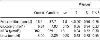
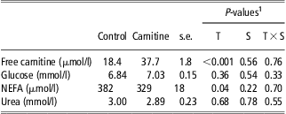
In response to carnitine supplementation, the concentration of free carnitine in blood plasma increased twofold (P < 0.001; Table 1). The concentrations of glucose and urea at day 28 of age were not different between carnitine-supplemented and control piglets. Piglets of the carnitine group had about 14% lower concentration of NEFA (P < 0.05). No significant treatment by sex interactions were observed for all traits.
Effect of carnitine supplementation from days 7 to 27 of age on concentrations of free carnitine and metabolic traits in blood plasma at day 28 of age (LSM ± s.e.)
LSM = least squares means; NEFA = non-esterified fatty acids.
1T = treatment; S = sex.
Growth performance and development of body composition
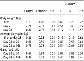
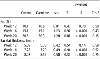
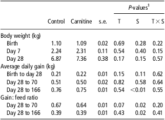
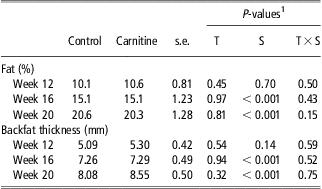
Because of the experimental design, BW at birth was not different between the control and the carnitine group (Table 2). Likewise, the groups did not differ in BW at the beginning of the experiment (day 7) and at weaning. No effect of treatment on average daily feed intake during the nursery phase or fattening period (data not shown), and on average daily gain from birth to weaning, during the nursery period (days 28 to 70), and during fattening (days 70 to 166) was observed. Gain : feed ratio tended to be lower in the carnitine group (P = 0.07) during the nursery phase, but was not different between groups during the whole growing-finishing period from weaning until slaughter. No effects of treatment on DXA-derived fat percentage of the whole body (Table 3), lean percentage, bone mineral content and bone mineral density (data not shown) were observed in weeks 12, 16 and 20 of age. Back fat thickness measured on biopsy samples at weeks 12, 16 and 20 was not influenced by treatment (Table 3). There were no significant treatment by sex interactions for all traits.
Effect of carnitine supplementation from days 7 to 27 of age on growth performance (LSM ± s.e.)
LSM = least squares means.
1T = treatment; S = sex.
Effect of carnitine supplementation from days 7 to 27 of age on fat percentage of the whole body derived by dual-energy X-ray absorptiometry and on back fat thickness (mm) measured on biopsy samples in weeks 12, 16 and 20 of age (LSM ± s.e.)
LSM = least squares means.
1T = treatment; S = sex.
Body composition and carcass quality
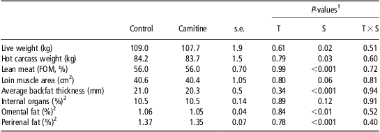
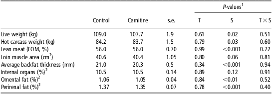
The control and carnitine group did not differ in LW at slaughter and hot carcass weight (Table 4). No effects of treatment on measures of meat yield and fat accretion in the half carcass were observed. Likewise, no differences between groups were found for the relative weights of internal fat depots, the proportion of total internal organs (Table 4) and the relative weights of internal organs such as heart, liver, kidneys and gut (data not shown).
Effect of carnitine supplementation from days 7 to 27 of age on carcass characteristics and relative tissue weights at market weight (day 166 of age; LSM ± s.e.)
LSM = least squares means.
1T = treatment; S = sex.
2Related to the whole empty body.
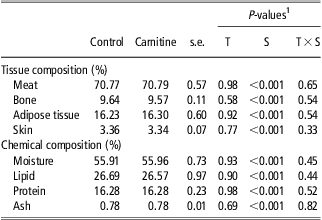
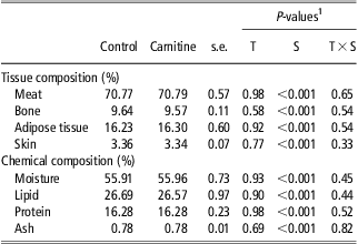
Tissue composition of four primal cuts (ham, shoulder, loin and neck) and chemical composition of the half carcass (Table 5) or of the whole empty body (data not shown) was not changed in response to treatment. Likewise, intramuscular fat concentration in ST (control: 4.10%, carnitine: 3.73%, s.e. 0.15; P = 0.15) and LM (control: 1.22%, carnitine: 1.16%, s.e. 0.08; P = 0.46) was not influenced by treatment. There were no significant treatment by sex interactions for all traits.
Effect of carnitine supplementation from days 7 to 27 of age on the tissue composition of four primal cuts (ham, shoulder, loin, neck) and on the chemical composition of the half carcass at market weight (day 166 of age; LSM ± s.e.)
LSM = least squares means.
1T = treatment; S = sex.
Structural and biochemical properties of skeletal muscle
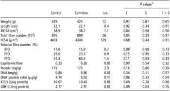
Weight, length and MCSA of ST were not affected by carnitine supplementation (Table 6). No effect of treatment on the average FCSA or the FCSA of individual fibre types (data not shown) could be observed. Similarly, the total fibre number in ST MCSA was unchanged. Fibre-type distribution was influenced by treatment in that the proportion of STO fibres tended to be smaller (P = 0.08) and the proportion of FTG fibres tended to be greater in the carnitine group (P = 0.11). In addition, the total number of capillaries per myofibre tended to be smaller in the carnitine group (P = 0.09), whereas the fibre area supplied by one capillary remained unchanged (P = 0.52; data not shown). Specific LDH activity (P = 0.09) and DNA : protein ratio (P = 0.06) tended to be increased in the carnitine group, whereas no effect of carnitine supplementation could be observed for DNA concentration, protein concentration and specific ICDH activity. In LM and RH, protein concentration and activities of LDH and ICDH were not affected by treatment (data not shown). There were no significant treatment by sex interactions for all traits.
Effect of carnitine supplementation from days 7 to 27 of age on structural and biochemical properties of m. semitendinosus at market weight (day 166 of age; LSM ± s.e.)

LSM = least squares means; MCSA = muscle cross-sectional area; FCSA = fibre cross-sectional area; STO = slow-twitch oxidative; FTO = fast-twitch oxidative; FTG = fast-twitch glycolytic; ICDH = isocitrate dehydrogenase; LDH = lactate dehydrogenase.
1T = treatment; S = sex.
Meat quality
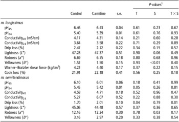
In LM, influences of treatment on conductivity, pH values, colour, drip loss, cook loss and Warner–Bratzler shear force could not be observed (Table 7). Similarly, colour and conductivity in ST muscle were not different between the groups. However, carnitine-supplemented pigs displayed a lower pH24 value in ST muscle (P = 0.05), which was associated with increased drip loss (P = 0.04). For drip loss an interaction occurred between treatment and sex (P = 0.01), with increased values in carnitine-supplemented castrated males compared with control (2.22 v. 1.53 ± 0.15%; P < 0.01) and no effect in females (1.79 v. 1.87 ± 0.15%; P = 0.71). No significant treatment by sex interactions were observed for the other meat quality traits.
Effect of carnitine supplementation from days 7 to 27 of age on meat quality traits in m. longissimus and m. semitendinosus at market weight (day 166 of age; LSM ± s.e.)

LSM = least squares means.
1T = treatment; S = sex.
Discussion
The long-term effects of carnitine persisting after the supplementation period were subject of this study. To confirm that a sufficient amount of carnitine was absorbed, carnitine in blood plasma was determined at day 28 of age (weaning). As a result from carnitine supplementation from days 7 to 27 of age, the carnitine concentration in blood plasma at weaning was doubled, which represents a similar extent as the increase in muscle carnitine observed in our previous study (Lösel et al., Reference Lösel, Kalbe and Rehfeldt2009). Because time of blood sampling was ∼24 h after the last carnitine administration, the increased plasma carnitine concentration is the result of a permanently increased carnitine pool. Moreover, the reduced NEFA concentration in blood plasma indicates that lipid metabolism was influenced during the period of carnitine supplementation. Owing to its essential role for the transportation of long-chain fatty acids into the mitochondrial matrix where β-oxidation of activated fatty acids takes place, carnitine is often associated with increased fatty acid utilization as shown in porcine mitochondria in vitro (Heo et al., Reference Heo, Lin, Odle and Han2000) and in liver mitochondria and hepatocytes of carnitine-supplemented growing pigs (Owen et al., Reference Owen, Jit, Maxwell, Nelssen, Goodband, Tokach, Tremblay and Koo2001). As a result, reduced body fat accretion has been observed in suckling, weaning and growing pigs in response to carnitine (Owen et al., Reference Owen, Nelssen, Goodband, Weeden and Blum1996 and Reference Owen, Jit, Maxwell, Nelssen, Goodband, Tokach, Tremblay and Koo2001; Lösel et al., Reference Lösel, Kalbe and Rehfeldt2009). However, the intensified utilization of fatty acids is not always reflected by decreased NEFA levels. The NEFA concentrations were not affected by carnitine in our previous study (Lösel et al., Reference Lösel, Kalbe and Rehfeldt2009) and in carnitine-supplemented weanling pigs (Owen et al., Reference Owen, Nelssen, Goodband, Weeden and Blum1996). In contrast, in gestating sows NEFA concentration was decreased by carnitine (Woodworth et al., Reference Woodworth, Tokach, Nelssen, Goodband, Dritz, Koo, Minton and Owen2007).
During the suckling period, which includes the period of supplementation from days 7 to 27 of age, average daily gain did not differ between the control and the supplemented group, resulting in similar BWs at weaning. This is in accordance with our previous study with piglets of low and medium birth weight using the same carnitine dosage during the same period (Lösel et al., Reference Lösel, Kalbe and Rehfeldt2009). Further, our results are comparable with experiments that increased carnitine supply to piglets in their first weeks of life by other ways. In piglets weaned at day 3 of age, the supplementation of the replacement diet with carnitine did not improve growth performance during the 21-day experimental period (Hoffman et al., Reference Hoffman, Ivers, Ellersieck and Veum1993). Likewise, litters suckled by sows supplemented with carnitine during lactation, which are known to exhibit greater carnitine concentrations in milk, did not differ in their weight gains until weaning from those suckled by unsupplemented sows (Musser et al., Reference Musser, Goodband, Tokach, Owen, Nelssen, Blum, Campbell, Smits, Dritz and Civis1999; Birkenfeld et al., Reference Birkenfeld, Kluge and Eder2006). In the present study, growth performance did not differ in the growth periods following carnitine supplementation, with the exception of a tendency for decreased gain : feed ratio in the period from weaning to day 70 of age. Regarding long-term effects of early-postnatal carnitine supplementation, no comparable experiments are known.
The most important finding from our pilot study representing an immediate effect of carnitine supplementation during the suckling period was that the total myofibre number was elevated in carnitine-treated piglets of low birth weight at day 28 of age. The results of the present study suggest that this difference was not maintained until market age. As a loss of fibres after weaning in carnitine-treated pigs seems very unlikely (Rehfeldt et al., Reference Rehfeldt, Fiedler and Wegner1987; Bérard et al., Reference Bérard, Kalbe, Lösel, Tuchscherer and Rehfeldt2011), the compensation in total fibre number is suggested to result from a different trajectory of the early-postnatal increase in fibre number in control and carnitine-supplemented piglets. An early-postnatal increase in total fibre number in pig muscle has been reported to occur in ST muscle cross sections at the mid-belly level (Rehfeldt et al., Reference Rehfeldt, Fiedler, Dietl and Ender2000 and Reference Rehfeldt, Tuchscherer, Hartung and Kuhn2008b; Bérard et al., Reference Bérard, Kalbe, Lösel, Tuchscherer and Rehfeldt2011) and can be attributed to two mechanisms. On the one hand, the prenatal formation of primary and secondary myofibres (Wigmore and Stickland, Reference Wigmore and Stickland1983; Lefaucheur et al., Reference Lefaucheur, Edom, Ecolan and Butler-Browne1995) is followed by a third wave of myofibre hyperplasia after birth and gives rise to new fibres. The frequency of these short and thin tertiary fibres decreases within the first weeks after birth (Mascarello et al., Reference Mascarello, Stecchini, Rowlerson and Ballocchi1992; Lefaucheur et al., Reference Lefaucheur, Edom, Ecolan and Butler-Browne1995; Bérard et al., Reference Bérard, Kalbe, Lösel, Tuchscherer and Rehfeldt2011). The second mechanism is related to the elongation of intrafascicularly terminating myofibres that already exist at birth. Some of them will appear in the plane of the section of the widest muscle girth resulting in an increase in fibre number in the cross-section (Bérard et al., Reference Bérard, Kalbe, Lösel, Tuchscherer and Rehfeldt2011). Our previous study (Lösel et al., Reference Lösel, Kalbe and Rehfeldt2009) revealed that carnitine may have accelerated the longitudinal growth of intrafascicularly terminating fibres and/or the formation of tertiary fibres during the period of supplementation in the piglets of low birth weight. However, fibre elongation and hyperplasia in control piglets could have proceeded slower but for a longer time compared with carnitine-supplemented piglets. Thus, control piglets could have caught up with carnitine-treated piglets in terms of myofibre number after weaning. This, however, remains to be investigated in detail.
Because an elevated fibre number was the prerequisite of hypothesized long term, persisting effects of carnitine on growth, body composition, carcass and meat quality, it is not surprising that we could not observe any improvement in these traits in the carnitine group. The relevance of total fibre number for these traits is obvious in pigs of low birth weight, which exhibit a smaller myofibre number at birth and throughout life compared with pigs of medium and heavy birth weight (Wigmore and Stickland, Reference Wigmore and Stickland1983; Rehfeldt and Kuhn, Reference Rehfeldt and Kuhn2006). These pigs have fatter carcasses and a smaller lean meat content (Bee, Reference Bee2004; Rehfeldt and Kuhn, Reference Rehfeldt and Kuhn2006; Rehfeldt et al., Reference Rehfeldt, Henning and Fiedler2008a), likely because the potential of lean accretion is limited and nutritional energy would be stored as fat instead. Because of the negative correlation between total fibre number and FCSA (Rehfeldt et al., Reference Rehfeldt, Fiedler, Dietl and Ender2000; Kim et al., Reference Kim, Lee, Choi, Kim, Yoo and Hong2008) piglets of low birth weight display highly hypertrophied fibres, which in turn is directly related to a poorer meat quality in terms of tenderness, water holding capacity and pH (Gondret et al., Reference Gondret, Lefaucheur, Juin, Louveau and Lebret2006; Rehfeldt and Kuhn, Reference Rehfeldt and Kuhn2006; Rehfeldt et al., Reference Rehfeldt, Henning and Fiedler2008a). However, in agreement with the unchanged total fibre number, FCSA was not different between groups in the present study. Therefore, the lower pH24 and the higher drip loss in ST muscle of the carnitine group cannot be explained by accelerated fibre hypertrophy.
Another important factor for meat quality is fibre-type composition. In fact, fibre-type distribution tended to be affected in ST muscle with an increased proportion of FTG and a decreased proportion of STO fibres in the carnitine group. An increasing proportion of glycolytic fibres negatively affected some meat quality traits, such as water holding capacity and pH decline (Larzul et al., Reference Larzul, Lefaucheur, Ecolan, Gogué, Talmant, Sellier, Le Roy and Monin1997). However, it remains unclear why drip loss in ST was increased in carnitine-supplemented castrated males, but not in females. The changed fibre-type distribution in ST was associated with an increased specific activity of LDH (a marker for the anaerobic glycolytic energy production) in carnitine-supplemented pigs. Because FTG fibres show higher LDH activities than STO fibres, an increased proportion of FTG fibres is associated with increased LDH activity in muscle tissue (Lefaucheur and Vigneron, Reference Lefaucheur and Vigneron1986; Lebret et al., Reference Lebret, Le Roy, Monin, Lefaucheur, Caritez, Talmant, Elsen and Sellier1999). The increase in glycolytic muscle metabolism in ST was accompanied by a decreased capillary density reflecting the known association between the abundance of capillaries and fibre-type distribution (Quiroz-Rothe and Rivero, Reference Quiroz-Rothe and Rivero2004). It has to be considered that muscles were affected differently, because changes in muscle metabolism, structural traits and meat quality were only observed in ST in our study.
It seems that carnitine treatment during the suckling period had a lasting effect on the metabolism of ST muscle in pigs at market age. Immediately after the supplementation period at day 28 of age, muscle metabolism was more oxidative in carnitine-treated piglets without changes in fibre-type composition (Lösel et al., Reference Lösel, Kalbe and Rehfeldt2009). Thus, the observed differences in fibre types seem to develop after the withdrawal of carnitine. The early-postnatal period is critical for both metabolic and contractile fibre-type differentiation (Lefaucheur and Gerrard, Reference Lefaucheur and Gerrard2000) because the greatest changes in fibre-type distribution occur during the first 5 to 8 weeks of life (Lefaucheur and Vigneron, Reference Lefaucheur and Vigneron1986; Rehfeldt et al., Reference Rehfeldt, Fiedler and Wegner1987; Wank et al., Reference Wank, Fischer, Walter and Bauer2006). In addition, weaning is a critical period with respect to carnitine supply, because piglets have to adapt to a diet that is relatively low in carnitine compared with milk. This change is even more challenging for carnitine-supplemented piglets, which had a greater carnitine status during suckling than control piglets. Moreover, endogenous biosynthesis of carnitine could be reduced in these piglets, as indicated by the depressed activity of γ-butyrobetaine dioxygenase (the enzyme catalysing the last step of carnitine biosynthesis) in kidney in carnitine-supplemented weaning piglets (Fischer et al., Reference Fischer, Varady, Hirche, Kluge and Eder2009). The observed intensification of the anaerobic-glycolytic pathway of energy generation in response to early-postnatal carnitine feeding may therefore be a long-term response to the withdrawal of carnitine that was available in much lower amounts after weaning to intensify oxidative muscle metabolism and therefore required some compensation to maintain energy balance for myofibre growth and contraction.
Conclusion
Carnitine supplementation during the suckling period, which has been shown previously to increase total muscle fibre number and oxidative muscle metabolism in piglets of low birth weight at weaning, exhibits only marginal lasting effects on muscle structure and metabolism. It does not permanently increase myofibre number and tends to change muscle metabolism including fibre-type composition towards the anaerobic-glycolytic pathway and to affect meat quality in pigs at market age in a muscle-specific manner.
Acknowledgements
The authors are grateful to Lohmann Animal Health, Cuxhaven, Germany, for financial support and for kindly providing the l-carnitine used in this study. They also thank H. Sievert and colleagues for animal care, Dr O. Bellmann for blood sampling and anaesthesia, R. Pfuhl and colleagues, Marie Jugert-Lund, Angela Steinborn, and M. Günther for technical assistance.