Introduction
The phenomenal success of Silicon technology hinged fundamentally on the ability to locally control its conductivity by doping. Today, semiconducting polymers have the potential to enable next-generation solution-processable and flexible optoelectronic devices.[Reference Arias, MacKenzie, McCulloch, Rivnay and Salleo1] Doping has been instrumental in enhancing the performance of organic light-emitting diodes, leading to their commercial success, but a well-developed doping technology that would enable controlled engineering of electronic properties is still lacking for instance, in polymeric thin film transistors. In organic semiconductors, the doping process is the result of a complex interplay of electronic and morphological effects caused by the interactions between the semiconductor matrix and the doping molecules. Therefore, fundamental studies of polymer–dopant interactions are necessary in order to develop a robust doping technology.
To date, studies of the model system consisting of the semiconducting polymer poly(3-hexylthiophene) (P3HT), and p-type dopant tetrafluoro-tetracyanoquinodimethane (F4TCNQ), have provided much useful information about the doping mechanism. Unlike with silicon, doping in organic semiconductors has traditionally involved the addition of sizeable amounts of dopants (upwards of one-tenth P3HT monomer mole fraction). These films are fabricated by co-mixing the dopants and polymer in a suitable solvent, yielding highly conductive P3HT:F4TCNQ films of up to 1 S/cm.[Reference Duong, Wang, Antono, Toney and Salleo2] The strong doping accessible through this method leads to significant changes in the macroscopic structural and optical properties of the doped P3HT,[Reference Méndez, Heimel, Opitz, Sauer, Barkowski, Oehzelt, Soeda, Okamoto, Takeya, Arlin, Balandier, Geerts, Koch and Salzmann3–Reference Gao, Stein, Thomas, Garcia-Galvez, Yang, Kirk and Grey5] with a new crystal structure emerging above a certain doping threshold.[Reference Duong, Wang, Antono, Toney and Salleo2] Other doping methods have been proposed to overcome these limitations.[Reference Scholes, Hawks, Yee, Wu, Lindemuth, Tolbert and Schwartz6–Reference Glaudell, Cochran, Patel and Chabinyc8]
At the opposite end of the spectrum, ultra-low doping (~10−4–10−6 dopant molar ratio) is of interest due to its potential to fill electronic traps. The expectation is that carrier mobilities in the semiconductor would increase because of trap filling, while the film would maintain its semiconducting properties because the charges introduced by doping are localized in the traps, as has been observed in the n-doping of certain polymers.[Reference Olthof, Mehraeen, Mohapatra, Barlow, Coropceanu, Brédas, Marder and Kahn9] Several recent independent reports[Reference Pingel, Schwarzl and Neher10–Reference Said, Zhang, Dasari, Anjum, Munir, Hu, Amassian, Barlow and Marder12] however, have shown that any trap-filling that does occur in P3HT, if at all, is insufficient to improve electrical transport in the polymer. For instance, hole mobility in thin films and solar cell power conversion efficiency either show minimal improvement or degradation with the addition of dopants.[Reference Said, Zhang, Dasari, Anjum, Munir, Hu, Amassian, Barlow and Marder12, Reference Pingel and Neher13]
Current computational evidence suggests that the failure of dopants to improve the transport properties of the polymer at low doping concentrations could be attributed to the very presence of the dopant molecules in the polymer matrix. These molecules could lead to dopant-induced disorder, resulting in the creation of additional trap states in the semiconductor matrix.[Reference Keum, Liu, Al-Shadeedi, Kaphle, Bunge and Lussem14] F4TCNQ undergoes integer charge transfer (CT) with P3HT, leading to high ionization efficiencies of almost 100%.[Reference Pingel and Neher13] Due to the intrinsically low dielectric constant of organic semiconductors,[Reference Arkhipov, Heremans, Emelianova and Bässler15] the anions formed by the CT process have the potential to strongly affect the local charge environment. This effect was first modeled by Arkhipov et al., who postulated the creation of Coulomb traps as a result of the ionized dopant, which leads to a broadening of the density of states, and thus a reduced hopping rate through the doped polymer.[Reference Arkhipov, Heremans, Emelianova and Bässler15]
In summary, ultra-low doping concentration can give rise to two opposing effects on transport: a beneficial one (trap passivation) and a detrimental one (additional disorder). Which effect prevails remains unclear. In one study, Pingel et al. showed that doping at low concentrations (~10−4–10−6 dopant molar ratio) can modulate the film conductivity by an order of magnitude.[Reference Pingel, Schwarzl and Neher10, Reference Pingel and Neher13] The conductivity increase was sub-linear, leading the authors to conclude that the carrier mobility was degraded as dopant concentration increased. Nevertheless, beyond characterizing the resulting electrical properties, the structural effect of doping at such low concentrations has been relatively unexplored to date. This is partially due to the fact that bulk characterization methods, such as optical absorption and x-ray diffraction,[Reference Ma, Lee, Park, Kim, Lee and Cho16] are unable to provide information about dopant-induced structural changes, making conclusive structure-property studies challenging.
Since the addition of trace dopants can lead to significant changes in the electrical properties of semiconducting polymers in the vicinity of the dopants, we investigate the structural mechanisms responsible for such effects. We resolve the structural effects of ultra-low doping on P3HT using charge modulation spectroscopy (CMS), providing direct experimental evidence for dopant-induced disorder in semiconducting polymers. Well-established theoretical models[Reference Spano17–Reference Brown, Thomas, Köhler, Wilson, Kim, Ramsdale, Sirringhaus and Friend19] are used to analyze the CM spectra, allowing us to quantify the changes in the P3HT local structure induced by F4TCNQ. Consistent with a previous work,[Reference Pingel, Schwarzl and Neher10] we observe that the carrier mobility decreases with doping concentration, while carrier concentration increases. By combining the spectral analyses with the electrical properties observed however, we develop a physical picture for polymer–dopant interaction at these ultra-low dopant concentrations, providing insight into the doping mechanism at play in polymeric semiconducting materials.
Experiment
Two forms of optical spectroscopy, ultraviolet–visible (UV–vis) absorption and CMS, were used to characterize the doped P3HT thin films. CMS is a technique that directly measures the optical absorption of charge carriers induced in the semiconductor by field effect,[Reference Österbacka, An, Jiang and Vardeny20, Reference Sirringhaus, Brown, Friend, Nielsen, Bechgaard, Langeveld-Voss, Spiering, Janssen, Meijer, Herwig and de Leeuw21] providing information on the local hole polaron environment. CMS has been used extensively to characterize disorder in neat polymer and small-molecule organic materials. As established in literature,[Reference Brown, Thomas, Köhler, Wilson, Kim, Ramsdale, Sirringhaus and Friend19] metal–insulator–semiconductor (MIS) diodes were used in both the CMS and electrical characterization measurements. The MIS diodes were fabricated by first spin-coating doped P3HT solutions onto pre-patterned ITO-glass substrates. A cross-linked poly-methyl methacrylate (CL-PMMA, MW = 100 kDa) insulator layer was then spin-coated at 4000 rpm onto P3HT using n-butyl acetate, an orthogonal solvent to P3HT according to the procedure outlined by Noh et al.[Reference Noh and Sirringhaus22] Finally, 15 nm-thick semitransparent aluminum electrodes were evaporated to complete the device. All electrical and CMS measurements were carried out in a controlled nitrogen atmosphere through the use of a custom-built sample chamber to prevent unwanted oxygen-doping.
Charge modulation (CM) spectra were measured by capturing small changes in the optical transmission of the MIS device using the FTIR spectrometer, while a fixed bias was applied to the device using a Keithley 2400 Sourcemeter. For each device, the CM spectra were calculated by taking the difference between transmission spectra of the sample biased in accumulation (e.g., V g = −5 V) and depletion (e.g., V g = 5 V) regimes, normalized by the sample transmission without bias. Ten thousand spectral averages were taken for each measurement to ensure accuracy, and box-car averaging was performed to reduce random noise in the spectra. The applied gate voltage modulates the amount of charge carriers present in the MIS device, allowing the weak polaron absorption signal and neutral chain “bleaching”, or loss in absorption, to be captured. As described in previous work, these polaronic absorption features observed below the P3HT band gap contain information about the local charge environment[Reference Sirringhaus, Brown, Friend, Nielsen, Bechgaard, Langeveld-Voss, Spiering, Janssen, Meijer, Herwig and de Leeuw21, Reference Zhao, Noh, Chang, Heeney, McCulloch and Sirringhaus23] that are otherwise difficult to obtain experimentally.
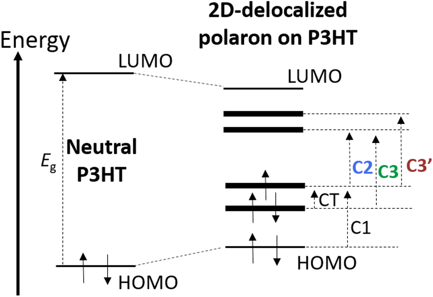
Ordered regions in regio-regular P3HT are known to display strong interchain interactions,[Reference Brown, Thomas, Köhler, Wilson, Kim, Ramsdale, Sirringhaus and Friend19] which enable charge carriers to take the form of two-dimensional (2D) polarons, where the polaron wave function delocalizes over neighboring π-stacked chains. The electronic transitions of this 2D-delocalized polaron are shown in Fig. 1: the original single polaronic intragap state splits due to delocalization giving rise to a new low-energy CT transition. The transitions observable within the near infrared spectral range studied in these experiments are highlighted in bold and color in Fig. 1. Other optical transitions such as CT and C1 have been observed by infrared spectroscopy and are the object of renewed interest;[Reference Ghosh, Pochas and Spano24] however, they are not the object of this manuscript. Although recent work by Winkler et al.[Reference Winkler, Amsalem, Frisch, Oehzelt, Heimel and Koch25] and Heimel[Reference Heimel26] have proposed a different energy-level assignment for polarons from that depicted in Fig. 1, the validity of both assignments is still being debated in the community. In addition, the model proposed by Winkler et al.[Reference Winkler, Amsalem, Frisch, Oehzelt, Heimel and Koch25] and Heimel[Reference Heimel26] has not been expanded to account for the 2D delocalization of polarons yet. As a result, its application to semicrystalline P3HT, where 2D delocalization has been extensively documented, remains questionable.
Energetic model for neutral P3HT compared with that of charged P3HT, with polarons delocalized over two neighboring chains. The associated optical transitions are depicted as well, with the key sub-band gap transitions studied in this paper highlighted in color.

Results
Spectroscopic characterization
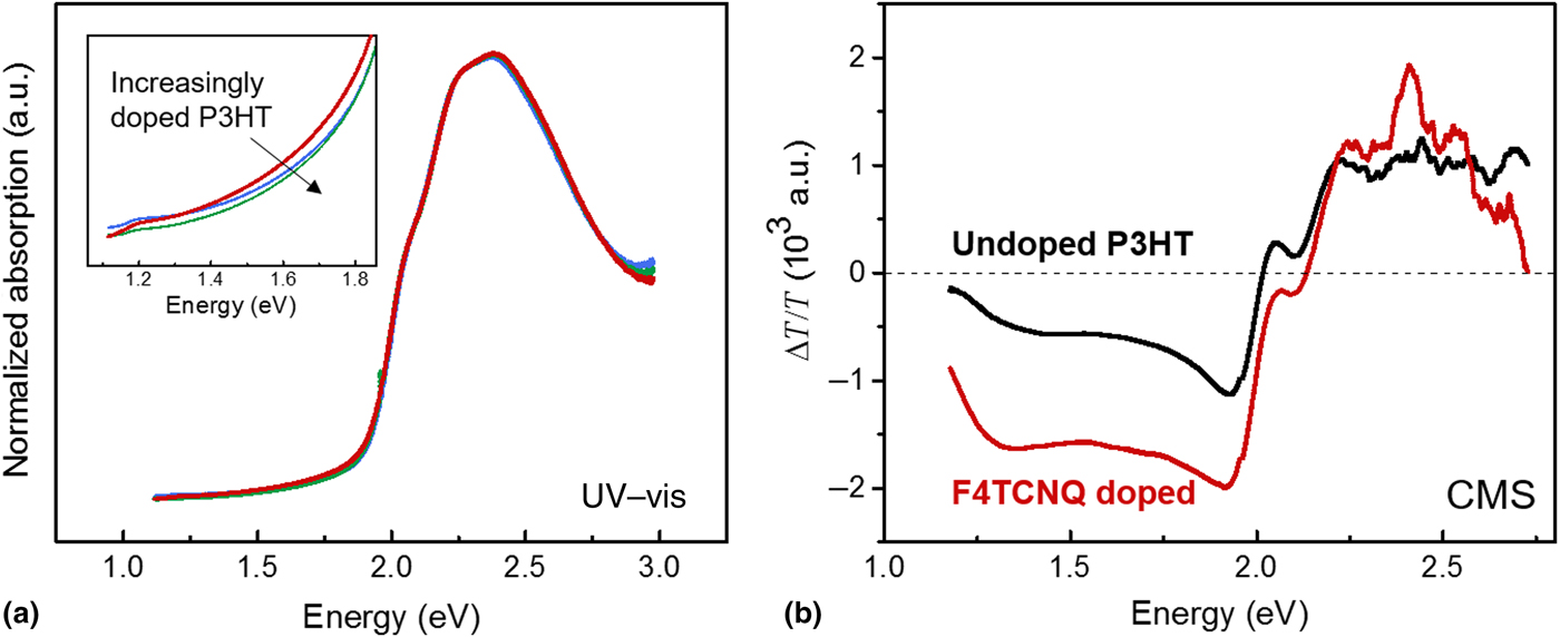
Absorption and CMS measurements of P3HT thin films at ultra-low doping concentrations are shown in Fig. 2. As expected, the absorption spectra in Fig. 2(a) of doped P3HT at various concentrations are indistinguishable from that of undoped P3HT. All spectra are normalized to the P3HT 0–0 transition. In addition, the key absorption features corresponding to the presence of ionized F4TCNQ molecules at 1.43 and 1.62 eV are below the detection limit, consistent with results of Duong et al.[Reference Duong, Wang, Antono, Toney and Salleo2] The CM spectra of doped P3HT however are clearly different from that of undoped P3HT [Fig. 2(b)], and can be analyzed using the following model.
Spectroscopic analysis of the effect of trace amounts of F4TCNQ dopants on P3HT thin films using (a) UV–visible (UV–Vis) absorption, normalized to the 0–1 transition, with the inset showing the zoomed sub-gap region where polaronic or F4TCNQ− features are absent, and (b) charge modulation spectroscopy (CMS).

Model for CMS analysis
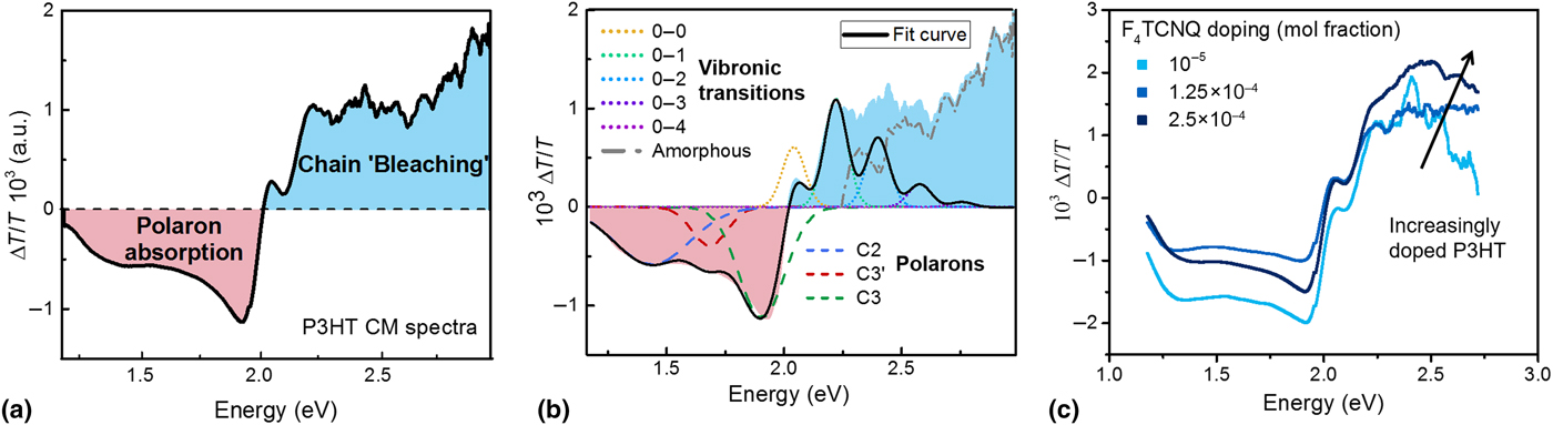
As established by Brown et al.,[Reference Brown, Thomas, Köhler, Wilson, Kim, Ramsdale, Sirringhaus and Friend19] the CM spectra obtained are a combination of (i) “bleaching” of neutral P3HT chains and (ii) polaron absorption upon the addition of a positive charge, with the different regions highlighted in the prototypical CM spectra for P3HT shown in Fig. 3.
Typical CMS spectrum obtained for regio-regular P3HT MIS devices depicting (a) the two major contributions to the spectrum–polaron absorption (ΔT/T < 0, shaded in red) and neutral chain bleaching (ΔT/T > 0, shaded in blue), and (b) the spectral decomposition carried out using the combined Spano–polaron absorption model. (c) Three representative P3HT CM spectra measured at various F4TCNQ doping concentrations.

(a) Bleaching of the absorption of P3HT neutral chains
For E > E g, the loss of neutral chain absorption dominates and ΔT/T > 0. The model proposed by Spano linking optical absorption of neutral polymer chains in the visible to their local microstructure can be applied:[Reference Spano17]
 $$\eqalign{\displaystyle{{\Delta T} \over T} &= C\mathop \sum \limits_{m = 0} \left( {\displaystyle{{{\rm e}^{ - S} S^m} \over {m!}}} \right)\left( {1 - \displaystyle{{W{\rm e}^{ - S}} \over {2E_p}} \mathop \sum \limits_{n \ne m} \displaystyle{{S^n} \over {n!\left( {n - m} \right)!}}} \right)^2 \cr & \quad\displaystyle{{{\rm e}^{\left( { - \left( {E - E_{0 - 0} - mE_p - 0.5WS^m e^{ - S}} \right)^2 /2\sigma ^2} \right)}} \over {\sigma \sqrt {2\pi}}}.} $$
S is the Huang Rhys parameter and assumed to be unity. The phonon energy of the C = C stretch, E p, is 0.179 eV for P3HT. The proportionality constant C, disorder parameter σ, excitonic bandwidth W, and energy of the intrachain 0–0 transition, E 0−0, are freely varying parameters in this model.
The Spano model allows key parameters of disorder and conjugation length to be extracted from the absorption spectra of P3HT by fitting vibronic transitions due to the optical absorption of the aggregates in the thin film. The spectra at ΔT/T > 0 thus provides information on the polymer chains on which the modulated holes reside, that is, the chains that ceased to be neutral upon modulation. This model was first established for neat P3HT but remains valid since the overall crystal structure of P3HT aggregates is retained[Reference Duong, Wang, Antono, Toney and Salleo2] at such low doping concentrations.
(b) Sub-band gap polaronic absorption
Where ΔT/T < 0 below the band gap of P3HT, the polaron absorption for each optical transition listed in Fig. 3(b) can be modeled as follows:
 $$\eqalign{\displaystyle{{\Delta T} \over T} &= - C_{{\rm ins}} \displaystyle{\sigma \over {eA}}\left( {V_{{\rm appl},1} - V_{{\rm appl},2}} \right) \cr & = - \displaystyle{1 \over {\sigma _{{\rm FWHM}} \sqrt {2\pi}}} {\rm e}^{ - \left( {x - \mu} \right)^2 /2\sigma _{{\rm FWHM}}^2}.} $$
C ins is the capacitance of the CL-PMMA insulator layer, σ is the absorption cross-section of the polaronic transition, and V appl,1 and V appl,2 refer to the different bias applied to the gate of the MIS device. Gaussians are used to model these polaronic features, in accordance with existing literature,[Reference Brown, Thomas, Köhler, Wilson, Kim, Ramsdale, Sirringhaus and Friend19] due to the strong electron–phonon coupling in P3HT.
Quantum-chemical calculations show that C3’ and C3 in Fig. 1 are two separate sub-band gap excitations,[Reference Beljonne, Cornil, Sirringhaus, Brown, Shkunov, Friend and Brédas18] while C2 is symmetry forbidden in 2D polarons. As a result, C2 has been interpreted in literature as originating from polarons that are localized on a single chain,[Reference Beljonne, Cornil, Sirringhaus, Brown, Shkunov, Friend and Brédas18, Reference Deng and Sirringhaus27] which is why previous CM spectra have largely been analyzed based on the relative intensity of the two main peak features[Reference Beljonne, Cornil, Sirringhaus, Brown, Shkunov, Friend and Brédas18, Reference Brown, Thomas, Köhler, Wilson, Kim, Ramsdale, Sirringhaus and Friend19, Reference Zhao, Noh, Chang, Heeney, McCulloch and Sirringhaus23] C2 and C3, which approximately correspond to 1.3 and 1.8 eV.
However, since symmetry selection rules governing optical transitions are not strictly valid[Reference Zhao, Noh, Chang, Heeney, McCulloch and Sirringhaus23] due to the presence of disorder in real polymer aggregates, a full fit to the CM spectra must consider polaron contributions from all three transitions. In accordance with previously established nomenclature, we model three polaronic transitions in our spectra; assigning the C2, C3′, and C3 polarons in order of increasing transition energy.
Therefore, CM spectra were decomposed using a linear combination of the Spano and polaron absorption models described above, as shown in Fig. 3(b). Combined, the two models provide a complementary framework to quantify the local disorder experienced by charge carriers in doped P3HT thin films. Initial parameters were obtained through a least-square regression, before using a Levenberg Marquardt iteration algorithm until fit convergence with a χ 2 tolerance value of 10−9 was reached.
CMS fits to analytical models
In the CMS measurements, charge density induced by field effect is 1.6 × 10¹⁸/cm3, assuming an accumulation layer of 5 nm. This figure is of the order of the concentration of ionized dopants but is over one order of magnitude higher than the estimated concentration of mobile charges (N d ≈ 5 × 10¹⁶/cm3) studied in this paper, given that only 5% of the dopant-induced charges are expected to be mobile.[Reference Pingel and Neher13] We note that the modulated mobile charges studied by CMS are induced by field effect and are distinct from those induced by doping. Assuming 100% dopant ionization,[Reference Pingel and Neher13] the total concentration of dopant ions is of the same order of magnitude as the concentration of charges induced by field effect.
In Fig. 3(c), we observe a qualitative increase in ΔT/T measured at energies >2.5 eV as a function of dopant loading in P3HT. The spectral deconstruction in Fig. 3(b) indicates that absorption at that spectral range is due to amorphous P3HT chains. The positive ΔT/T corresponds to a loss of absorption (or bleaching) in this spectral region, which increases with doping concentration. Since a fixed amount of charge is modulated in each spectrum, the increase in bleaching indicates that the addition of dopants leads to a greater tendency for mobile holes introduced by field effect to reside on amorphous P3HT chains, as opposed to P3HT aggregates. We recall that in an undoped film, injected charges tend to become confined to the ordered regions of the film.[Reference Noriega, Rivnay, Vandewal, Koch, Stingelin, Smith, Toney and Salleo28]
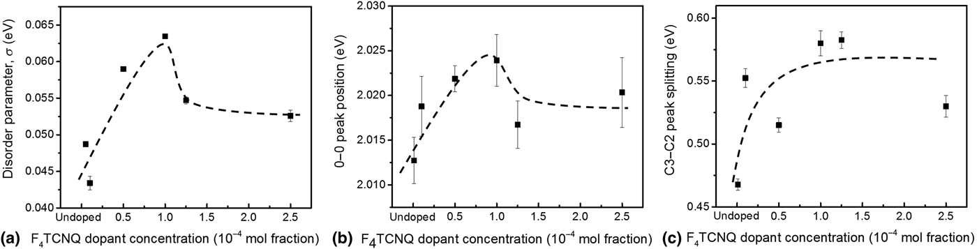
Spectral fitting using the Spano model parameters enables quantitative analysis as a function of doping, of the changes in the P3HT aggregates on which field-effect induced charge carriers reside. For example, the disorder parameter (σ) of the chains where polarons reside, which is a broad description related to both the intramolecular and intermolecular disorder in the doped P3HT thin films,[Reference Spano17, Reference Turner, Pingel, Steyrleuthner, Crossland, Ludwigs and Neher29] can be obtained from the fitted line widths. We observe an initial increase in disorder with increasing dopant concentration [Fig. 4(a)]. Beyond a critical doping concentration of 10−4 mol fraction F4TCNQ however, the trend reverses and σ decreases with increasing dopant concentration.
(a) Disorder parameter and (b) 0–0 peak position extracted from P3HT CM spectra as a function of F4TCNQ doping concentration. (c) Dependence of the splitting between C3 and C2 peak positions on the F4TCNQ doping concentration in P3HT, obtained from fits to the sub-band gap components of the CMS spectra shown in Fig. 4. Lines are drawn to guide the eye.

A similar trend is observed for the energy of the intrachain 0–0 transition, E 0−0 [Fig. 4(b)]. This transition has traditionally been interpreted as being due to mobile holes residing on more disordered P3HT chains.[Reference Brown, Thomas, Köhler, Wilson, Kim, Ramsdale, Sirringhaus and Friend19] Similar to how side-chain disorder increases the band gap and the E 0−0 transition energy in regio-random P3HT as compared with regio-regular P3HT, the increase in E 0−0 observed by CMS indicates that the hole local environment being probed undergoes an increase in disorder with doping. The trends in σ and E 0−0 thus provide evidence that adding dopants to P3HT does increase the local aggregate disorder experienced by mobile charges, which then appears to slightly reverse beyond a certain dopant concentration.
To reconcile the decrease in σ and E 0−0 beyond a critical doping mole fraction of 10−4, bleaching of both amorphous and crystalline P3HT must be considered. The gradual increase in amorphous P3HT chain bleaching is consistent with charged dopants located in the amorphous regions of the film exerting an attractive force on free holes in the material, making it progressively more favorable for polarons to localize on amorphous P3HT chains as doping increases. This effect is likely due to an increase in disorder caused by dopant loading, reducing the energetic driving force that causes charges to preferentially localize on P3HT crystallites.
On the other hand, σ and E 0−0 are extracted from the Spano model, which only evaluates the disorder in the local environment of the holes residing on P3HT aggregates. With increased doping concentration, the charges begin to populate more disordered regions, reducing the overall number of field-effect modulated charges probing the aggregates. Charges residing on more disordered aggregates are more likely to migrate onto amorphous chains with increased doping. As a result, the charges that remain in the aggregates are expected to reside in more ordered regions, leading to the slight decrease in σ and E 0−0 observed on average for field-induced charges. By considering both the P3HT crystalline and amorphous regions, the CMS data suggest that disorder steadily increases with increased dopant loading in the doped P3HT thin film. This overall increase in disorder is consistent with recent Monte-Carlo calculations by Zuo et al.,[Reference Zuo, Abdalla and Kemerink30] that show the addition of dopants lead to a gradual broadening of the intrinsic P3HT density of states.
Turning to the sub-band gap absorption, Fig. 4(c) shows the difference between the C2 and C3 polaron peak positions obtained from fits to the spectra, as a function of doping concentration. The splitting of the sub-band gap energy levels is dependent on the degree of interchain coupling, as shown by Fig. 1(b). With greater polaronic wave function overlap and 2D character, the magnitude of the C2 transition decreases, resulting in a larger difference between the magnitude of C2 and C3 transitions. This is what is observed in Fig. 4(c). As a result, the polaron peak positions measured by CMS demonstrate greater interchain coupling with increased dopant concentration of the chains where field-induced charges are located. We note that the splitting of energy levels (e.g., C3−C2 peak splitting studied here) is expected to correlate with charge delocalization regardless of the details of the energetic picture[Reference Beljonne, Cornil, Sirringhaus, Brown, Shkunov, Friend and Brédas18, Reference Winkler, Amsalem, Frisch, Oehzelt, Heimel and Koch25, Reference Heimel26] used to assign the single polaron optical transitions.
Although the simultaneous increase in intrachain disorder and increase in interchain coupling observed appear counter-intuitive, it is known that aggregates in polymeric semiconductors with longer conjugation lengths have weaker interchain coupling.[Reference Clark, Silva, Friend and Spano31] Our results show that the presence of dopants perturb the local structure, increasing disorder (σ). As dopants are added to the polymer matrix, intrachain disorder increases, enabling greater interchain delocalization and better wave function overlap across chains. Hence, polarons with greater 2D character are observed with increasing dopant concentration.
Electrical characterization
To confirm that the dopants present in the polymer matrix do dope the polymer, ionize, and contribute to an increase in free carriers despite what is observed by UV-vis spectroscopy, direct electrical characterization of the P3HT MIS devices fabricated at various doping concentrations was carried out (Supplementary Information). UV–vis spectroscopy is not a sensitive measurement, making it likely that absorption from the few F4TCNQ− present was overwhelmed by absorption from the P3HT tail, giving a false negative result that no ionization, and hence no doping took place. Consistent with what has been observed in literature[Reference Pingel and Neher13] at these low doping concentrations, the carrier concentration increases while mobility decreases upon doping. This observation is consistent with the increase in disorder observed by CMS, and clearly demonstrates the detrimental effect of trace dopants on charge transport in P3HT thin films.
Discussion
The CMS and electrical measurements show that even at ultra-low levels, doping induces significant changes to the local charge carrier environment, influencing charge transport and hence the electrical properties observed. CMS is a differential measurement that does not measure trapped holes. As such, the charge carriers captured by CMS provide information about the local environment that is directly relevant to charge transport in doped P3HT. The addition of dopant molecules leads to an increase in intrachain disorder, but interchain coupling improves. At the same time, an increasing number of field-induced charge carriers begins to reside on amorphous P3HT chains, rather than in the aggregates. This progressive shift in population distribution causes the hole mobility to drop as a function of doping concentration, while carrier concentration increases.
One unanswered fundamental question in the study of dopant–polymer interaction, especially when co-mixed at these low doping concentrations, is where do the dopants reside in the polymer matrix? A corresponding x-ray diffraction study at significantly higher doping concentrations by Duong et al.[Reference Duong, Wang, Antono, Toney and Salleo2] has demonstrated that below a critical doping concentration (0.03 dopant molar fraction), the overall crystal structure of P3HT is retained despite the addition of dopants. This has led to the prevailing assumption that for all doped films below that critical concentration, all the dopants must go in the amorphous P3HT regions, which are better able to accommodate the large dopant molecules. However, this assumption may be unwarranted due to the fact that x-ray diffraction may not be sensitive enough to monitor changes in the film at low doping concentrations. In this manuscript, CMS provides some evidence that a portion of the charged dopants do reside in the amorphous regions of the film: the gradual increase in amorphous P3HT chain bleaching observed with doping is likely due to coulomb attraction between the field-induced charges and ionized dopants.
The measured changes in σ and E 0−0, which describe P3HT aggregate absorption, indicate however that even ultra-low doping has a non-negligible effect on the crystalline P3HT domains. Since the polymer chain packing and disorder within the P3HT aggregates are affected, it is likely that some dopant molecules must reside in the aggregates, causing the changes observed. As a result, at these ultra-low doping concentrations, the dopants are more likely uniformly distributed between the crystalline and amorphous regions.
With the addition of more dopants to the P3HT matrix, the increase in amorphous bleaching observed could indicate that dopants increasingly reside in amorphous regions. This observation would be consistent with what has been found in previous work under similar processing conditions at much higher dopant concentrations.[Reference Duong, Wang, Antono, Toney and Salleo2] On the other hand, the increase in amorphous bleaching could be the result of increasing disorder in the crystalline regions, which reduce the energetic driving force that would lead to field-induced charges preferentially residing on the aggregates.
Combining the disorder observed in doped P3HT films by CMS with measured electrical properties, we can begin to develop a more complete picture for how the dopants affect polymer packing and charge transport. The increase in intermolecular coupling with doping allows efficient hopping between chains. Mobility drops with doping concentration however, and in fact previous CMS work,[Reference Zhao, Noh, Chang, Heeney, McCulloch and Sirringhaus23] did not find a correlation between increased delocalization and mobility. We postulate that the mobility decrease is due to a microstructural effect at the mesoscale. The increase in intramolecular disorder and σ with doping dominates the charge transport characteristics of doped P3HT. The overall increase in disorder reduces the energetic difference between the aggregate and amorphous regions, increasing the probability that polarons localize on amorphous P3HT chains, which led to the observed increase in amorphous chain bleaching with dopant concentration. The localization of charge carriers on amorphous chains results in overall less efficient charge transport as it weakens the formation of preferential transport paths through crystalline high-mobility regions. These results demonstrate that as conjugated polymers are doped, transport in the amorphous regions of the film becomes progressively more important, while in undoped polymers charges are nearly exclusively confined to the ordered regions of the film.
Conclusion
At “ultra-low” doping concentrations ranging from 10−5 to 10−4 mol fraction, changes in the charge carrier local environment are observed as ionized dopants perturb the polymer packing and chain-to-chain coupling, creating disorder in the polymer matrix. Our measurements provide structural insights into the dopant–polymer interaction, allowing us to rationalize how a few dopants can have such a large, albeit undesirable, impact on the polymer hole mobility. The added dopants induce disorder by acting as coulomb traps, drawing mobile holes to the amorphous regions while physically disrupting intrachain order in the polymer. These interactions result in localization of the charge carriers and the formation of more transport paths involving amorphous polymer, hindering efficient charge transport. Due to the reduction of intramolecular delocalization, intermolecular coupling increases. Hence, the P3HT chains become more oligomer-like with the addition of dopants, with stronger intermolecular interactions that could potentially lead to higher mobilities at significantly higher doping concentrations than those studied in this paper, as theoretically predicted.[Reference Arkhipov, Emelianova, Heremans and Bässler32] The use of sensitive techniques such as CMS enable the study of the effect of dopants in this model system, even at low concentrations. Such a measurement provides insight into the dopant–polymer interaction, allowing for the rational design of new dopants for polymeric semiconducting materials in the future.
Supplementary material
The supplementary material for this article can be found at https://doi.org/10.1557/mrc.2017.88.
Acknowledgments
The authors acknowledge financial support from the National Science Foundation under DMR—1533987 (DMREF).