




No CrossRef data available.
Article contents
Attention function after childhood stroke
Published online by Cambridge University Press: 01 November 2004
Abstract
We investigated attentional outcome after childhood stroke and orthopedic diagnosis in medical controls. Twenty-nine children with focal stroke lesions and individually matched children with clubfoot or scoliosis were studied with standardized attention and neuroimaging assessments. Stroke lesions were quite varied in location and commonly involved regions implicated in Posner's model of attention networks. Children with stroke lesions performed significantly more poorly regarding attention function compared with controls. Performance on the Starry Night, a test demanding alerting and sensory-orienting but not executive attention function, was significantly associated with lesion size in the alerting and sensory-orienting networks but not the executive attention network. Furthermore, earlier age at lesion acquisition was significantly associated with poorer attention function even when lesion size was controlled. These findings support the theory of dissociable networks of attention and add to evidence from studies of children with diffuse and focal brain damage that early insults are associated with worse long-term outcomes in many domains of neuropsychological function. In addition, these results may provide clues towards the understanding of mechanisms underlying attention in children. (JINS, 2004, 10, 976–986.)
Information
- Type
- SYMPOSIA ARTICLE
- Information
- Journal of the International Neuropsychological Society , Volume 10 , Issue 7 , November 2004 , pp. 976 - 986
- Copyright
- © 2004 The International Neuropsychological Society
INTRODUCTION
The literature on child outcomes following both focal and diffuse brain insult is replete with observations suggesting that these children have attentional problems (e.g., studies of meningitis: Taylor et al., 2000, early TBI: Kaufmann et al., 1993; hydrocephalus: Brewer et al., 2001; brain tumors: Radcliffe et al., 1994; and acute lymphoblastic leukemia: Reddick et al., 2003; Taylor et al., 1987). Unfortunately, we know little about brain mechanisms responsible for these deficits. The extant data are also lacking in regard to the relationships between the location of lesions and corresponding attention deficits. Furthermore, the degree to which lesions in specific brain networks are associated with attention deficits per se, as opposed to more general cognitive deficits, has not been investigated. Children with focal stroke lesions provide a potentially useful model for the investigation of attention after brain injury.
Only two studies have examined attention in children with single focal unilateral childhood strokes (Aram & Ekelman, 1986; Block et al., 1999). The first study found that children with right-sided lesions displayed reduced attention and task persistence as evidenced in their relatively low scores on the Freedom From Distractibility factor on the WISC–R (Aram & Ekelman, 1986). The second study examined divided attention using the Symbol Digit Modalities Test (Block et al., 1999), and found that children with left-sided lesions exhibited a significant deficit relative to controls, while those with right-sided lesions only had a marginally significant impairment compared with controls.
The literature reviewed above suggests links between pediatric stroke and attentional problems. Consequently, in the study reported here we pursued a hypothesis-driven investigation of attention associated with stroke, based on an attention networks model. We hypothesized that the type of attentional disturbance will vary in relation to the specific network damaged. Posner's model of attention, which is grounded in cognitive neuroscience, provides a useful framework for the study of attention and brain relations in children, although we acknowledge that there are competing models of attention. Included among these is Mirsky's model which is based on the associations among neuropsychological test scores in various patient populations (Mirsky et al., 1991; Mirsky & Duncan, 2001). These models are actually quite similar (Mirsky et al., 1991) but we have chosen Posner's model because it has been more extensively validated.
A brief review of Posner's three-component attention networks model follows: The alerting network is involved in maintaining the alert state. Intrinsic alertness represents the cognitive control of wakefulness and arousal and is typically assessed by simple reaction time tasks without a preceding warning stimulus. Phasic alertness refers to the process operative in reaction time tasks in which a warning stimulus precedes the target. It reflects the ability to increase response readiness subsequent to external cueing (Sturm & Willmes, 2001). This network is subserved by the locus coeruleus, right frontal cortex (superior region of Brodmann area 6), and right parietal cortex (Posner & Petersen, 1990; Posner & Raichle, 1994). Patients with right parietal lesions have difficulty sustaining attention and are deficient in using warning signals to improve alertness (Robertson & Jehknen, 1977; Rothbart & Posner, 2001). Recent findings suggest that the thalamus and brain stem are also activated during alerting tasks (Sturm & Willmes, 2001). Alerting attention is present by 3 months in most individuals (Posner & Raichle, 1994) and has a protracted course of development extending beyond childhood (Rueda et al., 2004).
The sensory-orienting network is responsible for covert orienting to sensory, particularly visual, signals (Posner & Dehaene, 1994). This network includes the parietal lobes, temporo-parietal junction, frontal eye fields, superior colliculus, and thalamus. These areas have been implicated in functional neuroimaging (Corbetta, 1998; Corbetta et al., 2000; Posner et al., 1988) and lesion (Friedrich et al., 1998; Karnath et al., 2001; Parasuraman et al., 1992; Posner, 1988; Rafal, 1998; Sapir et al., 1999) studies. The superior parietal lobule is critical for voluntary shifts of attention following a cue even without eye movements to the target (Corbetta et al., 2000). The frontal eye fields and superior colliculus appear to participate in the processes of covert shift of attention and eye movements to targets (Corbetta, 1998). Patients with thalamic lesions show subtle deficits in visual orienting tasks that may be related to the access of the ventral information processing stream (Posner & Fan, in press). Lesions of the temporoparietal junction or superior temporal lobe disrupt the processing of a novel stimulus (Friedrich et al., 1998; Karnath et al., 2001). The sensory-orienting network develops early in infancy. By age 4 months children can have attention captured by a cue without executing an eye movement (Rothbart & Posner, 2001).
The executive network is required during tasks when conflict is present and when the production of a non-habitual response is required (Posner & Fan, in press), i.e., executive attention requires the mobilization of inhibitory attentional processes which are elements of “cognitive control” (Casey, 2001; Casey et al., 1997, 2000, 2002; Filoteo et al., 2002; Miller & Cohen, 2001). Tasks tapping executive attention require suppression of attention to a salient stimulus or suppression of a competing response choice. Examples of such tasks include the Stroop (Stroop, 1935), the flanker task (Eriksen & Eriksen, 1974), Attention Network Task (Fan et al., 2002), stop signal task (Logan, 1994), and go/no-go tasks (Vaidya et al., 1998). Evidence from fMRI (Casey et al., 1997, 2000), PET (Lee et al., 2001), and structural imaging (Casey et al., 2000) studies in normal adults and children suggest that the executive attention network is subserved by the anterior cingulate, prefrontal cortex, and the basal ganglia (Beauregard et al., 2001; Casey, 2001; Casey et al., 1997, 2000, 2002; Filoteo et al., 2002; Ochsner et al., 2002). Striatal dysfunction, specifically, has been associated with impairment in a variety of tasks that are demanding of attentional resources (Filoteo et al., 2002). These areas are part of a closely connected cortico–striatal–pallidal–thalamic network. Cognitive control mechanisms mediated by this network develop over a prolonged period through at least 12 years of age (Casey et al., 2000; Rueda et al., 2004).
Developmental aspects are important in any study of outcome after childhood stroke. Therefore we were interested in studying the relationship of attention function to age at injury. The so-called Kennard effect refers to the milder deficits and greater recovery of function that purportedly occur after early brain damage (see a review, Finger, 1991). Kennard's animal experiments supported such a conclusion particularly for the motor system. However, this observation was not universal in her work as other results suggested more problematic behavioral outcomes after early damage to association cortices that included the prefrontal region. Kennard also noted progressive onset of deficits at later ages. Thus the Kennard effect is not a consistent finding of early cerebral damage.
In fact, children do not “escape” from early focal insults (e.g., Stiles' research on effects of right hemisphere disease on spatial skills (Stiles, 2000; Stiles & Nass, 1991; Stiles et al., 1997)). We (Chapman et al., 2003; Lansing et al., 2004) and others (Aram & Eisele, 1994; Riva & Cazzaniga, 1986; Vargha-Khadem et al., 1985; Woods, 1980) have shown that, if anything, early stroke lesions have greater effects on subsequent development than do later lesions. Neurobiologically, correlates of developmental outcomes after early insults may include damage to integrative neural regions (such as prefrontal and parietal cortices) and changes that can be found in interconnected cerebral regions (Goldman & Galkin, 1978; Kolb et al., 1996, 2000) that affect cellular connectivity, brain weight, dendritic arborization, and spine density.
Given evidence of only limited forms of plasticity, we hypothesize that insults at younger ages will have similar, though more exaggerated, effects on attention compared with insults at later ages. To the degree that attention is required for development of other cognitive skills, earlier lesions may also impede development of outcomes other than attention (e.g., IQ, higher order language, memory) to a greater extent than later lesions. Age at testing and time between insult and testing are other relevant factors in predicting outcomes (Taylor & Alden, 1997). However, the latter age factors have not been examined in relation to specific attention deficits in children with focal stroke.
In summary, we hypothesize that (1) children with stroke lesions will have deficits in attentional skills when compared with matched controls; (2) alerting and sensory-orienting attention disturbance will be related to damage to the alerting and sensory-orienting attention networks rather than the executive network. Furthermore, the relationship will persist even when controlling for general intellectual functioning. Thus the type of attentional disturbance will vary in relation to the specific networks damaged and this disturbance will not be attributable to general cognitive deficits; 3) stroke lesions acquired earlier in life (e.g., before age 1 year) will be associated with greater attentional disturbances than lesions acquired later in child development, and the relationship will persist even when controlling for lesion size.
METHODS
Research Participants
Stroke cases
The research design, previously reported in detail (Max et al., 2002b), is a cross-sectional study of children with a history of a single stroke and a medical control group. The study focused on psychiatric, neuropsychological, academic, adaptive, and family function outcomes in children with strokes. In accordance with previous studies (Riva & Cazzaniga, 1986; Woods, 1980)., stroke subjects were considered to have early lesions if their brain lesion occurred prenatally or up to 12 months of postnatal life. The late lesion group consisted of children who acquired their stroke at age 12 months or later. We matched early stroke subjects with children with clubfoot, with the rationale that physical deformity in both groups was an early, and frequently congenital, condition. We matched late stroke subjects with children who had scoliosis because these children were without physical deformity prior to their acquired disorders.
Inclusion criteria for stroke cases were (1) neuroimaging documentation of a focal, non-recurrent and non-progressive supratentorial brain parenchymal lesion caused by a stroke before age 14; (2) subjects aged 5–19 years at the time of the assessment; (3) 1 year or more since stroke; and (4) English as first language. The following exclusions were applied: (1) neonatal bleeds (e.g., intraventricular hemorrhages, germinal matrix hemorrhages) potentially associated with prematurity; (2) neonatal watershed infarcts associated with hypoxia; (3) hemoglobinopathies; (4) progressive neurometabolic disorders; (5)Down's syndrome and other chromosomal abnormalities; (6) malignancy; (7) congenital hydrocephalus; (8) shunts; (9) congenital and acquired CNS infections; (10) clotting factor deficiency; (11) stroke in a pregnant minor; (12) previous organ or bone marrow transplant; (13) cerebral cysts; (14) trauma; (15) transient ischemic attack; (16) moya-moya; (17) severe and profound mental retardation; (18) quadriplegia, triplegia, or diplegia diagnoses; (19) syndromic vascular malformations (excluding A-V aneurysm ruptures); (20) systemic lupus erythematosis; and (21) multiple lesions (unless in close proximity).
Stroke subjects evaluated included 17 with early lesions and 12 with late lesions. The strokes were ischemic in 21 cases and hemorrhagic in 8 cases. Etiology included 15 idiopathic occlusive cases, 2 idiopathic hemorrhagic cases, 4 cases occurred in subjects with congenital heart disease (3 after cardiac surgery or catheterization and 1 after varicella zoster infection), 5 cases of arteriovenous malformation rupture, 1 case of ruptured angioma, 1 case possibly linked to comorbid ulcerative colitis, and 1 case followed a varicella infection. Both cases associated with varicella infections were presumed to be due to vasculitis and there was no evidence for encephalitis (Roach & Riela, 1995). The distribution of the brain lesions included 7 cases of predominantly putamen lesions (4 right/3 left lesions), 9 large middle cerebral artery (MCA) distribution infarcts including deep gray structures (4 right/5 left lesions), 10 smaller MCA distribution frontotemporal or temporoparietal lesions sparing the deep gray (6 right/4 left lesions), and 3 cases of parietal or parieto–occipital strokes (2 right/1 left lesion).
Eleven of 29 (38%) subjects had a history of seizures, but only 5 were receiving anticonvulsant medication (carbamazepine monotherapy in 3, carbamazepine plus primidone in 1, phenytoin in 1) at the time of the assessment, and all were in good control. Only 3 children were treated with medication for psychiatric indications (1 subject with methylphenidate, clonidine, clomipramine, and risperidone; 1 subject with Imipramine; 1 subject with nefazodone hydrochloride and clonazepam). Forty-eight subjects (including all stroke subjects) were recruited from one university hospital while 10 subjects were recruited from a second university hospital due to the relocation of the first author.
Control participants
Children with congenital clubfoot and children with scoliosis were individually matched to subjects with stroke according to age of onset of stroke (i.e., early vs. late). Matching was based on gender, ethnicity, socio-economic status (SES; see under measures below), and age within 1 year. Age matching had to be extended to 16 months in 3 cases. Orthopedic controls were excluded when they had evidence in the chart of acquired or congenital CNS injury that may be part of broader (e.g., neuromuscular) syndromes unrelated to the common idiopathic syndromes. Matching was possible for all but 2 children with late stroke lesions. These 2 late-onset stroke subjects were matched with children with clubfoot. Comparisons of the two control groups failed to reveal differences in background characteristics, hence these groups were combined for analysis into a single control group.
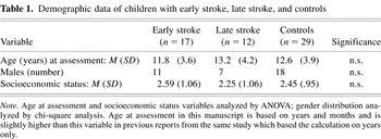
The stroke and orthopedic groups were not significantly different on matching variables of age and SES. Respective age means (SD) of stroke and orthopedic subjects were 12.6 (3.9) and 12.4 (3.9), df = 56, t = −.186, p > .8. The early and late onset stroke groups were not significantly different in age at assessment (11.84 ± 3.60 vs. 13.15 ± 4.22 years respectively; p > .37) but as may be anticipated were significantly different in time from onset to assessment (11.77 ± 3.52 vs. 5.40 ± 3.23 years respectively; p < .0005). In the early stroke group, prenatal onset occurred in 12 children and postnatal onset occurred in 5 children (Day 1 in 3 children; 2.5 months in 1 child; 9 months in 1 child). Age at stroke for the late onset group was 7.8 ± 3.2 years. Respective SES means (SD) of stroke and orthopedic subjects were 2.45 (.95) and 2.45 (1.06), df = 56, t = 0, p = 1.0. There were 18 males, 27 White and 2 biracial children in each of the stroke and orthopedic groups. Table 1 provides demographic data for the early stroke, late stroke, and control groups. Poststroke psychiatric disorder was more common than postorthopedic diagnosis psychiatric disorder (17/29; 59% vs. 4/29; 14%; Fisher Exact Test ≤ .001; Max et al., 2002b).
Demographic data of children with early stroke, late stroke, and controls

Measures
Attention function
Attention was assessed by administering the Starry Night (Rizzo & Robin, 1990, 1996), a task in which children respond to the appearance and disappearance of a white light (event) embedded in other “stars” on the computer monitor at unpredictable intervals. In this task, children are instructed to fixate on a cross in the center of the computer screen. Trials began after the children were dark adapted and indicated a readiness to proceed. For this study, stimulus presentations included 250 stars on the screen and 200 events, with the children instructed to push a computer key as quickly as possible when they detected the appearance of a new event or the disappearance of an existing event. Responses were scored such that any response falling between 100–2000 ms was considered a hit, no response in that interval a miss, and a response following a hit or a miss when a stimulus had not occurred, a false positive. The primary dependent variable was accuracy (d′) in detecting the appearance and disappearance of stars. d′ assesses the difference between the probability that a subject will report an event given noise alone versus signal-plus-noise. It reflects the distance between the means of the noise and signal-plus-noise distributions in terms of standard deviation units (Rizzo & Robin, 1996). Reaction time (RT) and Beta (β, response criterion) were also recorded. β is the cut-off point whereby a decision of a yes signal is present versus a no, indicating that the decision is not made. β of 1 is an equal probability of a yes or no. Less than 1 is a bias to yes (greater false positives), greater than 1 a more stringent bias, that is, less like to say yes. d′ is an observer's true sensitivity (independent of bias). Whereas “percent correct” targets responded to changes as a function of bias, d′ does not. These concepts are dealt with in greater detail elsewhere (Gescheider, 1985; Rizzo & Robin, 1990, 1996). This test taps alerting attention primarily, as sustained attention is required during the test interval to detect the appearance and disappearance of stars. Orienting attention is also be required for detection of newly appearing or disappearing stars because these events occur in quadrants of the screen peripheral to the central cross. However, cues which are typically provided in orienting tasks are not offered by Starry Night (Lodge-Miller et al., 1993; Rizzo & Robin, 1990). Executive attention is not tapped by this task, as conflict resolution is not required. We acknowledge that the use of a test that does not specifically provide separate scores for alerting, sensory-orienting, and executive attention represents an important constraint on our use of the Posner model.
Three children with stroke had partial visual field cuts detected by neurological exam completed by J.E.M. or K.M. Attention outcome measures in these 3 subjects ranged from .33 to 1.12 standard deviations below the mean for the stroke cohort. The pattern of results (see below) was similar whether or not these subjects were excluded. Therefore these subjects were included in the analyses. The inclusion of children with congenital heart disease and varicella infection did not significantly affect the attention outcome data. All these subjects had attention outcome measures above the mean for the stroke cohort except one child with the third largest lesion.
Neuroimaging
Acquisition
To assess lesion location and the proportion of the attention networks of interest that were damaged, MRI scans were obtained for the children with stroke (T1-weighted volumetric mode, SPGR/40°, TR/TE = 26/7 ms, NEX = 2, X/Y/Z = 1 × 1.5 × 1 mm thickness with no skip; T2-weighted dual-echo, FSE/V, TR = 2350, TE = 17/102, NEX = 1, X/Y/Z = 1 × 1 × 5 mm thickness with 1 mm skip). Twenty-six of 29 stroke subjects underwent research scans that were the basis of their lesion location analyses. The other 3 subjects (1 early-onset and 2 late-onset stroke) who could not have a research MRI (due to refusal, concern about intracerebral metallic clips, and equipment failure, respectively) had lesion location determined from previous clinical CT scans (2) or MRI scan (1).
Lesion characterization
A neurologist (F.F.M.) who was blind to the neuropsychological data marked the lesions on hard copy films and represented the lesions on two-dimensional templates (Damasio & Damasio, 1989). The regions identified included 14 frontal lobe regions (F01–F14; five constituting the mesial aspect, five constituting the lateral aspect, four constituting the orbital aspect), 12 temporal lobe regions (nine lateral/superior aspect areas, three mesial aspect areas), six parietal lobe areas (P01–P06; two inferior parietal lobule, four superior parietal lobule), seven occipital lobe regions, two insula regions, four basal ganglia regions (BG1–BG4; two caudate, two lenticular nucleus), four thalamic areas (TH1–TH4), three internal capsule areas, the hypothalamus, and three corpus callosum areas. The proportion of each of the areas damaged was coded as follows: zero = no damage; 1 = < 25% damaged; 2 = 25–75% damaged; 3 = >75% damaged.
MRI analyses also generated three-dimensional (volume) measures. Guided by the lesion markings on hard copy films made by the neurologist (FFM), an experienced neuroanatomist painted each lesion using a 3-D brain-morphometrics package (Paus et al., 1996) under supervision of P.T.F. and J.L.L. Lesion volume was computed in native and Talairach coordinate systems for intersubject differences in brain size (Lancaster et al., 1997). The computerized Talairach atlas (Lancaster et al., 2000) was used to obtain anatomical labels for painted lesions. The Talairach Region Extraction (TREX) software (Lancaster et al., 2000; available at http://ric.uthscsa.edu/projects/) was used to calculate volumes of anatomical structures that were covered by painted lesions in an automated manner. Lesion volume was highly skewed (M ± SD: 39.1 ± 63.0 cm3; range: .21–256.8 cm3; MDN: 8.03 cm3). We have tabulated lesion volume and laterality of the individuals within this cohort in previous publications (Max et al., 2002a, 2003).
Anatomical definition of Posner's attention networks: 2-D measures
Areas for each of the attention networks were defined as follows: alerting network = right inferior parietal lobule (P01–P02), right superior regions of Brodmann area 6 (F03 and F08), and right thalamus (TH1–TH4); sensory-orienting network = bilateral parietal lobes (P01–P06), bilateral thalamus (TH1–TH4), and bilateral frontal eye fields (F08); executive attention network = frontal midline structures (F01–F05) bilaterally including anterior and posterior cingulate, supplementary motor area, prefrontal region, Rolandic region, and bilateral basal ganglia (BG1–BG4). Damage totals (0–3) in each of the defined areas of each anatomical attention network were summed to derive lesion scores for the respective networks. For example, the maximum possible score for the alerting network (eight regions × 3) was 24, sensory-orienting network (11 regions × 3) was 33, and executive network (nine regions × 3) was 27.
Anatomical definition of Posner's attention networks: 3-D measures
Three-dimensional volumes were computed based on Talairach Daemon structures as follows: alerting network = right inferior parietal lobule, right precentral and superior frontal gyri, and right thalamus; sensory-orienting network = bilateral parietal lobes, bilateral sublobar areas, bilateral thalamus, and bilateral precentral gyri; executive attention network = bilateral anterior cingulum, posterior cingulum, superior frontal gyrus, paracentral lobule, caudate, lentiform, and claustrum. The bivariate correlations between the two-dimensional and three-dimensional lesion measures were quite high for the alerting, sensory-orienting, and executive attention networks (rs = .76, .79, and .91, respectively).
General Intellectual Functioning
The Wechsler Intelligence Scale for Children, Third Edition (WISC–III; Wechsler, 1991) was used to assess general intellectual function. An estimate of Verbal IQ (VIQ) was based on prorated Information and Similarities subtests. Proration was used to limit testing burden associated with the larger battery used in this study. All children were verbal and no adjustments were necessary to avoid those with lesions affecting the language areas.
Socioeconomic Status
SES was measured by the Four Factor Index (Hollingshead, 1975). Classification depends on scores derived from a formula involving both mother's and father's educational levels and occupational levels. Level I (scores 55–66) refers to the major business and professional class, Level II (scores 40–54) refers to the medium business, minor professional, and technical class, Level III (scores 30–39) refers to the skilled craftsmen, clerical, and sales workers class, Level IV (scores 20–29) refers to the machine operators and semiskilled workers, and Level V (scores 8–19) refers to the unskilled laborers, and menial service workers class. Roman numeral classes were recoded to corresponding whole numbers.
Statistical Analysis
Attention measures in children with stroke versus controls were compared with independent sample t tests. The relationship between attentional outcome and lesion size within each of the three attention networks was described by bivariate correlations. Partial correlations were then used to test the uniqueness of this relationship by controlling for general intellectual function (Verbal IQ). The relation of developmental variables (age at insult, age at assessment, time since injury) to attentional outcome was assessed by bivariate correlations in analyses in which these variables were treated as continuous measures. ANOVAs comparing children with early lesions, later lesions, and the aggregate control group (children with clubfoot combined with children with scoliosis) were used in group comparisons. When the analysis was significant (p < .05), the post-hoc Tukey b test was used to determine which groups differed significantly. Attentional outcome in children with early versus late lesions were further analyzed by linear regression analyses controlling separately for total lesion size, alerting network lesion size, and sensory-orienting network lesion size. These analyses were conducted to determine whether the relationship between attention and age at insult was confounded by the lesion distribution.
RESULTS
Effects of Stroke
The distribution of lesion size mean scores (SD; range) was as follows: alerting attention network 1.83 (3.21; 0–12); sensory-orienting attention network 5.41 (6.82; 0–26); executive attention network 2.86 (3.54; 0–14). The 29 stroke participants could be individually classified according to the distribution of lesioned attention networks as follows: executive attention network alone (n = 8), combined executive attention and sensory orienting (n = 6), executive attention and sensory orienting and alerting (n = 4), sensory orienting and alerting (n = 6), sensory orienting alone (n = 2), no attention network (n = 3). These data confirm that stroke lesions in children are quite varied and are frequently localized within the proposed attention networks.
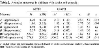
Table 2 summarizes attention outcomes for children with stroke compared with controls. Accuracy in detection of both appearing and disappearing stars was significantly worse in the children with stroke. RT was also significantly longer for this group compared with the control group, although only for disappearing stars. Group differences in β were not significant.
Attention measures in children with stroke and controls

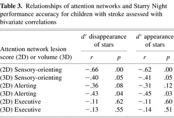
Analysis of associations of lesion size with accuracy (d′) scores on the Starry Night task yielded findings consistent with our expectations of specific brain–behavior relationships. As summarized in Table 3, accuracy performance on the Starry Night task was related to lesion size (both area and volume) in both the sensory-orienting and alerting networks, but not in the executive network. To further investigate the uniqueness of these lesion-attentional network relationships, we conducted partial correlations between lesion size in these networks and performance accuracy on the Starry Night test, controlling for intellectual function as assessed by VIQ. The results confirmed that both Starry Night measures (appearance and disappearance d′) remained significantly associated with lesion size in the sensory-orienting network (p values ≤.007 and ≤.015, respectively), but not with lesion size in the alerting network. The results suggest that lesions in the sensory-orienting network result in attention deficits that are relatively independent of deficits in other cognitive functions.
Relationships of attention networks and Starry Night performance accuracy for children with stroke assessed with bivariate correlations

Effects of Age at Insult
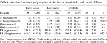
To investigate age at insult on performance on the Starry Night task, early and later stroke subgroups were compared with controls (Table 4). The results revealed significantly poorer detection of both appearing and disappearing stars in the early onset stroke subgroup when compared with either the controls or the later onset stroke subgroup. Analysis failed to reveal significant differences between the later onset and control groups. The groups did not differ on the response criterion measure (β).
Attention function in early-acquired stroke, later-acquired stroke, and control children

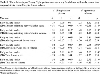
Table 5 demonstrates that differences between the early onset stroke subgroup and later onset stroke group on the Starry Night task remained significant in separate analyses that controlled for measures of total lesion size (2-D and 3-D), alerting network lesion size (2-D and 3-D), and 3-D sensory-orienting network lesion size. The differences were only marginally significant controlling for 2-D sensory-orienting lesion size.
The relationship of Starry Night performance accuracy for children with early versus later acquired stroke controlling for lesion indices

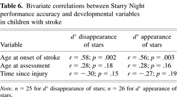
We further examined the relationship between attention outcome and age at stroke onset with the latter treated as a continuous variable. The bivariate correlation with age at onset was .56 (p = .002) for detection of appearing stars and .58 (p = .003) for detection of disappearing stars. This could not be attributed to chronological age or time since injury because the respective bivariate correlations with accuracy measures were not significant (Table 6).
Bivariate correlations between Starry Night performance accuracy and developmental variables in children with stroke

DISCUSSION
We found that stroke lesions in children are quite varied and commonly involve the widely distributed brain regions defined by Posner's model of attention networks (Posner & Petersen, 1990). Alerting and sensory-orienting attention (tapped by means of the Starry Night test) were associated with the extent of lesions in corresponding brain networks but not with the extent of lesions in the executive attention network. We additionally found that the degree of damage within the sensory-orienting brain network was significantly related to performance on a measure of sensory-orienting attention, even when taking verbal IQ into account. Therefore the lesion–behavioral finding appears to be specific and not merely a reflection of general cognitive impairment.
We have documented in a separate series of analyses that a behavioral expression of attention problems, attention-deficit/hyperactivity disorder and traits of the disorder (ADHD/traits), is associated with lesions within the executive attention network (putamen; Max et al., 2002a) and with lesion size within the executive attention network and not with corresponding measures of the alerting or sensory-orienting networks (Max et al., 2004). Furthermore, Starry Night d′ scores were not significantly associated with the presence of ADHD/traits (unpublished data). Thus although the psychiatric diagnosis of ADHD/traits is not generated on the basis of a neuropsychological test, the results taken as a whole suggest that psychiatric manifestations of poststroke attention deficit are distinct from alerting and sensory-orienting attention deficits and may be a reflection of executive attention deficits.
Developmental considerations were important in accounting for attentional outcome. Children with focal stroke lesions, particularly those with lesions acquired early (i.e., before age 1 year), performed at significantly lower levels than controls in the domains of alerting and sensory-orienting attention. A similar relationship was found when age at brain insult was treated as a continuous variable demonstrating that the finding was not dependent on the cut-off (1 year) used to define the two age at insult subgroups. Younger age at insult was associated with poorer attention even when controlling for total lesion size, or for the extent of lesions in the alerting attention and sensory-orienting attention networks. These results suggest that age at insult effects were not an artifact of subgroup differences in lesion type or severity. Furthermore, the findings could not be attributed to other potentially important developmental variables such as age at assessment or time interval since injury. The findings are consistent with mounting evidence of worse neuropsychological function in association with earlier lesions in a variety of conditions characterized by diffuse brain injury (Taylor & Alden, 1997) and even focal brain injury (Aram & Eisele, 1994; Chapman et al., 2003; Lansing et al., 2004; Riva & Cazzaniga, 1986; Vargha-Khadem et al., 1985; Woods, 1980).
This study of children with focal strokes enhances knowledge regarding the specific neural mechanisms that underlie attention problems. The methodology is unique not only in childhood stroke research but also with respect to adult focal lesion research. Cognitive neuroscientists commonly investigate a group of patients with circumscribed lesions in a specific area that is believed to contribute to a component of attention and then determine if this group is different than a control group (often another group of patients with circumscribed lesions in areas thought not to contribute to the attentional process under study; Baldo et al., 2001; Barcelo & Knight, 2002; Barrash et al., 2000; Eslinger & Grattan, 1993; Filoteo et al., 2002; Gehring & Knight, 2002; Swick & Knight, 1998). The approach used in this study was to examine patients with damage to different brain regions, characterize the extent of damage, and determine if the damage contributed to deficits in attention. This approach takes advantage of variability in lesion size and lesion location to further knowledge of brain–behavior relationships underlying attentional disturbances.
The conclusions reached here must be regarded as tentative, given shortcomings related to the samples and the assessment battery. Our assessment of attention was narrow and the attention test placed demands on both alerting and sensory-orienting attention. Further, because we did not administer tests targeting the executive component of attention, we could not determine if lesions in the executive attention brain network were associated with a corresponding weakness in executive attention. Given the wide variations in age at insult and age at assessment, a larger sample would provide a more rigorous test of the moderating influences of these factors. The small sample size also limited the power of the group comparisons to detect stroke effects. We were careful to select participants with congenital heart disease uncomplicated by hypoxic events and found that they did not confound attention outcome measures, yet some may argue that hypoxic events may be subtle and could go undetected.
Future studies should include a multi-site recruitment of a larger sample of children with stroke as well as controls. A larger study would increase statistical power for tests of the moderating effects of age at insult and age at assessment. Further investigations are also needed to examine more specific associations between lesion location within the attention network and attentional skills, as well as to determine the effects of stroke and of stroke-related attention deficits on learning and behavior.
ACKNOWLEDGMENTS
We would like to thank Michael Posner and Paul Eslinger for their helpful comments. We would like to acknowledge the support of Amy Lansing for data management, Damien Ihrig for data collection, and Michelle Barney for editing. This study was supported by a National Alliance for Research in Schizophrenia and Affective Disorders (NARSAD) Young Investigator Award and (K-08) MH 01800-04 (Dr. Max); and UTHSCSA portion of 5 PO2 MH52176-07 (NIMH, NIDA, NCI) (Dr. Fox).
References
REFERENCES
Aram, D.M. &
Eisele, J.A.
(1994).
Intellectual stability in children with unilateral brain lesions.
Neuropsychologia,
32,
85–95.CrossRefGoogle Scholar
Aram, D.M. &
Ekelman, B.L.
(1986).
Cognitive profiles of children with early onset of unilateral
lesions.
Developmental Neuropsychology,
2,
155–172.CrossRefGoogle Scholar
Baldo, J.V.,
Shimamura, A.P.,
Delis, D.C.,
Kramer, J., &
Kaplan, E.
(2001).
Verbal and design fluency in patients with frontal lobe lesions.
Journal of the International Neuropsychological Society,
7,
586–596.CrossRefGoogle Scholar
Barcelo, F. &
Knight, R.T.
(2002).
Both random and perseverative errors underlie WCST deficits in
prefrontal patients.
Neuropsychologia,
40,
349–356.CrossRefGoogle Scholar
Barrash, J.,
Tranel, D., &
Anderson, S.W.
(2000).
Acquired personality disturbances associated with bilateral damage
to the ventromedial prefrontal region.
Developmental Neuropsychology,
18,
355–381.CrossRefGoogle Scholar
Beauregard, M.,
Levesque, J., &
Bourgouin, P.
(2001).
Neural correlates of conscious self-regulation of emotion.
Journal of Neuroscience,
21,
161–166.Google Scholar
Block, G.W.,
Nanson, J.L., &
Lowry, N.J.
(1999).
Attention, memory and language after pediatric ischemic stroke.
Child Neuropsychology,
5,
81–91.Google Scholar
Brewer, V.R.,
Fletcher, J.M.,
Hiscock, M., &
Davidson, K.C.
(2001).
Attention processes in children with shunted hydrocephalus versus
attention deficit-hyperactivity disorder.
Neuropsychology,
15,
185–198.CrossRefGoogle Scholar
Casey, B.J.
(2001).
Disruption of inhibitory control in developmental disorders: A
mechanistic model of implicated frontostriatal circuitry. In
J.L. McClelland &
R.S. Siegler (Eds.),
Mechanisms of cognitive development: Behavioral and neural
perspectives.
(Vol. 14, pp.
327–349).
Hillsdale, NJ:
Lawrence Erlbaum Associates.
Casey, B.J.,
Giedd, J.N., &
Thomas, K.M.
(2000).
Structural and functional brain development and its relation to
cognitive development.
Biological Psychology,
54,
241–257.CrossRefGoogle Scholar
Casey, B.J.,
Tottenham, N., &
Fossella, J.
(2002).
Clinical, imaging, lesion, and genetic approaches toward a model of
cognitive control.
Developmental Psychobiology,
40,
237–254.Google Scholar
Casey, B.J.,
Trainor, R.,
Giedd, J.,
Vauss, Y.,
Vaituzis, C.K.,
Hamburger, S.,
Kozuch, P., &
Rapoport, J.L.
(1997).
The role of the anterior cingulate in automatic and controlled
processes: A developmental neuroanatomical study.
Developmental Psychobiology,
30,
61–69.Google Scholar
Chapman, S.,
Max, J.E.,
McGlothlin, J.,
Cliff, S., &
Gamino, J.
(2003).
Discourse plasticity in children after stroke: Age at injury and
lesion effects.
Pediatric Neurology,
29,
34–41.CrossRefGoogle Scholar
Corbetta, M.
(1998).
Frontoparietal cortical networks for directing attention and the
eye to visual locations: Identical, independent, or overlapping neural
systems?
Proceedings of the National Academy of Sciences of the United
States of America,
95,
831–838.Google Scholar
Corbetta, M.,
Kincade, J.M.,
Ollinger, J.M.,
McAvoy, M.P., &
Shulman, G.
(2000).
Voluntary orienting is dissociated from target detection in human
posterior parietal cortex.
Nature Neuroscience,
3,
292–297.Google Scholar
Damasio, H. &
Damasio, A.R.
(1989).
Lesion analysis in neuropsychology.
New York:
New York University Press.
Eriksen, B.A. &
Eriksen, C.W.
(1974).
Effects of noise letters upon the identification of a target letter
in a nonsearch task.
Perception and Psychophysics,
16,
143–149.CrossRefGoogle Scholar
Eslinger, P.J. &
Grattan, L.M.
(1993).
Frontal lobe and frontal–striatal substrates for different
forms of human cognitive flexibility.
Neuropsychologia,
31,
17–28.CrossRefGoogle Scholar
Fan, J.,
McCandliss, B.D.,
Sommer, T.,
Raz, A., &
Posner, M.I.
(2002).
Testing the efficiency and independence of attentional networks.
Journal of Cognitive Neuroscience,
14,
340–347.Google Scholar
Filoteo, J.V.,
Rilling, L.M., &
Strayer, D.L.
(2002).
Negative priming in patients with parkinson's disease:
Evidence for a role of the striatum in inhibitory attentional
processes.
Neuropsychology,
16,
230–241.CrossRefGoogle Scholar
Finger, S.
(1991).
Brain damage, development, and behavior: Early findings.
Developmental Neuropsychology,
7,
261–274.CrossRefGoogle Scholar
Friedrich, F.J.,
Egly, R.,
Rafal, R.D., &
Beck, D.
(1998).
Spatial attention deficits in humans: A comparison of superior
parietal and temporal-parietal junction lesions.
Neuropsychology,
12,
193–207.CrossRefGoogle Scholar
Gehring, W.J. &
Knight, R.T.
(2002).
Lateral prefrontal damage affects processing selection but not
attention switching.
Brain Research. Cognitive Brain Research,
13,
267–279.CrossRefGoogle Scholar
Gescheider, G.A.
(1985).
Psychophysics: Method, theory, and application.
Hillsdale, NJ:
Lawrence Erlbaum Associates.
Goldman, P.S. &
Galkin, T.W.
(1978).
Prenatal removal of frontal association cortex in fetal rhesus
monkey: Anatomical and functional consequences in postnatal life.
Brain Research. Cognitive Brain Research,
12,
451–485.Google Scholar
Hollingshead, A.
(1975).
Four factor index of social status.
New Haven, CT:
Yale University, Department of Sociology.
Karnath, H.O.,
Ferber, S., &
Himmelbach, M.
(2001).
Spatial awareness is a function of the temporal not the posterior
parietal lobe.
Nature,
411,
950–953.CrossRefGoogle Scholar
Kaufmann, P.M.,
Fletcher, J.M.,
Levin, H.S.,
Miner, M.E., &
Ewing-Cobbs, L.
(1993).
Attentional disturbance after pediatric closed head injury.
Journal of Child Neurology,
8,
348–353.Google Scholar
Kolb, B.,
Gibb, R., &
Gorny, G.
(2000).
Cortical plasticity and the development of behavior after early
frontal cortical injury.
Developmental Neuropsychology,
18,
423–444.CrossRefGoogle Scholar
Kolb, B.,
Petrie, B., &
Cioe, J.
(1996).
Recovery from early cortical damage in rats, vii. Comparison of the
behavioural and anatomical effects of medial prefrontal lesions at
different of neural maturation.
Behavioral Brain Research,
79,
1–14.Google Scholar
Lancaster, J.L.,
Rainey, L.,
Summerlin, J.,
Freitas, C.,
Fox, P.T.,
Evans, A.C.,
Toga, A.W., &
Mazziotta, J.
(1997).
Automated labeling of the human brain: A preliminary report on the
development and evaluation of a forward-transform method.
Human Brain Mapping,
5,
238–242.3.0.CO;2-4>CrossRefGoogle Scholar
Lancaster, J.L.,
Woldorff, M.G.,
Parsons, L.M.,
Liotti, M.,
Freitas, C.,
Rainey, L.,
Kochunov, P.,
Nickerson, D.,
Mikiten, S.A., &
Fox, P.T.
(2000).
Automated talairach atlas labels for functional brain mapping.
Human Brain Mapping,
10,
120–131.3.0.CO;2-8>CrossRefGoogle Scholar
Lansing, A.E.,
Max, J.E.,
Delis, D.,
Fox, P.T.,
Lancaster, J.L.,
Manes, F.F., &
Schatz, A.M.
(2004).
Verbal learning and memory after childhood stroke.
Journal of the International Neuropsychological Society,
10,
742–752.Google Scholar
Lee, T.M.,
Liu, H.L.,
Feng, C.M.,
Hou, J.,
Mahankali, S.,
Fox, P.T., &
Gao, J.H.
(2001).
Neural correlates of response inhibition for behavioral regulation
in humans assessed by functional magnetic resonance imaging.
Neuroscience Letters,
309,
109–112.Google Scholar
Lodge-Miller, K.A.,
Robin, D.A., &
Schum, R.L.
(1993).
Attentional impairments following closed head injury.
Journal of Medical Speech-Language Pathology,
1,
133–146.Google Scholar
Logan, G.D.
(1994).
On ability to inhibit thought and action: A user's guide to
the stop signal paradigm. In
D. Dasenbach &
T.H. Carr (Eds.),
Inhibitory processes in attention, memory, and language (pp.
189–239).
San Diego, CA:
Academic Press.
Max, J.E.,
Fox, P.T.,
Lancaster, J.L.,
Kochunov, P.,
Mathews, K.,
Manes, F.F.,
Robertson, B.A.,
Arndt, S.,
Robin, D.A., &
Lansing, A.E.
(2002a).
Putamen lesions and the development of
attention-deficit/hyperactivity symptomatology.
Journal of the American Academy of Child and Adolescent Psychiatry,
41,
563–571.Google Scholar
Max, J.E.,
Manes, F.,
Robertson, B.A.M.,
Mathews, K.,
Fox, P.T., &
Lancaster, J.L.
(2004).
Prefrontal and executive attention network lesions and the development
of attention-deficit/hyperactivity symptomatology.
Manuscript submitted for publication.
Max, J.E.,
Mathews, K.,
Lansing, A.,
Robertson, B.,
Fox, P.,
Lancaster, J.,
Manes, F., &
Smith, J.
(2002b).
Psychiatric disorders after childhood stroke.
Journal of the American Academy of Child and Adolescent Psychiatry,
41,
555–562.Google Scholar
Max, J.E.,
Mathews, K.,
Manes, F.F.,
Robertson, B.A.,
Arndt, S.,
Fox, P.T.,
Lancaster, J.L.,
Lansing, A.E.,
Schatz, A.M., &
Collings, N.
(2003).
Attention deficit hyperactivity disorder and neurocognitive
correlates after childhood stroke.
Journal of the International Neuropsychological Society,
9,
815–829.Google Scholar
Miller, E.K. &
Cohen, J.D.
(2001).
An integrative theory of prefrontal cortex function.
Annual Review of Neuroscience,
24,
167–202.CrossRefGoogle Scholar
Mirsky, A.F.,
Anthony, B.J.,
Duncan, C.C.,
Ahearn, M.B., &
Kellam, S.G.
(1991).
Analysis of the elements of attention: A neuropsychological
approach.
Neuropsychology Review,
2,
109–145.CrossRefGoogle Scholar
Mirsky, A.F. &
Duncan, C.C.
(2001).
A nosology of disorders of attention.
Annals of the New York Academy of Sciences,
931,
17–32.Google Scholar
Ochsner, K.N.,
Bunge, S.A.,
Gross, J.J., &
Gabrieli, J.D.E.
(2002).
Rethinking feelings: An fMRI study of the cognitive regulation of
emotion.
Journal of Cognitive Neurosciences,
14,
1215–1229.CrossRefGoogle Scholar
Parasuraman, R.,
Greenwood, P.M.,
Haxby, J.V., &
Grady, C.L.
(1992).
Visuospatial attention in dementia of the alzheimer type.
Brain,
115,
711–733.Google Scholar
Paus, T.,
Otaky, N.,
Caramanos, Z.,
MacDonald, D.,
Zijdenbos, A.,
D'Avirro, D.,
Gutmans, D.,
Holmes, C.,
Tomaiuolo, F., &
Evans, A.C.
(1996).
In vivo morphometry of the intrasulcal gray matter in the human
cingulate, paracingulate, and superior-rostral sulci: Hemispheric
asymmetries, gender differences and probability maps.
Journal of Comparative Neurology,
376,
664–673.3.0.CO;2-M>CrossRefGoogle Scholar
Posner, M.I.
(1988).
Structures and functions of selective attention. In
T. Boll &
B. Bryant (Eds.),
Master lectures in clinical neuropsychology and brain function:
Research, measurement, and practice (pp.
171–202).
Washington, DC:
American Psychological Association.
Posner, M.I. &
Dehaene, S.
(1994).
Attentional networks.
Trends in Neurosciences,
17,
75–79.CrossRefGoogle Scholar
Posner, M.I. &
Fan, J.
(in press).
Attention as an organ system. In
J. Pomerantz (Ed.),
Neurobiology of perception and communication: From synapse to
society.
The IVth de Lange conference.
Cambridge, UK:
Cambridge University Press.
Posner, M.I. &
Petersen, S.E.
(1990).
The attention system of the human brain.
Annual Review of Neuroscience,
13,
25–42.Google Scholar
Posner, M.I.,
Petersen, S.E.,
Fox, P.T., &
Raichle, M.E.
(1988).
Localization of cognitive operations in the human brain.
Science,
240,
1627–1631.Google Scholar
Posner, M.I. &
Raichle, M.E.
(1994).
Images of mind.
New York:
Scientific American Books.
Radcliffe, J.,
Bunin, G.R.,
Sutton, L.N.,
Goldwein, J.W., &
Phillips, P.C.
(1994).
Cognitive deficits in long-term survivors of childhood
medulloblastoma and other noncortical tumors: Age-dependent effects of
whole brain radiation.
International Journal of Developmental Neuroscience,
12,
327–334.CrossRefGoogle Scholar
Rafal, R.D.
(1998).
Neglect. In
R. Parasuraman (Ed.),
The attentive brain (pp.
711–733).
Cambridge, MA:
MIT Press.
Reddick, W.E.,
White, H.A.,
Glass, J.O.,
Wheeler, G.C.,
Thompson, S.J.,
Gajjar, A.,
Leigh, L., &
Mulhern, R.K.
(2003).
Developmental model relating white matter volume to neurocognitive
deficits in pediatric brain tumor survivors.
Cancer,
97,
2512–2519.CrossRefGoogle Scholar
Riva, D. &
Cazzaniga, L.
(1986).
Late effects of unilateral brain lesions sustained before and after
age one.
Neuropsychologia,
24,
423–428.Google Scholar
Rizzo, M. &
Robin, D.A.
(1990).
Simultanagnosia: A defect of sustained attention yields insights on
visual information processing.
Neurology,
40,
447–455.Google Scholar
Rizzo, M. &
Robin, D.A.
(1996).
Bilateral effects of unilateral visual cortex lesions in human.
Brain,
119,
951–963.Google Scholar
Roach, E.S. &
Riela, A.R.
(1995).
Inflammatory vascular disorders. In
E.S. Roach &
A.R. Riela (Eds.),
Pediatric cerebrovascular disorders (pp.
121–139).
Armonk, NY:
Futura Publishing Company Inc.
Robertson, B.A. &
Jehknen, M.
(1977).
Auditory sustained attention is a marker of unilateral spatial
neglect.
Neuropsychologia,
35,
1527–1532.Google Scholar
Rothbart, M.K. &
Posner, M.I.
(2001).
Mechanism and variation in the development of attentional
networks. In
C.A. Nelson &
M. Luciana (Eds.),
Handbook of developmental cognitive neuroscience Vol. 24, (pp.
353–363).
Cambridge, MA:
MIT Press.
Rueda, M.R.,
Fan, J.,
McCandliss, B.D.,
Halparin, J.D.,
Gruber, D.B.,
Lercari, L.P., &
Posner, M.I.
(2004).
Development of attentional networks in childhood.
Neuropsychologia,
42,
1029–1040.Google Scholar
Sapir, A.,
Soroker, N.,
Berger, A., &
Henik, A.
(1999).
Inhibition of return in spatial attention: Direct evidence for
collicular generation.
Nature Neuroscience,
2,
1053–1054.Google Scholar
Stiles, J.
(2000).
Neural plasticity and cognitive development.
Developmental Neuropsychology,
18,
237–272.CrossRefGoogle Scholar
Stiles, J. &
Nass, R.
(1991).
Spatial grouping activity in young children with congenital right
or left hemisphere brain injury.
Brain and Cognition,
15,
201–222.CrossRefGoogle Scholar
Stiles, J.,
Trauner, D.,
Engel, M., &
Nass, R.
(1997).
The development of drawing in children with congenital focal brain
injury: Evidence for limited functional recovery.
Neuropsychologia,
35,
299–312.CrossRefGoogle Scholar
Stroop, J.
(1935).
Studies of interference in serial verbal reactions.
Journal of Experimental Psychology,
18,
643–662.Google Scholar
Sturm, W. &
Willmes, K.
(2001).
On the functional neuroanatomy of intrinsic and phasic alertness.
Neuroimage,
14,
S76–84.CrossRefGoogle Scholar
Swick, D. &
Knight, R.T.
(1998).
Cortical lesions and attention. In
R. Parasuraman (Ed.),
The attentive brain (pp.
143–162).
Cambridge, MA:
The MIT Press.
Taylor, H.G.,
Albo, V.C.,
Phebus, C.K.,
Sachs, B.R., &
Bierl, P.G.
(1987).
Postirradiation treatment outcomes for children with acute
lymphocytic leukemia: Clarification of risks.
Journal of Pediatric Psychology,
12,
395–411.Google Scholar
Taylor, H.G. &
Alden, J.
(1997).
Age-related differences in outcomes following childhood brain
insults: An introduction and overview.
Journal of the International Neuropsychological Society,
3,
555–567.Google Scholar
Taylor, H.G.,
Schatschneider, C., &
Minich, N.M.
(2000).
Longitudinal outcomes of haemophilus influenzae meningitis in
school-age children.
Neuropsychology,
14,
509–518.Google Scholar
Vaidya, C.J.,
Austin, G.,
Kirkorian, G.,
Ridlehuber, H.W.,
Desmond, J.E.,
Glover, G.H., &
Gabrieli, J.D.
(1998).
Selective effects of methylphenidate in attention deficit
hyperactivity disorder: A functional magnetic resonance study.
Proceedings of the National Academy of Sciences of the United
States of America,
95,
14494–14499.CrossRefGoogle Scholar
Vargha-Khadem, F.,
O'Gorman, A.M., &
Watters, G.V.
(1985).
Aphasia and handedness in relation to hemispheric side, age at
injury and severity of cerebral lesion during childhood.
Brain,
108,
677–696.CrossRefGoogle Scholar
Wechsler, D.
(1991).
Wechsler intelligence scale for children—third edition.
San Antonio, TX:
The Psychological Corporation.
Woods, B.T.
(1980).
The restricted effects of right-hemisphere lesions after age one:
Wechsler Test data.
Neuropsychologia,
18,
65–70.CrossRefGoogle Scholar
Demographic data of children with early stroke, late stroke, and controls
Attention measures in children with stroke and controls
Relationships of attention networks and Starry Night performance accuracy for children with stroke assessed with bivariate correlations
Attention function in early-acquired stroke, later-acquired stroke, and control children
The relationship of Starry Night performance accuracy for children with early versus later acquired stroke controlling for lesion indices
Bivariate correlations between Starry Night performance accuracy and developmental variables in children with stroke
 You have
Access
- 28
- Cited by