INTRODUCTION
This study is part of a larger research programme aimed at evaluating the impact on biota of the construction and functioning of the ‘Adriatic Liquefied Natural Gas’ (ALNG), an European common interest project (G.U.C.E. n. L176 of 15/7/2003) operating an offshore receiving and re-gasification LNG plant located along the coast of the Veneto region, northern Adriatic Sea, Italy.
Our main research focuses on the diversity and density of marine meiofauna that includes microscopic benthic invertebrates with body sizes comprising between 1.0 and 0.045 mm (Higgins & Thiel, Reference Higgins and Thiel1988). The meiofauna community contributes substantially to the biodiversity and biomass of marine environments, being characterized by high diversity, including at least 20 of the 35 recognized phyla of the Animalia, and high abundance, with density of ~103 ind./10 cm2 (10 sq. cm, not 10 cm sq.), being most commonly encountered in different sedimentary environments from littoral to the deep-sea (Giere, Reference Giere2009).
During the ‘blank’ phase of our survey (i.e. prior the construction of the ALNG), we found several major meiofaunal taxa, among which are the Gastrotricha, represented by several species belonging to both the orders Macrodasyida and Chaetonotida. All the gastrotrich species were represented by a low number of specimens, with the noticeable exception of the generally rare Musellifer profundus Vivier, Reference Vivier1974, which here was fairly abundant.
Beside M. profundus, the genus Musellifer (Chaetonotida: Paucitubulatina) includes another two formally described taxa: the North Pacific M. sublitoralis Hummon, Reference Hummon1969, the type species, originally reported from the San Juan Archipelago, Washington, USA (Hummon, Reference Hummon1969), but apparently present also in Alaska and in Japan (see Hummon, Reference Hummon2007) and the Mediterranean M. delamarei (Renaud-Mornant, 1968), originally described from the island of Ischia (Tyrrhenian Sea), but subsequently reported from other Italian seas, except the Adriatic (cf. Todaro et al., Reference Todaro, Hummon, Balsamo, Fregni and Tongiorgi2001; Guidi et al., Reference Guidi, Marotta, Pierboni, Ferraguti, Todaro and Balsamo2003; Leasi & Todaro, Reference Leasi and Todaro2008).
Musellifer profundus was reported at first from the north-western Mediterranean basin, near Marseille, France (Vivier, Reference Vivier1974); subsequently it has been found in the Baltic Sea and along the Scottish coast of the North Sea (Hummon & Brient, Reference Hummon and Brient1991). The original description was based on light microscopy (very likely bright field and perhaps phase contrast) observation of a single specimen found at 370 m water depth. Later specimens, three from Poland and 30 from Scotland, were studied with differential interference contrast optics and the external traits have been clarified by scanning electron microscopy. Unfortunately no morphological details were provided in the short abstract provided by the authors (Hummon & Brient, Reference Hummon and Brient1991).
Within an evolutionary framework, the rare species of Musellifer bears particular relevance. Historically affiliated with the family Chaetonotidae, Musellifer shows a set of characteristics perceived to be plesiomorphic, at least from a morpho-functional point of view. In fact, in contrast with their putative kin, mostly freshwater and parthenogenetic, species of Musellifer are found exclusively in the sea and possess well-structured spermatozoa and some accessory reproductive structures, which probably contribute to a bi-parental reproduction (Guidi et al., Reference Guidi, Marotta, Pierboni, Ferraguti, Todaro and Balsamo2003).
Recently, a cladistic analysis based mostly on new information regarding the muscular system, acquired under confocal laser scanning microscopy, showed Musellifer on a clade separated from other Chaetonotidae; consequently, the genus was removed from the original family and affiliated with a new family named the Muselliferidae (Leasi & Todaro, Reference Leasi and Todaro2008). The latter comprises the sister genera Musellifer and Diuronotus and the clade appears in the most basal position along the Paucitubulatina suborder lineage (Leasi & Todaro, Reference Leasi and Todaro2008). While a basal position of Musellifer also has been suggested in other previous analysis (e.g. Hochberg & Litvaitis, Reference Hochberg and Litvaitis2000; Marotta et al., Reference Marotta, Guidi, Pierboni, Ferraguti, Todaro and Balsamo2005; Todaro et al., Reference Todaro, Telford, Lockyer and Littlewood2006a), the putative sister taxon relationships between Musellifer and Diuronotus has recently gained support from a comparative analysis based on the ultrastructure of their respective spermatozoa (Kristensen et al., Reference Kristensen, Guidi, Pierboni and Balsamo2008; see also Todaro et al., Reference Todaro, Balsamo and Kristensen2005).
It should be emphasized however, that these recent hypotheses about the evolution of Musellifer are based on information obtained from a single species, i.e. M. delamarei, which, we know to differ from the other two congeners, for example with respect to its ecology. In fact, M. delamarei is an interstitial taxon, while M. sublitoralis and M. profundus are mud dwellers. Consequently, differences in traits known to bear strong phylogenetic signals, which are also thought to be linked to the habitat in which the animals live, e.g. reproductive features, muscular system, etc, cannot be ruled out.
In this regard it is a pity that information about the genital apparatus of Musellifer species is extremely scanty (e.g. for M. sublitoralis) or non-existent (e.g. for M. profundus). Despite the detailed information on the spermatozoa, also the reproductive biology of M. delamarei is imperfectly understood (Guidi et al., Reference Guidi, Marotta, Pierboni, Ferraguti, Todaro and Balsamo2003).
The main scope of this study is to re-describe a rare species, providing for the first time information on its reproductive apparatus. In a larger framework this information will be useful in future studies focusing on taxonomy, zoogeography and phylogeny of Gastrotricha. As quantitative studies on gastrotrichs are very scarce, here we also provide information on the gastrotrich community structure in an area characterized by very fine sediments, usually neglected by people working on marine Gastrotricha.
MATERIALS AND METHODS
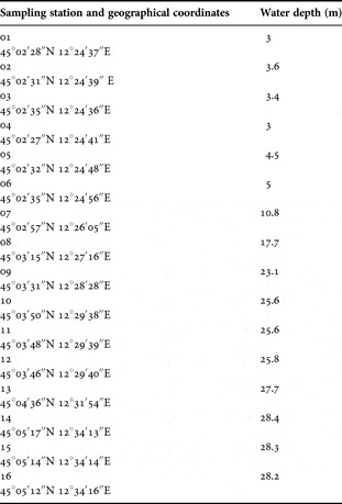
The investigated area is located in the upper Adriatic Sea, at about 45 km south of Venice (latitude 45°02′28″–45°05′12″N; longitude 12°24′37″–12°34′16″E, Table 1). Quantitative samples in three replicates were collected in February 2006 from 16 stations at 3–28 m water depth using a gravity box corer. For meiofaunal analysis, the top 5 cm sediment layer from each box corer was subsampled using a hand-held piston corer (2.37 cm i.d.×10 cm h); subsequently each sub-sample was transferred to a 0.5 l plastic jar and the fauna narcotized using a 7% magnesium chloride solution; after 10 minutes animals were fixed on site with a 10% buffered formalin solution, pre-stained with rose Bengal, and stored for later checking.
Location of the sampling stations and related water depth.

In the laboratory, meiofauna were extracted by the centrifugation decantation technique using Ludox-A30 colloidal silica, d = 1.210 (Pfannkuche & Thiel, Reference Pfannkuche, Thiel, Higgins and Thiel1988). The supernatant was filtered through a 45 µm mesh size sieve and the retained fauna were sorted by major taxa and counted under a Wild M8 stereomicroscope. The microscope inspection of the sandy pellets allowed us to ascertain that the efficiency of the extraction was almost 100% as far as all the taxa were concerned.
The sorted Gastrotricha were transferred to a water–glycerine mixture for 1–2 hours, and the water allowed to evaporate overnight. Later, individuals were placed in a tiny drop of pure glycerin between two coverslips placed in an H-S slide, for microscopic observation from both sides. The specimens were studied using a Nikon Eclipse 90i microscope equipped with DIC (Nomarski) optics and a DS-5Mc Nikon digital camera; morphometric data were obtained directly or derived from photographs using the UCT-2U Nikon software. Additional worms were prepared for a SEM survey. To this end, fixed specimens were rinsed in distilled water, dehydrated through a graded ethanol series, critical point-dried using CO2, mounted on aluminium stubs, sputter coated with gold-palladium and observed with a Philips XL 30 scanning electron microscope. The positions of anatomical structures are expressed in reference to percentage body units; total body = 100 units (U).
RESULTS
Gastrotrich fauna
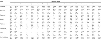
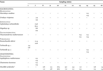
In the 48 examined samples, there were 494 gastrotrich individuals, with an overall density of 8.7±28.4 ind./10 cm2 (mean±SD), equal to 0.7% of the total meiofauna. Gastrotrichs were found in 11 out of the 16 investigated stations, with densities ranging from 0.6±0.5 to 97.7±71.2 ind./10 cm2 (Table 2). The taxonocoenosis was formed of 11 species distributed in 10 genera and six families (Table 3). Eight species are included in the order Macrodasyida (i.e. Cephalodasys turbanelloides, Macrodasys sp., Megadasys sp., Paraturbanella dohrni, Thaumastoderma mediterraneum, Turbanella sp.1, Turbanella sp.2 and Urodasys viviparus) and three in the order Chaetonotida (i.e. Aspidiophorus mediterraneus, Chaetonotus lacunosus and Musellifer profundus).
Mean density±standard deviation (ind./10 cm2) of major taxa and total meiofauna found in the 16 sampling stations; the relative abundance (%) of single taxa are derived from total density.

Mean density ± standard deviation (ind./10 cm2) of gastrotrich species found during the study; only the sampling stations hosting gastrotrichs are reported.

*, where present Musellifer profundus was the only gastrotrich living there except at Station 13 where the species made up 71.4% of all the gastrotrich community.
The community was dominated by M. profundus, which made up 24.5% of the entire gastrotrich assemblage. The species was found in eight out of the 11 locations that hosted gastrotrichs; where present, M. profundus constituted anything from 75% to 100% of the total Gastrotricha (Table 3). Peaks of abundance, 18.3±26.0 and 9.0±7.0 ind./10 cm2, were found in samples from Stations 10 and 11 respectively; in both stations the sedimentary bottom is at 25.6 m water depth. All the other species were present sporadically (e.g. Stations: 1, at −3 m; 7, at –10.8 m; 13 at –27.7 m; 14 at –28.8 m) and in low numbers, with the exception of Cephalodasys turbanelloides that in Station 7 reached the density record of 73.6±62.5 ind./10 cm2 (Table 3).
While we were able to identify as known species most of the specimens found, in some cases a full identification (i.e. to species) was not possible due to fixation artefacts, which altered the morphometry of key taxonomic characters. On the other hand, the good preservation of many specimens of Musellifer profundus allowed for a more comprehensive description of this rare, phylogenetically important species, the details of which are reported below.
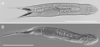
Musellifer profundus. Habitus, (A) dorsoventral view; (B) lateral view. DIC micrographs. Scale bars: 100 µm.

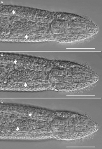
Musellifer profundus. Anterior region of an adult specimen, showing the male reproductive apparatus, arrowheads, at different focal planes, dorsal to ventral. (A) Testes and anteriorly directed deferens; (B) deferens jointing the middle of the glandular organ; (C) glandular organ showing the tube-like structure at its posterior portion. DIC microphotographs. Scale bars: 25 µm.

Musellifer profundus. Posterior region of adult specimens, showing the female reproductive apparatus, arrowheads. (A) Allosperm filling-up the region posterior to a mature egg; (B, C) maturing eggs on the right and left side respectively; (D) maturing eggs on the right side different view. DIC micrographs; (A–C ) dorsoventral view, (D), lateral view. Scale bars: 20 µm.

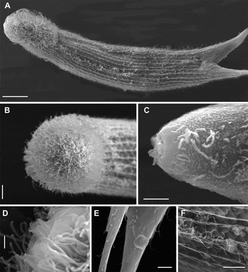
Musellifer profundus. (A) Habitus, ventral view; (B) close-up of the head showing thick ciliation; (C) head shaved, showing the cuticular muzzle and insertion of the cilia; (D) close up of the mouth, showing the moth cone; (E) close up of the furca covered by scales; (F), close up of the cuticle of the trunk in ventral view showing the scales and a portion of the ventral ciliature. SEM microphotographs. Scale bars: A, 50 µm; B, C, 20 µm; D–F, 10 µm.

DESCRIPTION
General morphology
The body appears compact, slightly spiny, averaging 320 µm in total length (range 310–340 µm, N = 15), with a relatively small head, bearing a pronounced muzzle and surrounded by a broad and dense ciliature; neck short; trunk robust ending with a furcate caudum comprising 29% of the total body length (Figures 1 & 4). Widths of head, neck, trunk and caudal base are as follows: 24/27.7/41/31 µm at U5.5/U19/U48/U71 respectively. Caudum elongate, with a distal furca that indents medially to U82 and has parallel exterior borders; each furcal branch is 93 µm in length and tapers quite rapidly from thick base to very thin tip.
The muzzle resembles a shallow frustum of cone; the proximal lower base, 6.5 µm in diameter, shows 12 triangular, 1.5 µm deep indentations; the distal upper base, 4 µm in diameter, bears the mouth at its centre; the oral opening, 3 µm in diameter, is armed with some rod-like structures that protrude by 1 µm from it; SEM images indicate that rods are fused to each other to form a sort of mouth tube. The pharynx is 54 µm in length and of increasing diameter from anterior, 10.5 µm, to posterior 18 µm; pharyngeal–intestinal junction at about U19; the intestine is straight, slightly broader at its middle, 18 µm, ending at U67, presumably with a ventral anus (not seen).
Cuticular armature
The body is completely enveloped by spined scales, except for the head's anterior ciliary areas and the most distal end of the furcal branches. Scales are arranged in 26–28 columns (11 dorsal+4×2 lateral+7–9 ventral) of 46–49 scales each dorsally and 38–40 ventrally. Generally, scales overlap strongly, except for the distal half of the furca, where the anterior margin of scales is barely hidden by the posterior margin of the preceding elements.
The basic shape of the scales is reminiscent of a pentagonal arrowhead, with a broadly rounded tip and spiny posterior corners; however, locally length and width appear allometrically decoupled from size. Scales of largest size, wider than long (up to 6.5 × 6 µm), cover the dorsal posterior side of the trunk whereas relatively small scales, longer then wide (down to 2 × 3 µm ), cover the ventral side and the furcal branches; further, scales of mid- to small-size, as wide as long (4 × 4 µm), cover the dorsal and lateral areas just posterior to the ciliary band on the head. With few exceptions, scales each bear a simple spine, which emerges as the continuation of a poorly-defined keel originating from the central region of the scale; spines appear very delicate and range 2–10 µm in length according to the size of the carrying scale, the longer being present dorsally on the posterior region of the trunk.
Ciliature
A broad, dense band of cilia encircle the anterior region of the body, most cilia are 4–6 µm, but some reach 13–14 µm in length. The ciliated area is much broader on the ventral side resembling a beard that extends from the muzzle almost to the neck constriction (to U17) and becomes abruptly narrower while covering the lateral and the dorsal side of the head, where it extends to a maximum at U8.5. On the ventral side, the peculiar cephalic ciliary band unites with the two more usual longitudinal bands; these are especially narrow (~1 µm) and run at maximum to U51, there terminating much before the presumed anal opening; ventral locomotor cilia are 4–5 µm in length.
Genital apparatus
Musellifer profundus is a proterandrous, then simultaneous hermaphrodite, with paired testes and ovaries. The male system is located along the anterior third of the intestine, lateral and ventral to it. Testes are lateral, pear-like in shape and small in size, about 5 µm in diameter; they begin wider at U32.5 and get narrower anteriorly, while flowing into the vasa deferentia (Figure 2). The latter are rather short, about 15 µm length and 2–3 µm in diameter and extend ventrolaterally and to the fore, terminating in the middle of a common, ovoid, glandular organ, centred at U25. Size and appearance of the ventral, glandular structure is variable, in accordance with the specimen's age (sexual status). It becomes visible in young adults (i.e. animals without ova) where it is small and contains granules of similar size; in fully mature specimens (i.e. with a large egg inside), the glandular organ reaches its largest size, 20 µm in length and 12 µm in width. At this stage, inside its anterior margin (i.e. toward the pharyngeal–intestinal junction) there is a small area filled with minute, round, refringent granules, whereas the rest of the organ is filled with variously shaped granules, of comparatively larger size. Also at this stage, a tube like-structure becomes visible, 9 µm in length and 2–3 µm in diameter, that originates from the posterior margin of the glandular organ while its distal end remains open. We were unable to ascertain whether the tube opens to the outside.
The female system is located along the posterior half of the intestine, lateral and ventral to it (Figure 3). Ovaries are lateral (from U55 to U60) and contain several oocytes maturing anteriorly and alternatively in the two ovaries. In a fully mature specimen, a large egg, 34 µm in length and 21 µm in width, was seen ventrolaterally at U47. In fully mature individuals (i.e. showing a large egg inside) the area in between the two ovaries, ventral and dorsal to the intestine, is filled with spermatozoa. Some spermatozoa are coiled over whereas others appear as free, filiform elements. The discontinuity from male gonads suggests these gametes to be allosperm. No accessory sexual structures (e.g. frontal and/or caudal organ) could be detected in this region.
DISCUSSION
Gastrotrich fauna
With regard to the gastrotrich fauna, Italy is one of the best studied countries, with about 160 species known from over 180 localities (Todaro et al., Reference Todaro, Balsamo and Tongiorgi2008; Hummon & Todaro, Reference Hummon and Todaroin press); however, no previous data were available from the area subject to this study. Here we found 11 species from 16 stations, constituting less than 1% of the local total meiobenthos. Both the low number of species and the low density indicated that the area is not particularly suitable for supporting large gastrotrich communities; likely, this has much to do with the very fine sediments (muddy sand) that characterize the area. Marine gastrotrichs are known to be strictly interstitial animals; therefore it is expected that their numbers would drop when the mean particle size of the sediment falls below 125 µm (Coull, Reference Coull, Higgins and Thiel1988). Indeed, it is not by coincidence that previous studies, conducted in other areas characterized by fine sediments yielded scanty gastrotrich communities (e.g. Todaro et al., Reference Todaro, Fleeger and Hummon1995; Todaro & Rocha, Reference Todaro and Rocha2004). The finding in several stations of a relatively high number of specimens belonging to the genus Musellifer does not come as a surprise as these exceptional, marine gastrotrichs are known to live in muddy substrata (e.g. Todaro & Hummon, Reference Todaro and Hummon2008). On the other hand, the high density of Cephalodasys turbanelloides at Station 7 may be due to the occurrence of a localized, superficial patch of coarser sediment (mean diameter > 125 µm). Although samples were mixed soon after the sampling, and therefore there is no direct observation of this, the presence at the same time of another five interstitial species in these samples (Table 3), supports the hypothesis that restricted patches of coarser sediment occur in the area.
Notwithstanding the low number of taxa present in the area, the finding of some species bears relevance within a biogeographical/taxonomic framework. For instance, among the Macrodasyida, the geographical distribution of Thaumastoderma mediterraneum in the Adriatic Sea appears now broader then previously thought, since earlier studies indicated the species to be confined to the southern part of the basin i.e. Apulian coasts (see Todaro et al., Reference Todaro, Hummon, Balsamo, Fregni and Tongiorgi2001); moreover, the distribution of Urodasys viviparus within the Adriatic Sea appears now less patchy than previously reported, since the species was only previously known from the southern and northernmost part of the Adriatic Sea (Trieste) but not for the Veneto region (Todaro et al., Reference Todaro, Hummon, Balsamo, Fregni and Tongiorgi2001).
Musellifer profundus
With little doubt, the finding of Musellifer profundus is the most important result of our study, for several reasons.
The species was described based on a single specimen found off Marseille (Vivier, Reference Vivier1974); although the microscopic examination was carried out without DIC and/or SEM technologies, the specimen's external morphology appears accurately reported; however, the author could not detect any trace of the reproductive apparatus. Considering that the holotype measured 329 µm in total length its reproductive apparatus should have been fully developed, at least it is so in specimens from the northern Adriatic Sea that measure >300 µm in total length.
There are at least two other noticeable morphometric discrepancies between specimens of the two populations (Marseille versus northern Adriatic); one regards the length of the furca and its extension in relation to the total body length. The French specimen had a furca 123 µm long, equal to 37% of total body length whereas, on average, the Italian specimens have a furca which is 93 µm long, equal to 29% of total body length. The other difference pertains to the presence in the French animal of two columns of ventral scales each bearing two spines, which are absent in the Adriatic gastrotrichs.
These differences raise the question of whether Vivier's specimen and the individuals found in the northern Adriatic Sea belong to the same species; after all, the northern Adriatic Sea is rather far from the type locality and the two populations appear well isolated from each other. At present, we consider the specimens from both locales to be conspecifics for the following reasons: first, recently we have found in Sicily (Gulf of Trapani) several specimens of Musellifer morphologically similar to the specimens from the northern Adriatic Sea (M.A. Todaro & M. Dal Zotto, unpublished), indicating a wide geographical distribution for this taxon.
Second, the measurements reported by Vivier (Reference Vivier1974) appear affected by errors, which make the metrics reported for his specimen somewhat dubious. In fact, according to her figure 1A and related scale, the specimen measures 297.2 µm in total length (not 329 µm as stated) and the furca only 100 µm, equal to 33% of the total body length; the situation gets even more confusing considering Vivier's figure 1C, which depicts an animal 334 µm in total body length, having furcal appendages 102 µm long, which represents just 30% of the total length. Consequently it is difficult in this framework to establish the real size of the French specimen and to exclude for example that the animal surveyed by Vivier (Reference Vivier1974) was at its sub-adult stage, a condition that would account for the lacking of a reproductive apparatus.
Third, the distribution and arrangement of the ventral scales, which Vivier (Reference Vivier1974) reports as bearing two spines, coincides with the topographic distribution of the two ventral ciliary bands in our gastrotrichs. Further, SEM pictures of our specimens show that the two narrow ciliary columns emerge from tightly appressed, side by side, overlapped scales, and what Vivier interpreted (her words) as a scale with two spines are in fact the keels of two flanking scales.
All possible doubts regarding these differences could be solved by surveying the French specimens with modern microscopy techniques; unfortunately, the type material, deposited at the Muséum National d'Histoire Naturelle, Paris under the accession number GAS-MHV-1, has vanished from its repository, making first hand comparison impossible.
In our opinion, while there is no doubt about the taxonomic status of the north Adriatic specimens records of M. profundus from locations outside the Mediterranean need confirmations. An authoritative identification of this species cannot be achieved without taking into account the shape and layout of its reproductive apparatus. This statement gets strength by considering for example that while differences in the reproductive apparatus can allow an easy discrimination between congeneric species (e.g. M. profundus and M. sublitoralis) the same does not hold true by using as a key trait, the morphology of the cuticular armature (see SEM pictures of putative M. profundus and M. sublitoralis at the website http://hummon-nas.biosci.ohiou.edu/MarGastPics/Chaetonotidae/Musellifer).
CONCLUDING REMARKS
Despite the fact that the gastrotrich fauna of the Italian coastline is well documented (Todaro & Leasi, Reference Todaro and Leasi2006; Todaro et al., Reference Todaro, Hummon, Balsamo, Fregni and Tongiorgi2001, Reference Todaro, Matinato, Balsamo and Tongiorgi2003, Reference Todaro, Guidi, Leasi and Tongiorgi2006b, Reference Todaro, Leasi, Bizzarri and Tongiorgic, Reference Todaro, Balsamo and Tongiorgi2008; Hummon & Todaro, in press), the present study has revealed new details on the biogeography of several taxa, and further revealed the importance of muddy sediment as a previously overlooked substratum of ecological importance. The detailed description of the reproductive apparatus of a phylogenetically relevant taxon (i.e. Musellifer profundus) will certainly be useful in evolutionary studies aimed at shedding light on the relationships within Gastrotricha, while offering at the same time additional morphological traits to be used in reliable species identification. In this regard it should be emphasized that although only three species of Musellifer have been described so far, several others are awaiting formal affiliation, a task that will definitely benefit from this new information.
ACKNOWLEDGEMENTS
We are grateful to C. Virno-Lamberti of ISPRA, Rome, for permitting our involvement in the general project. Our research benefited also from a MIUR grant to M.A.T. (PRIN-2007—Approccio integrato all'identificazione dei Gastrotrichi marini). Comments by referees improved the readability of this paper.