Enteroviruses are a common cause of illness in young infants, with neonates accounting for 11.4–13% of all enteroviral infections each year. Reference Chuang and Huang1–Reference Schlapbach, Ersch and Balmer3 While many infected neonates remain asymptomatic, 47–63% of neonatal rule out sepsis cases and 11.6–24% of neonates presenting with signs of shock have a positive enteroviral test. Reference Chuang and Huang1,Reference Schlapbach, Ersch and Balmer3,Reference Jenista, Powell and Menegus4 Enteroviruses are also the most common cause of viral myocarditis in neonates, and, in spite of aggressive supportive strategies, a diagnosis of enteroviral myocarditis has been associated with a high risk of in-hospital mortality. Reference Schlapbach, Ersch and Balmer3,Reference Freund, Kleinveld, Krediet, van Loon and Verboon-Maciolek5
Mimicking non-cardiac causes of neonatal shock, enteroviral myocarditis starts as a febrile illness that can progress to cardiovascular collapse. Reference Abzug6–Reference Inwald, Franklin, Cubitt, Peters, Goldman and Burch8 Cardiomegaly is sometimes incidentally noted on chest X-ray, but echocardiography is the primary diagnostic tool that demonstrates the hallmark left ventricular systolic dysfunction with or without dilation and/or wall thinning. Reference Freund, Kleinveld, Krediet, van Loon and Verboon-Maciolek5 Amongst survivors, the risk of long-term cardiac injury is significant, specifically chronic systolic dysfunction and presence of basal posterior left ventricular aneurysms, which have been reported in children with this disease process. Reference Freund, Kleinveld, Krediet, van Loon and Verboon-Maciolek5,Reference Wittekind, Allen and Jefferies9–Reference Chan and Lun11
Multi-organ system dysfunction has been shown to be a harbinger of mortality; however, the utility of echo indices in predicting outcomes has not been investigated in neonates diagnosed with enteroviral myocarditis. Reference Madden, Thiagarajan, Rycus and Rajagopal2,Reference Freund, Kleinveld, Krediet, van Loon and Verboon-Maciolek5,Reference Inwald, Franklin, Cubitt, Peters, Goldman and Burch8,Reference Chan and Lun11 Myocardial strain imaging has shown promise for the detection of myocardial injury prior to changes in left ventricular ejection fraction in other cohorts such as patients monitored after receiving cardiotoxic chemotherapy. Reference Thavendiranathan, Poulin, Lim, Plana, Woo and Marwick12 Strain analysis in children has noted abnormal findings in children with HIV and Kawasaki disease despite normal ejection fraction, suggesting these measures can detect early myocardial changes prior to functional decline. Reference Sims, Frank and Cross13,Reference Sanchez, Tejtel, Almeida-Jones, Feagin, Altman and Pignatelli14 We sought to determine whether any echo-derived left ventricular strain measures, obtained at time of diagnosis, could help stratify infants with enteroviral myocarditis at highest risk for poor outcome. A secondary aim was to further characterise long-term sequelae, including frequency of left ventricular aneurysms.
Materials and methods
We conducted a retrospective chart review of all patients diagnosed with neonatal enteroviral myocarditis at Children’s Wisconsin, from 1999 to 2019. Patients born prior to 1999 were excluded because medical records and echocardiograms were not available in a digital format. Inclusion criteria included patients with a structurally normal heart, age less than thirty days of life at time of diagnosis, an echocardiogram at the time of diagnosis demonstrating abnormal left ventricular systolic function, and positive real-time enteroviral polymerase chain reaction test in one or more body fluids (blood, cerebrospinal fluid, nasopharyngeal swab, and/or stool). Patients were excluded if estimated gestational age at birth was less than 35 weeks and/or genetic testing documented variants known to be associated with cardiomyopathies.
Echocardiograms at the time of diagnosis and at most recent follow up were retrospectively analysed. In the parasternal short-axis plane, left ventricular end-diastolic and end-systolic diameters, as well as shortening fraction, were measured, at the level of the papillary muscles. In the apical four-chamber plane, left ventricular end-diastolic and end-systolic volumes were measured and left ventricular ejection fraction was calculated using Simpson’s method. The presence and degree of mitral and/or tricuspid regurgitation was qualitatively assessed using colour Doppler from the apical four-chamber and parasternal long-axis planes. Two-dimensional global left ventricular peak systolic longitudinal and circumferential strain were measured in the apical four-chamber and parasternal short-axis planes, respectively, using speckle tracking technology (TomTec Imaging Systems GmbH, Unterschleissheim, Germany). Reference Mor-Avi, Lang and Badano15,Reference Voigt, Pedrizzetti and Lysyansky16 Presence of a left ventricular aneurysm was defined as a focal area of ventricular wall thinning/dilation with paradoxical motion (expansion in systole, collapse in diastole). Laboratory values during the initial hospitalisation and hospital clinical course and outcomes were also collected.
Statistical testing was performed to compare variables at presentation between survivors and those who were transplanted or died using Fisher’s exact and Mann–Whitney non-parametric tests for ordinal measures and chi-square linear trend tests for ordered measures. Analysis was performed using IBM SPSS Statistics v.20. This study was approved by the local Institutional Review Board.
Results
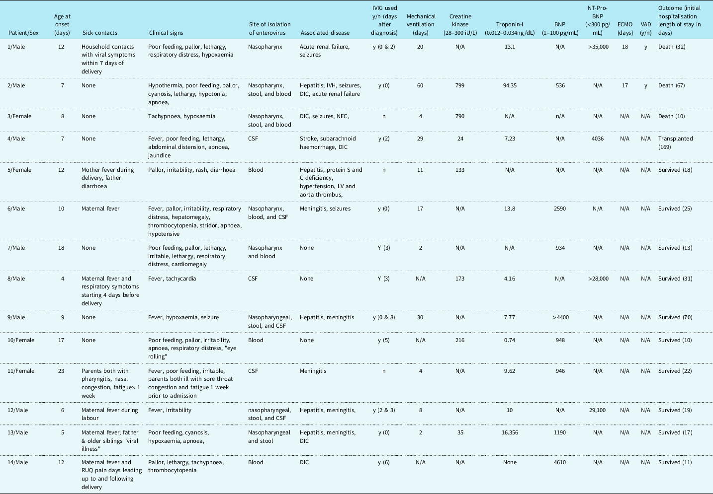
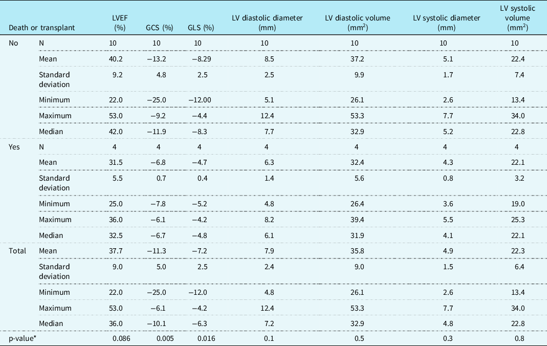
Fourteen infants (4 females and 10 males) were diagnosed with neonatal enteroviral myocarditis at an average age of 10.7 days (range 4–23 days). All patients presented with signs and symptoms of shock, and 50% of patients had a first degree relative with infectious symptoms suggestive of enteroviral infection (Table 1). All patients, at the time of diagnosis, had documented cardiac dysfunction, with an average left ventricular ejection fraction of 37.7%, an average global circumferential strain of −11.3%, and an average global longitudinal strain of −7.2% (Table 2).
Cohort demographic and laboratory variables

Cohort echocardiogram characteristics at diagnosis

GCS = global circumferential strain; GLS = global longitudinal strain; LV = left ventricle; LVEF = Left ventricular ejection fraction.
* Mann–Whitney U-test.
Three patients died, and one required transplantation during the initial hospitalisation after diagnosis. One patient died of rapidly progressive multisystem organ failure 2 days after diagnosis. The other two patients who died required extracorporeal membrane oxygenation support for an average of 17.5 days and eventually ventricular assist device placement; both died from complications related to mechanical support. None of the survivors, including the one transplant patient, needed any form of mechanical support; the transplant was performed because of persistent need for intravenous inotropic support. Average length of stay for survivors who did not require transplant was 24 days (range 11–70 days) with 7/10 requiring mechanical ventilation for an average of 10.5 days (range 2–30 days). Eleven patients had documented supraventricular and/or ventricular arrhythmias during their initial hospitalisation. Non-specific/diffuse ST changes were present on electrocardiogram for 8 of the 14 patients at presentation, but neither arrhythmias nor ST changes on electrocardiogram were associated with outcome.
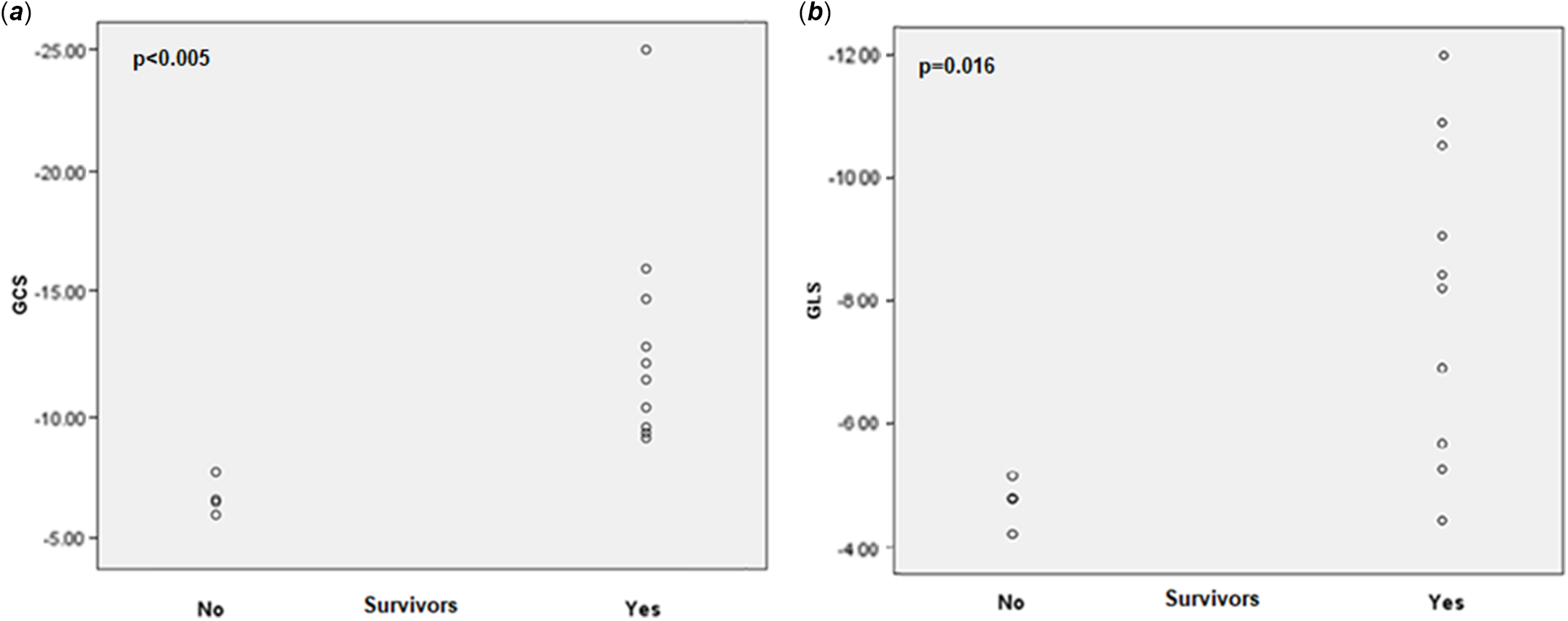
Transplant-free survivors on average had a better left ventricular ejection fraction at presentation than non-survivors (40.2% versus 31.5%) but this difference did not reach statistical significance (p = 0.086). Left ventricular peak systolic strain values, however, were found to be statistically significantly different between groups. Transplant-free survivors had better global circumferential strain (−13.25% [range −9.2 to −25%] versus −6.8% [range −6.1 to −7.8%], p = 0.005) and global longitudinal strain (−8.2% [range −4.4 to −12%] versus −4.7% [range −4.2 to −5.2%], p = 0.016). No other echocardiographic measures were different between the two groups (Table 2). A scatter plot of global circumferential strain and global longitudinal strain values between survivors and patients who experienced death or transplant is shown below in Figures 1a and 1b, respectively. No demographic (age, sex, birth weight, gestational age, age at presentation, or birth month) or laboratory variables (troponin-I, brain natriuretic peptide, NT-Pro-brain natriuretic peptide, or creatine kinase) were different between the two groups (Table 1). Eight survivors, two of the three patients that died, and the transplanted patient, received intravenous immunoglobulin, which was not found to have a statistically significant impact on outcome.
(a) A scatter plot comparing global circumferential strain at presentation between survivors and patients who experienced death or transplant. (b) A scatter plot comparing global longitudinal strain between survivors and patients who experienced death or transplant. GCS = global circumferential strain.

Of the eight survivors who had follow-up data available over a mean interval of 8.3 years (range 4–14 years), three were felt to have completely recovered and were discharged from cardiology care. The other five had chronic cardiac complications. All survivors, including those discharged from cardiology care, had persistently abnormal left ventricular systolic function (as defined by persistently abnormal global longitudinal strain; Video 1), with four having abnormal left ventricular ejection fraction and three with abnormal global circumferential strain. Three of the eight survivors continued to receive heart failure medications (beta-blocker and/or angiotensin-converting enzyme inhibitor). Finally, 5 of 14 patients developed a focal posterior basal left ventricular wall aneurysm (Fig 2; Video 2), 4 of the late survivors and the 1 transplanted patient. These aneurysms were remarkably similar in location in all the patients. Despite these complications, no late arrhythmias were identified in survivors, neither clinically or by Holter surveillance. There were no late deaths.
Parasternal long-axis image of a left ventricular aneurysm in a survivor of neonatal enteroviral myocarditis. The aneurysm is in the posterior basal LV (arrows) with myocardial wall thinning and paradoxical bulging of the aneurysm during ventricular systole. Ao = aorta; LA = left atrium; RV = right ventricle.

Discussion
This case series highlights the significant early mortality and long-term morbidity associated with neonatal enteroviral myocarditis and the potential utility of left ventricular peak systolic strain in identifying those at highest risk of poor outcome. Using previously described normal paediatric strain values, we found that longitudinal and circumferential strain values were abnormal in infants with enteroviral myocarditis. Reference Levy, Mejia, Machefsky, Fowler, Holland and Singh17–Reference Marcus, Mavinkurve-Groothuis and Barends19 These measurements were the only echo parameters that identified patients at increased risk of in-hospital mortality. While limited by a small sample size, this study is the first to demonstrate that two-dimensional left ventricular strain imaging has potential prognostic utility for infants diagnosed with neonatal enteroviral myocarditis. Given the mortality risk associated with neonatal enteroviral myocarditis, we speculate that identification of high-risk patients using this tool may improve outcomes by prompting earlier initiation of advanced therapies in those with the most concerning strain measures. Reference Madden, Thiagarajan, Rycus and Rajagopal2,Reference Schlapbach, Ersch and Balmer3,Reference Freund, Kleinveld, Krediet, van Loon and Verboon-Maciolek5,Reference Inwald, Franklin, Cubitt, Peters, Goldman and Burch8,Reference Goudevenos, Parry and Gold10,Reference Chan and Lun11
Our report again documents the poor prognosis of infants with enteroviral myocarditis. A diagnosis of severe neonatal enteroviral infection is known to be associated with poor outcomes, with the highest mortality rates seen amongst patients diagnosed with myocarditis. Reference Chan and Lun11 In 2010, Freund et al reported that 31% of patients diagnosed with neonatal enteroviral myocarditis did not survive to hospital discharge and of those that did 66% developed “severe [cardiac] damage.” Reference Freund, Kleinveld, Krediet, van Loon and Verboon-Maciolek5 Development of multi-organ system dysfunction, prompting initiation of advanced therapies (extracorporeal membrane oxygenation, ventricular assist device, dialysis, etc.), confers a worse prognosis in patients with enteroviral myocarditis. Several case series have shown that in-hospital mortality experienced by patients requiring extracorporeal membrane oxygenation cannulation ranges from 67 to 80%. Reference Madden, Thiagarajan, Rycus and Rajagopal2,Reference Freund, Kleinveld, Krediet, van Loon and Verboon-Maciolek5,Reference Inwald, Franklin, Cubitt, Peters, Goldman and Burch8,Reference Chan and Lun11 The patients within our cohort experienced similar rates of early mortality to those seen by Freund et al (28 versus 31%, respectively). In our cohort, 100% of patients requiring support with extracorporeal membrane oxygenation and/or renal replacement therapy died. Importantly, 62.5% of survivors had evidence of cardiac injury on follow up, while only 37.5% of survivors recovered fully. Although survivors in our cohort have done well at intermediate follow up, long-term surveillance is prudent to observe for late complications including, but not limited to, development of heart failure and/or arrhythmias. A 2021 American Heart Association scientific statement Reference Law, Lal and Chen20 reviewing the diagnosis and management of viral myocarditis in children states that while normalisation of ventricular systolic function, as measured by left ventricular ejection fraction, occurs in 52–54% of individuals Reference Foerster, Canter and Cinar21 adverse remodelling can continue and may be an aetiology of idiopathic dilated cardiomyopathy in later in life. To date, none of our patients have died or required transplant after discharge from their initial hospitalisation, and all are clinically stable.
Seemingly unique to children with enteroviral myocarditis, and first described by Goudevenos et. al in 1989, is the development of focal left ventricular posterior basal free wall aneurysms. Reference Goudevenos, Parry and Gold10 It has not been reported in other forms of childhood myocarditis. Survivors in Freund et al’s cohort all developed dilated cardiomyopathy with “mild” to “severe” left ventricular aneurysms located in the posterior basal left ventricular-free wall. Reference Freund, Kleinveld, Krediet, van Loon and Verboon-Maciolek5 Within our cohort, four of eight survivors at most recent follow-up (range 15 months to 6 years) had left ventricular aneurysms in the posterior basal left ventricular-free wall, identical in location to those reported previously. Reference Freund, Kleinveld, Krediet, van Loon and Verboon-Maciolek5,Reference Wittekind, Allen and Jefferies9–Reference Chan and Lun11 In all, 5 of the 14 patients in our cohort had a left ventricular aneurysm identified, including the transplanted patient, who developed a persistent aneurysm and failed to wean from inotropes during his hospitalisation prior to transplantation. The aetiology of left ventricular injury is unclear, and the mechanism for this consistently similar and focal site of myocardial injury is unknown. The transcription factor nuclear factor kappa B is one of many intracellular signalling molecules responsible for regulating inflammatory cytokine production within myocytes. By dysregulating “anti- and pro-apoptotic” cellular pathways, viruses stand to gain a survival advantage, either by facilitating the release of viral particles or by preventing cell death before the end of the virus’s life cycle. Reference Esfandiarei, Boroomand, Suarez, Si, Rahmani and McManus22,Reference Matsumori, Nunokawa and Yamaki23 Dysregulation of pathways in which transcription factor nuclear factor kappa B is the downstream target, which occurs with enteroviral infection, is thought to be a potential mechanism by which myocardial cell death occurs Reference Esfandiarei, Boroomand, Suarez, Si, Rahmani and McManus22 but a definitive mechanism is not known.
Conclusion
Neonatal enteroviral myocarditis carries a high risk of early mortality and late morbidity. Echo-derived left ventricular global circumferential and longitudinal strain at time of diagnosis are potentially useful for risk stratifying infants at presentation. Most survivors continue to have late left ventricular systolic dysfunction necessitating cardiology surveillance and medical therapy, and the development of chronic left ventricular basal posterior wall aneurysms is a common sequelae that is not seen in other forms of paediatric cardiomyopathy.
Supplementary material
To view supplementary material for this article, please visit https://doi.org/10.1017/S1047951122001512
Acknowledgements
None.
Financial support
This research received no specific grant from any funding agency, commercial or not-for-profit sectors.
Conflicts of interest
None.