INTRODUCTION
Epibiosis is a facultative association of two organisms: the epibiont and the basibiont. The term ‘epibiont’ includes organisms that, during the sessile phase of their life cycle, are attached to the surface of a living substratum, while the basibiont lodges and constitutes a support for the epibiont. Both concepts describe ecological functions (Wahl, Reference Wahl1989). Many groups of Crustacea, i.e. Cladocera, Copepoda, Cirripedia, Isopoda, Amphipoda and Decapoda, include forms that are hosts for macroepibiont invertebrates (Ross, Reference Ross and Bliss1983), e.g. Porifera, Cnidaria, Platyhelminthes, Nemertea, Rotifera, Nematoda, Polychaeta, Cirripedia, Decapoda, Gastropoda, Bivalvia, Phoronida, Bryozoa, Ascidiacea and others, as well as for microepibionts from the Protozoa (Corliss, Reference Corliss1979; Small & Lynn, Reference Small, Lynn, Lee, Hunter and Bovee1985).
Epibiosis is a common phenomenon on marine and freshwater Crustacea, and there are a number of records about this kind of association in species of the majority of the crustacean taxa (Morado & Small, Reference Morado and Small1995; Fernandez-Leborans & Tato-Porto, Reference Fernandez-Leborans and Tato-Porto2000a, Reference Fernandez-Leborans and Tato-Portob; Fernandez-Leborans, Reference Fernandez-Leborans2001). There has been a noticeable increase in reports of new animal groups and species involved in epibiosis in recent years, and their relevance is probably mainly attributable to the fact that epibiotic relationships are important at many levels: physiological, ecological, evolutionary as well as on those related to biodiversity and bioconservation (Fernandez-Leborans, Reference Fernandez-Leborans2009).
The complex shell-crab of the hermit crab species has been subject of numerous surveys about their epibiont communities (Reiss et al., Reference Reiss, Knäuper and Krönche2003; Williams & McDermott, Reference Williams and McDermott2004). The studies include diverse aspects such as the presence of different epibiont species, the distribution of the epibiont species, the epibiosis and the life-cycle of hermit crabs, and the interactions between epibiont and basibiont (Jensen & Bender, Reference Jensen and Bender1973; Partridge, Reference Partridge1980; Karlson & Shenk, Reference Karlson and Shenk1983; Brooks & Gwaltney, Reference Brooks and Gwaltney1993; Van Winkle et al., Reference Van Winkle, Longnecker and Blackstone2000). These communities may include species of a number of invertebrate phyla, as well as other groups, e.g. bacteria, microalgae and ciliate protozoans. Among the hermit crabs, Pagurus bernhardus is one of the most studied species, and epibionts of diverse groups (Foraminifera, Cnidaria, Plathyhelminthes, Nemertea, Mollusca, Polychaeta, Crustacea, Nematoda, Phoronida, Bryozoa and Tunicata) had been described on this hermit crab (Reiss et al., Reference Reiss, Knäuper and Krönche2003).
On specimens of Pagurus bernhardus, sampled on the west coast of Scotland, ciliate hyperepibionts were found on the hydrozoan Dicoryne conferta, attached to the shell occupied by the hermit crab. The aim of this study is to describe the epibiont species present on the crab and the shell occupied by the crab, as well as the hyperepibiont species living on the hydrozoan found on the shell. The spatial distribution of epibionts, as well as the morphology, taxonomic position and distribution of the ciliate hyperepibionts are described below.
MATERIALS AND METHODS
Specimens of Pagurus bernhardus were collected on the west coast of Scotland, in the Firth of Clyde. The coastal area used was between Largs and the Isle of Cumbrae (55°48′N 4°50′W). The sampling was carried out using a beam trawl. An initial examination of crabs was made, in order to observe the epibionts and select the crabs with them. Crabs and their shells with epibionts were fixed in 4% formaldehyde in seawater. In the laboratory, crabs were dissected and each anatomical unit was observed under a stereoscopic microscope. All shells belonged to Buccinum undatum. The shell surface was measured and divided into nine zones of the same area (A, B, C, D, E, F, G, H and I) (Brooks, Reference Brooks1989). The location of the different invertebrate epibionts (i.e. specific zone occupied) on the shell occupied by P. bernhardus was recorded. Epibionts on the shell and surface of the anatomical units of the crabs were observed and counted under a stereoscopic microscope. The number of the hydrozoans on the surface of the shell was estimated from the area occupied by them. In order to identify the protozoan hyperepibionts, they were isolated and treated using the silver carbonate technique, according to the procedure described by Fernandez-Leborans & Castro de Zaldumbide (Reference Fernandez-Leborans and Castro de Zaldumbide1986), and also with methyl green and neutral red. Measurements of the epibionts were calculated using an ocular micrometer. Light microscope images were obtained using Image Analysis (KS300 Zeiss) and the diverse morphological features of the images were used to obtain the schemes of the epibiont species. Statistical analysis was performed using the program Statgraphics.
RESULTS
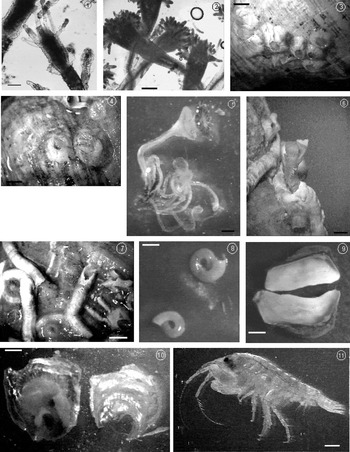
The specimens with epibionts presented the following species: the hydrozoan cnidarians Hydractinia echinata and Dycorine conferta, the cirriped crustaceans Balanus balanus and Balanus crenatus, the polychaete annelids Hydroides norvegica, Pomatoceros triqueter and Circeis armoricana, the bivalve molluscs Hiatella arctica and Anomia ephippium, and the amphipod crustacean Podoceropsis nitida (Figures 1–11).
Invertebrate epibiont species found in the study: (1) Hydractinia echinata. Scale bar: 300 µm; (2) Dicoryne conferta. Scale bar: 200 µm; (3) Balanus balanus. Scale bar: 5 mm; (4) Balanus crenatus. Scale bar: 10 mm; (5) Hydroides norvegica. Scale bar: 10 mm; (6) Hydroides norvegica. Detail of the anterior area. Scale bar: 10 mm; (7) Pomatoceros triqueter. Scale bar: 10 mm; (8) Circeis armoricana. Scale bar: 1 mm; (9) Hiatella arctica. Scale bar: 7 mm; (10) Anomia ephippium. Scale bar: 60 mm; (11) Podoceropsis nitida. Scale bar: 10 mm.

Hyperepibionts
The hyperepibionts attached to the hydrozoan Dicoryne conferta, which was found on the shell inhabited by Pagurus bernhardus belong to 7 ciliate protozoan species: 6 suctorian species (Conchacineta constricta, Corynophrya anisostyla, Actinocyathula homari, Actinocyathula crenata, Acineta sulcata and Acineta corophii), and one peritrich species (Zoothamnium sp.).
SYSTEMATICS (LYNN AND SMALL, Reference Lynn, Small, Lee, Leedale and Bradbury2000)
Phylum CILIOPHORA Doflein, 1901
Subphylum INTRAMACRONUCLEATA Lynn, 1998
Class PHYLLOPHARYNGEA De Puytorac et al., 1974
Order ENDOGENIDA Collin, Reference Collin1912
Family ACINETIDAE Stein, 1859
Genus Conchacineta Jankowski, 1978
Conchacineta constricta Jankowski, 1978
DESCRIPTION
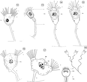
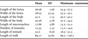
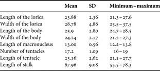
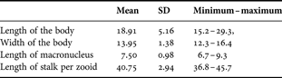
These ciliates were laterally flattened, of oval to circular contour, and the body was surrounded by a lorica compacted. The stalk was long, reaching 2–3 times the length of the body. The macronucleus was oval, located in the middle of the body. The lorica was cup-shaped, and the body projects above the anterior edge of the lorica forming an entrant characteristic above it. The anterior part of the body contained two fascicles of capitate tentacles arranged in a row (Table 1; Figures 12 & 19).
Schemes of the ciliate hyperepibiont species (cv, contractile vacuole; ma, macronucleus; my, muyoneme; p. peristome; s, stalk; t, tentacles): (12) Conchacineta constricta; (13) Corynophrya anisostyla; (14) Actinocyathula homari; (15) Actinocyathula crenata; (16) Acineta sulcata; (17) Acineta corophii; (18) Zoothamnium sp.

Photomicrographs of the ciliate hyperepibiont species: (19) Conchacineta constricta. Scale bar: 20 µm; (20) Corynophrya anisostyla. Scale bar: 20 µm; (21) Corynophrya anisostyla. Scale bar: 30 µm; (22) Actinocyathula homari. Scale bar: 20 µm; (23) Actinocyathula crenata. Scale bar: 20 µm; (24) Acineta sulcata. Scale bar: 10 µm; (25) Acineta corophii. Scale bar: 10 µm; (26) Zoothamnium sp. Scale bar: 10 µm.

Biometric features of Conchacineta constricta (measures in µm) (N = 60).

REMARKS
As this species they showed a laterally flattened body, without actinophores. The tentacles were arranged in two fascicles forming each a single row. The lorica was compressed laterally, and in its anterior aperture the body protruded exhibiting a waist-like indentation in each lateral edge. The macronucleus was located centrally (Curds, Reference Curds1985).
DISTRIBUTION
Number of individuals: 0–17 per hydrocaulus. This species has been observed previously on Pagurus cuanensis in Cette (France) (Collin, Reference Collin1909).
SYSTEMATICS (LYNN AND SMALL, Reference Lynn2000)
Phylum CILIOPHORA Doflein, 1901
Subphylum INTRAMACRONUCLEATA Lynn, 1998
Class PHYLLOPHARYNGEA De Puytorac et al., 1974
Order EXOGENIDA Collin, Reference Collin1912
Family CORYNOPHRYIDAE Jankowski, 1981
Genus Corynophrya Kahl, 1934
Corynophrya anisostyla Fernandez-Leborans and Gomez del Arco, Reference Fernandez-Leborans and Gomez del Arco1996
DESCRIPTION
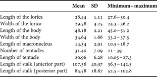
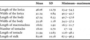
These ciliates showed a body ovoid to spherical, which was surrounded by a lorica that was prolonged posteriorly, and present a depression at its end where it was joined to the stalk. The surface of the body was not covered by the lorica, corresponding to the anterior area, and showed tentacles arranged radially. The macronucleus was spheroid and located in the body towards the posterior region. The stalk had two parts, an anterior wider with strong furrows and a posterior part narrow and tubular-shaped (Table 2; Figures 13, 20 & 21).
Biometric features of Corynophrya anisostyla (measures in µm) (N = 60).

REMARKS
The features of the ciliates observed coincide with those of this species: body spherical to ovoid, circular in transverse section, lorica surrounded the posterior half of the body, capitates tentacles from the anterior part of the body, remain outside the lorica. Stalk was divided in two parts: one anterior, close to the body, thick and flexible, and a posterior tubular, narrower than the anterior. There was a spherical macronucleus located in the posterior half of the body (Fernandez-Leborans & Gomez del Arco, Reference Fernandez-Leborans and Gomez del Arco1996).
DISTRIBUTION
Number of individuals: 0–51 per hydrocaulus. This species has been described on the hermit crabs Paguristes eremita (as Pagurus oculatus), P. prideaux, and on the braquiures Goneplax romboides and Liocarcinus maculates, on the Catalonian coast of the Mediterranean Sea (Fernandez-Leborans & Gomez del Arco, Reference Fernandez-Leborans and Gomez del Arco1996).
SYSTEMATICS (LYNN AND SMALL, Reference Lynn2000)
Phylum CILIOPHORA Doflein, 1901
Subphylum INTRAMACRONUCLEATA Lynn, 1998
Class PHYLLOPHARYNGEA De Puytorac et al., 1974
Order EXOGENIDA Collin, Reference Collin1912
Family PARACINETIDAE Jankowski, 1978
Genus Actinocyathula Corliss, 1960
Actinocyathula homari Curds, Reference Curds1987
DESCRIPTION
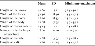
These ciliates were surrounded by a conical or triangular lorica that only covered the half of the body, while the anterior part of the body projects out from the lorica. The body was ovoid with a spherical macronucleus located in the median or posterior region of the body. The capitates tentacles are disposed from the anterior area of the body arranged radially. The stalk was located from the posterior end of the lorica, and was slender, long, reaching at least two times the length of the body (Table 3; Figures 14 & 22).
Biometric features of Actinocyathula homari (measures in µm) (N = 60).

REMARKS
The ciliates showed the features of this species: marine loricate suctorian of body ovoid protruding from the apical part of the lorica. Tentacles arranged radiating out from the anterior area of the body. Lorica mounted on a robust rigid stalk (Curds, Reference Curds1987). The only difference concerns the length of the stalk that, in our specimens, is greater.
DISTRIBUTION
Number of individuals: 0–27 per hydrocaulus. This species had been observed as epizoic on a variety of crustaceans: on the asellote isopods Munna acanthifera and Heteromesus frigidus in Icelandic waters (Ólafsdóttir & Svavarsson, Reference Ólafsdottir and Svavarsson2002); and on Paguristes eremita, Pagurus excavatus, Pagurus prideaux, Paguristes oculatus, Medorippe lanata, Parthenope angulifrons, Corystes cassivelaunus, Liocarcinus depurator, Goneplax rhomboides and Diogenes pugilator (north-western Mediterranean Sea) (Fernandez-Leborans, Reference Fernandez-Leborans2003a). Actynocyathula sp. had been found previously on Munna hansenii and Heteromesus frigidus in Icelandic waters (Ólafsdóttir & Svavarsson, Reference Ólafsdottir and Svavarsson2002).
SYSTEMATICS (LYNN AND SMALL, Reference Lynn2000)
Phylum CILIOPHORA Doflein, 1901
Subphylum INTRAMACRONUCLEATA Lynn, 1998
Class PHYLLOPHARYNGEA De Puytorac et al., 1974
Order EXOGENIDA Collin, Reference Collin1912
Family PARACINETIDAE Jankowski, 1978
Genus Actinocyathula Corliss, 1960
Actinocyathula crenata Curds, Reference Curds1987
DESCRIPTION
The ciliates showed a crenulated lorica with its surface covered by several transversal striations. From the anterior area of this lorica the body projects out and exhibits, radiating from the apical area, capitate tentacles. The lorica was cup-shaped, triangular in lateral view, and the body did not fill completely the lorica. The macronucleus was oval, located in the middle of the body. The stalk was narrow, tubular and of variable length (Table 4; Figures 15 & 23).
Biometric features of Actinocyathula crenata (measures in µm) (N = 60).

REMARKS
The characteristics of this species coincide with those of the ciliates observed: crenulated lorica with three to many transverse striations, triangular to elongate in outline, rounded in cross-section. There is a thin cup-like platform where the ovoid zooid is located and protrudes from the anterior region. Stalk slender 3–4 times the lorica length. There is a spherical and central macronucleus (Curds, Reference Curds1987).
DISTRIBUTION
Number of individuals: 0–28 per hydrocaulus. This suctorian had been recorded previously as epizoic in a variety of marine invertebrates including the hydroids Clytia volubilis, Leptoscyphus grigoriewi and Perigonimus repens and on the polychaete Aphrodite aculeata (Curds, Reference Curds1987).
SYSTEMATICS (LYNN AND SMALL, Reference Lynn2000)
Phylum CILIOPHORA Doflein, 1901
Subphylum INTRAMACRONUCLEATA Lynn, 1998
Class PHYLLOPHARYNGEA De Puytorac et al., 1974
Order ENDOGENIDA Collin, Reference Collin1912
Family ACINETIDAE Stein, 1859
Genus Acineta Ehrenberg, 1833
Acineta sulcata Dons, 1928
DESCRIPTION
The suctorians showed a crimpy lorica, orthogonal-shaped, laterally flattened, with numerous transversal furrows or striations. Inside the lorica, the body was oval to triangular in shape, narrowing to its posterior end. At the anterior region there were capitates tentacles arranged in two fascicles located in two not well-developed actinophores situated in the lateral angles. Near the posterior end of the lorica there was the stalk, narrow and tubular, its area of junction to the lorica was enlarged in a discoidal plate. The macronucleus was ovoid, located towards the posterior half of the body (Table 5; Figures 16 & 24).
Biometric features of Acineta sulcata (measures in µm) (N = 60).

REMARKS
The specimens observed showed the features of this species: small marine loricate species, with corrugated almost rectangular outline, laterally compressed. Heavily striated or transversally ridged lorica. Body with two actinophores, each bearing a fascicle of capitates tentacles. Short stalk. Spherical macronucleus (Curds, Reference Curds1985). This species can be confused with A. branchicola, but the last present two well developed actinophores, which were inconspicuous in A. sulcata. The lorica in A. branchicola is ribbed only in the posterior half, while in A. sulcata it is heavily striated or transversally ribbed along the entire length. The size of A. branchicola is up to 100 µm, and in A. sulcata up to 50 µm.
DISTRIBUTION
Number of individuals: 0–52 per hydrocaulus. This species had been previously found as epibiont on the halacarid mite Copidognathus fabriciusi, on the ostracods Hemicythere villosa and Cythereis tuberculata, in the Kiel Bat (Germany) (Curds, Reference Curds1985; Fernandez-Leborans & Tato-Porto, Reference Fernandez-Leborans and Tato-Porto2000b).
SYSTEMATICS (LYNN AND SMALL, Reference Lynn2000)
Phylum CILIOPHORA Doflein, 1901
Subphylum INTRAMACRONUCLEATA Lynn, 1998
Class PHYLLOPHARYNGEA De Puytorac et al., 1974
Order ENDOGENIDA Collin, Reference Collin1912
Family ACINETIDAE Stein, 1859
Genus Acineta Ehrenberg, 1833
Acineta corophii Collin, Reference Collin1912
DESCRIPTION
These ciliates are loricate suctorian oval to triangular-shaped, wider than long, and they were laterally compressed. The actinophores protrude from the apical region of the body in the corners, and each giving rise to a bundle of capitate tentacles. The actinophores were located laterally and the central front of the body, rounded, protruding outward. The macronucleus was ovoid and located centrally in the body. The stalk was short, narrow and generally curved, with a wide plate in the junction with the body (Table 6; Figures 17 & 25).
Biometric features of Acineta corophii (measures in µm) (N = 60).

REMARKS
They coincide in the features of this species: small marine loricate, oval to triangular in outline, always wider than deep. Laterally flattened. Two anterior lobe-like actinophores each with a fascicle of capitate tentacles. Stalk short (5–10 µm). An ovoid macronucleus (Curds, Reference Curds1985). They differ only in the length of the stalk, which was longer in the specimens observed. This species can be confused with A. branchicola, but the size of A. corophii is smaller (up to 30 µm), and the body is always wider than long in A. corophii.
DISTRIBUTION
Number of individuals: 0–11 per hydrocaulus. This species had been found previously attached to crustaceans such as copepods and on the branchiae of Corophium volutaror (as C. longicorne) in Roscoff (France) (Collin, Reference Collin1912; Curds, Reference Curds1985; Fernandez-Leborans & Tato-Porto, Reference Fernandez-Leborans and Tato-Porto2000b).
SYSTEMATICS (LYNN AND SMALL, Reference Lynn2000)
Phylum CILIOPHORA Doflein, 1901
Subphylum INTRAMACRONUCLEATA Lynn, 1998
Class OLIGOHYMENOPHOREA De Puytorac et al., 1974
Subclass PERITRICHIA Stein, 1859
Order SESSILIDA Kahl, 1933
Family ZOOTHAMNIIDAE Sommer, 1951
Genus Zoothamnium Bory de St Vincent, 1826
Zoothamnium sp.
DESCRIPTION
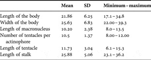
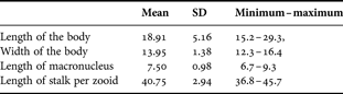
These ciliates were colonial, with an oval body when contracted, and a branched stalk with a spasmoneme that continued in the entire colony. The macronucleus is ‘C' shaped located in transversal position in the body (Table 7; Figures 18 & 26).
Biometric features of Zoothamnium sp. (measures in µm) (N = 60).
REMARKS
The ciliates coincide with the features of the genus: zooids with contractile myoneme (spasmoneme) that compresses on contraction into zigzag folds in one plane. Zooids arranged in colonial forms, sharing continuous spasmoneme that runs throughout the entire colony, so that the entire colony is contractile. Zooids slenderly campanulate, peristomial disc slightly slanting, constricted beneath peristomial collar, smooth pellicle; often bent over or held at angle to stalk. Macronucleus elongate C- to horseshoe-shaped, transverse to main body axis. One contractile vacuole at ventral wall of vestibulum. Colonies with few (about 10) zooids on dichotomously branched stalk, up to 550 µm high. Main stalk long, lateral stalks short (1 zooid/branch), without longitudinal striations (Kahl, Reference Kahl and Dahl1935; Lynn, Reference Lynn2008).
DISTRIBUTION
Number of individuals: 0–12 per hydrocaulus.
Spatial distribution of the epibionts on the basibiont surface
In the specimens infested the higher colonization was observed on the shell, which showed the majority of the species (90%). The shell also presented the highest values of number of individuals, belonging to the hydrozoan epibionts (Table 8).
Number of individuals of each epibiont species (minimum–maximum) on the shell and crab (N = 60).

EPIBIONTS ON THE SHELL
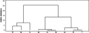
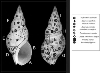
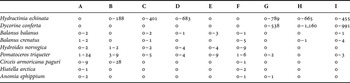
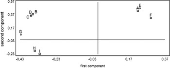
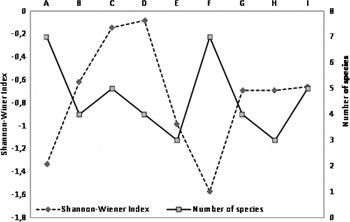
The epibionts on the shell were distributed on all the areas, although the more abundant species, the hydrozoans, were only observed on the 66.6% of the areas (Table 9). From these hydrozoan species the species with more wide location was Hydractinia echinata, which appeared in 6 of the 9 areas, whilst Dicoryne conferta only was recorded in 33.3% of the areas. Figure 27 shows the dendrogram of the hierarchical cluster analysis with the different areas of the shell (according to the mean number of individuals of the epibiont species) grouped in three clusters: (1) it included 33.3% of the areas (A, E and F), those presented the high abundance of epibionts (mean 1.6 individuals per shell); (2) a cluster with 33.3% of areas B, C and D, counting those with a mean abundance of epibionts (mean 24.68 individuals per shell); and (3) a cluster with areas (also 33.3%) showing high number of epibionts (mean 85.37 individuals per shell): these areas were G, H and I. Figure 28 shows the presence of the different epibiont species on the areas of the shell. The highest number of species was recorded at areas A and F, which were areas where no hydrozoan species were observed. All the species of the other groups appeared represented in these areas, which were located near the aperture of the shell (A), and separated from that (F). With great difference the highest number of epibionts corresponded to the hydrozoan species, and the major abundance was observed in areas G, H and I, located along the edge from the aperture to the apex of the shell. Other areas with a remarkable number of individuals were C and D, whereas in hydrozoans only Hydractinia was observed.
Dendrogram of the hierarchical cluster analysis showing the areas of the shell and performed with the mean number of individuals of the different epibiont species (metric distance: City Block (Manhattan); clustering method: Ward).

Image of the shell with the presence of the epibiont species on the different areas.

Number of epibionts of each species on the different areas of the shell (minimum–maximum) (N = 60).

The principal component analysis, performed with the mean values of the number of epibionts of each species on the different areas of the shell, indicates that there were three clusters in which the areas included revealed a similar distribution of the epibiont species: (1) the areas A, E and F; (2) the areas C, B, and D; and (3) the areas H and I (Figure 29). In each cluster the areas correlated (P ≤ 0.05).
Graphic of the principal component analysis performed with the number of individuals of the epibiont species, showing the different areas of the shell.

Figure 30 shows the diversity index in the different areas of the shell. There was not a significant correlation between the number of species and the diversity (−0.56; P ≤ 0.05). There was a statistically significant difference between the means of the two variables (t – 10.30; P ≤ 0.05). On the other hand, the analysis of variance indicated that there was no statistically significant relationship between the variables (P: 0.1117). The areas with high diversity were C and D, while in the case of areas A and F diversity and species number showed an opposite trend with high number of species and low diversity.
Shannon–Wiener diversity index and fluctuation of the number of species in the areas of the shell.

EPIBIONTS ON THE CRAB
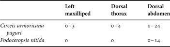
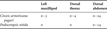
Table 10 shows the number of epibionts on the colonized areas of the crab. Circeis armoricana was the species most widely distributed, whereas Pododeropsis nitida only was recorded on the abdomen of the crab.
Number of epibionts of each species on the crab (minimum–maximum) (N = 60).
Spatial distribution of ciliate hyperepibionts on Dicoryne conferta
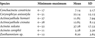
On the surface of the shells and crabs, ciliate epibionts were not observed, which were found only on the surface of the hydrozoan Dicoryne conferta located on the shell. Table 11 shows data about the number of ciliates on the surface of Dicoryne conferta. The dendrogram of the principal component analysis (Figure 33) performed with the abundance of ciliate epibionts indicated the existence of three clusters: (1) included the ciliate species with low number of individuals (mean 6.21 ciliates per hydrocaulus) and accounted for 42.86% of the species (Acineta corophii, Conchacineta constricta and Zoothamnium sp.); (2) 20.57% of the species were integrated in this cluster, with a median number of epibionts (mean 12.35 individuals per hydrocaulii) (the two species of Actinocyathula: A. crenata and A. homari); and (3) in this cluster were located the species with high abundance (mean 29.71 ciliates per hydrocaulus), also 20.57% of species (Acineta sulcata and Corynophrya anisostyla).
Number of ciliate epibionts on Dicoryne conferta (N = 60).

DISCUSSION
The hydrozoans found in this study (Hydractinia echinata and Dicoryne conferta), as well as the polychaetes (Hydroides norvegica, Pomatoceros triqueter and Circeis armoricana) had been well described previously as epibionts on Pagurus bernhardus (Reiss et al., Reference Reiss, Knäuper and Krönche2003). Also, Balanus crenatus has been found on P. bernhardus, although Balanus balanus had not been found previously as epibiont on this species, nor on other hermit crabs (Reiss et al., Reference Reiss, Knäuper and Krönche2003; Williams & McDermott, Reference Williams and McDermott2004), with the exception of a study of this crab in the same geographical area (Fernandez-Leborans & Gabilondo, Reference Fernandez-Leborans and Gabilondo2006). Anomia ephippium, Hiatella arctica and Podoceropsis nitida had been also found before in this crab (Reiss et al., Reference Reiss, Knäuper and Krönche2003; Williams & McDermott, Reference Williams and McDermott2004).
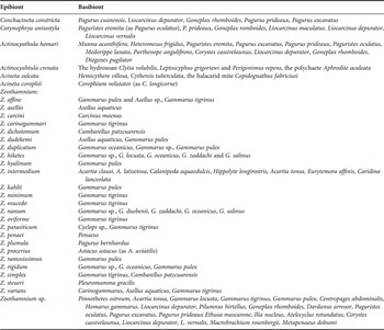
Table 12 shows the basibionts indicated in the literature of the hyperepibiont species recorded in the present study. The genus with highest number of basibiont species is Zoothamnium. Only the species Zoothamnium plumula had been found on P. bernhardus. Also, one species, Actinocyathula crenata, has been recorded on hydrozoan species (Clytia volubilis, Leptoscyphus grigoriewi and Perigonimus repens), but not attached to a hermit crab. Other ciliate hyperepibiont species had not been found previously on hydrozoans, or in Pagurus bernhardus. Similar secondary epibionts of the genera Zoothamnium and Corynophrya had been described on the bryozoans Triticella flava, located on the decapod Goneplax rhomboides from the north-western Mediterranean coast (Fernandez-Leborans, Reference Fernandez-Leborans2003b).
Hyperepibiont species found in the study and their basibiont species from the literature (Curds, Reference Curds1985, Reference Curds1987; Fernanadez-Leborans & Tato-Porto, Reference Fernandez-Leborans and Tato-Porto2000a, b; Fernandez-Leborans, Reference Fernandez-Leborans2009).

The abundance of the different epibiont species on the shell is similar to that shown in previous surveys. It is the case of the polychaetes Circeis armoricana and Hydroides norvegica, which present a number of epibionts comparable to that of previous studies (Reiss et al., Reference Reiss, Knäuper and Krönche2003), although the number of cirripeds is lower in the present study, and the amphipod Podoceropsis nitida was not recorded on the shell, only on the crab. There are few papers about the distribution of epibionts in different areas of the shell. In a previous study (Fernandez-Leborans & Gabilondo, Reference Fernandez-Leborans and Gabilondo2006) the most colonized areas of the shell were, in one year E, H and G, and in another year G, H and A, while in the present study these areas were H, I and G. An important difference corresponds to the area I, which in previous studies was little colonized, with a low abundance of hydrozoans, which, in contrast, were present in high numbers in the present study. The colonization on the shell seems to follow a trend consisting in location of an important number of epibionts on the edge above and near the aperture of the shell, in the areas G, H and I, and this tendency was accompanied by the occupation of the areas surrounding the aperture and near the edge indicated (D, C, and B). The preference in the colonization according to an edge of the shell, or with respect to the aperture, may be related to the comparative position of the crab and shell, and the proximity of the different parts of the shell to the substrate.
The high values of the diversity index in areas C and D of the shell was due to a moderate number of species in these areas coinciding with a relatively high number of epibionts of the hydrozoan Hydractinia echinata. These areas are located near the edge of the shell, where an important abundance of epibionts was observed. The areas A and F, showing an opposite trend between diversity and number of species, were characterized by their relative presence of a great number of species, but the species (hydrozoan species) with the highest number of individuals on other areas, were absent on areas A and F.
With respect to Podoceropsis (Gammaropsis) nitida we have not observed in any case these amphipods living in the tubes secreted by the third and fourth pereiopods (Dixon & Moore, Reference Dixon and Moore1997). The amphipods found were located always laterally attached to the abdomen of P. bernhardus.
It is difficult to explain the reasons for the presence of the hyperepibiont ciliates on the surface of the hydrozoan Dicoryne conferta, especially when all these species, excepting Actinocyathula crenata, were previously observed as epibionts on crustaceans. In the present study they only appear on the hydrozoan and not in the surface of the shell or the crab. This ciliate community seems to grow without apparent predation from the hydroids, and possibly the high colonization by Dicoryne on wide areas of the shell arises from the lack of available space for ciliates, and the change of location of ciliates to a hyperepibiosis on the hydrozoan. Epibiont species of diverse groups (bacteria, microalgae, foraminifera and polychaetes) have been described on hydrozoans (Bavestrello et al., Reference Bavestrello, Cerrano, Di Camillo, Puce, Romagnoli, Tazioli and Totti2008), and among them some ciliate protozoan species, such as the peritrich ciliate Vorticella, the suctorian Ephelota, the hypotrich ciliate Kerona pediculus and the licnophorid Licnophora chattoni (Warren & Robson, Reference Warren and Robson1998; Bavestrello et al., Reference Bavestrello, Cerrano, Di Camillo, Puce, Romagnoli, Tazioli and Totti2008; Silva-Neto et al., Reference Silva-Neto, Silva, Pedroso, Alexandre and Esteves2012). However, there are no data in the literature about the presence of the ciliate species found in the present study on Dicoryne. With the exception of Actinicyathula crenata, observed on Clytia, none of these species had been observed on other cnidaria. With the data from the literature, it seems that the ciliates found in the study had not been observed previously as hyperepibiont on cnidarians attached to crustacea.
ACKNOWLEDGEMENTS
We thank Dr Fiona Hannah and the personnel of the University Marine Biological Station Millport (Isle of Cumbrae, Scotland) for their help in technical work in sampling, initial identification of crabs, and for the kind welcome and warm hospitality enjoyed in their laboratory; Dr Regina Gabilondo for her collaboration in the sampling; Dr Peter Schuchert (Muséum d'Histoire Naturelle, Geneva, Switzerland) for his indispensable and very generous help with the identification of the hydroid epibionts; Dr Henning Reiss (Senckenberg Institute, Department for Marine Research, Germany) for the identification of polychaete and bivalve species; and Dr Wim Vader (Tromsø Museum, Norway) for his help in the identification and additional information on the amphipod epibiont).