Introduction
Otosclerosis is a focal bone disease that affects the otic capsule. It is a prevalent cause of hearing loss and, less frequently, of other audio-vestibular symptoms in adults.Reference Lee, Aviv, Chen, Nedzelski, Fox and Symons1,Reference Vicente, Yamashita, Luiz, Albernaz, Penido and Paulo2 The diagnosis is usually clinical and supported by audiometry, which usually shows a conductive hearing loss in the affected ear.
High-resolution computed tomography (CT) scans of temporal bones are capable of demonstrating suggestive findings of otosclerosis in many patients.Reference Vicente, Yamashita, Luiz, Albernaz, Penido and Paulo2 There is also growing evidence of the relationship between some high-resolution CT data and the clinical manifestation of the disease. However, the studies are very heterogeneous, and the literature on this subject remains scarce.Reference Lee, Aviv, Chen, Nedzelski, Fox and Symons1,Reference Min, Chung, Young, Sun, Hwa and Jin3 Investigating the relationship between high-resolution CT findings and clinical features in otosclerosis might contribute to the current understanding of the pathophysiology of the disease and potentially improve the follow up of patients in terms of prognostic prediction and management.
This study aimed to determine the impact of the radiological findings on the audiometric results in treatment-naïve patients with otosclerosis.
Materials and methods
This was a retrospective case-control study, in which medical records of patients undergoing stapedotomy for the treatment of hearing loss secondary to otosclerosis between 2018 and 2020 in a tertiary care hospital were reviewed. General, audiometric and high-resolution CT data of eligible patients were systematically collected. The obtained information was gathered, examined and analysed, considering each ear separately. The study was approved by the Institutional Ethics and Research Committee (approval certificate number: 11483219.1.0000.0096).
Ears of patients over 18 years old, with clinical suspicion of otosclerosis based on audiometric and stapedial reflex testing results, who underwent primary stapedotomy and whose surgery confirmed only stapes fixation were included. All ears were operated on by the same surgeon under local anaesthesia and sedation using a microscope-guided transcanal approach.
Ears of patients whose high-resolution CT scans were not obtained following the predefined protocol for imaging acquisition and processing and in the same institution of the study were excluded. In addition, those who did not undergo tonal audiometry at the predetermined time points, or did it in institutions other than that of the clinical and surgical follow-up institution were also withdrawn from analysis.
The ears of the included patients were all submitted to audiometry in the same institution pre- and post-operatively, with the same speech therapist. We also used the same audiometer (Diagnostic Audiometer, model AD 229; Interacoustics, Middlefart, Denmark). The equipment was calibrated according to the manufacturer's guidelines, and at most six months before use. Only ears whose pre-operative audiometric assessments were performed within three months of the surgery were considered eligible.
As for the audiometric examination, we systematically searched air-conduction pure-tone hearing thresholds at 0.5, 1, 2, 3, 4, 6 and 8 kHz and bone-conduction pure-tone hearing thresholds at 0.5, 1, 2, 3 and 4 kHz for each ear and with the due contralateral masking, according to the Hood technique.Reference Wolfovitz and Luntz4 The reference values for pure-tone audiometry in this study followed the criteria established by Silman and Silverman in 1997.Reference Silman and Silverman5 The simple pure tone average (PTA) was calculated as the simple mean of the air-conduction thresholds at 0.5, 1 and 2. The high-frequency PTA was calculated as the simple mean of the air-conduction thresholds at 4, 6 and 8 kHz. For bone-conduction pure-tone thresholds, the simple mean of the results at 0.5, 1 and 2 was used.
The high-resolution CT images analysed for this study should have been obtained within three months prior to stapedotomy. They were obtained in a matrix of 512 × 512 pixels, using 256-channel equipment and with a slice thickness of 1 mm. Each side was considered separately. After being obtained, they were transferred to a workstation for post-processing. The set of images was then reformatted in axial, coronal or oblique planes. The dataset from the examinations was filed and encrypted before passing them on to a radiologist experienced in temporal-bone evaluation. Thus, to ensure the blinding of the imaging analysis, each set of images was given a unique random number for identification. The correlation of these numbers with the corresponding ear was kept under the knowledge of the main researcher only. For the evaluation of each radiological parameter studied, specific and standardised protocols were used, which are indicated below. The image viewer used was the RadAnt DICOM viewer 2020.1 (Medixant, Poznan, Poland).
In general, disease foci were considered based on the presence of regions of decreased radiological density (otospongiotic or active focus), or abnormalities in the contour or volume of the structures of the otic capsule (otosclerotic or inactive focus). First, according to the general evaluation of the images, the radiologist considered the examination compatible or not with the otosclerosis diagnosis. Then, some objective parameters were systematically evaluated and used to stratify the sample. These parameters are dissected below.
Number of regions affected by disease
Twelve sites were systematically searched for disease foci: fissula ante fenestram, stapes footplate, basal turn of the cochlea, middle turn of the cochlea, round window, promontory, labyrinthine segment of the facial nerve, tympanic segment of the facial nerve, semicircular canals, vestibule, cochlear aqueduct and internal auditory canal. The number of disease sites for each ear was then determined by the summation of affected regions. Then the ears were divided into three categories according to the results: less than or equal to three, between four and six, or equal to or more than seven regions of involvement.
Disease pattern according to foci location
The ears without pathological findings in the high-resolution CT were excluded from this analysis. The remaining ears were then grouped according to the location of the observed foci into those with isolated fenestral otosclerosis, those with isolated retrofenestral disease and those with mixed involvement.
Disease pattern according to foci characteristics
Likewise, those without pathological high-resolution CT findings were excluded from this analysis. In the presence of radiological disease, the ears were divided according to the type of foci. Thus, in this analysis three patterns were possible: an active-only (otospongiosis), inactive-only (otosclerosis) or a mixed pattern, with concomitant findings of both radiological types of foci.
Extension to the cochlear endosteum
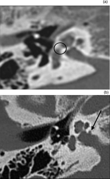
Endosteal extension was considered when the ante fenestral focus, if present, reached the cochlear internal lumen. In order to evaluate this finding, the radiologist used an axial section at the level of the oval window (Figure 1a). The ears were then grouped into those with or without radiological cochlear extension of ante fenestral disease.
High-resolution computed tomography scan sections of the right temporal bone in the axial plane at the level of the oval window. (a) Shows the presence of endosteal extension of the ante fenestral focus of the disease (black circle). (b) Shows the presence of an internal auditory meatus diverticulum, the so-called ‘nipple sign’ (black arrow)

Presence of internal auditory canal diverticula
This finding was sought in two sections: in an axial section at the level of the anteroposterior axis of the internal auditory canal (Figure 1b) and in a coronal section including the craniocaudal axis of this structure. A diverticulum was considered in the presence of a focus of decreased density in continuity with the wall of the internal auditory canal (‘nipple sign’). Then, the ears were divided according to the presence of the finding.
For each sample grouping listed above, the effect of the high-resolution CT variable on each audiometric parameter (PTA, high-frequency PTA and bone conduction PTA) was assessed.
The numerical data obtained were summarised according to their mean and standard deviation (SD). For the investigation of the normal distribution by the numerical data of the different statistical evaluations performed, the Shapiro–Wilk test was used. The t- and Mann–Whitney tests were used for statistical comparison of quantitative variables (PTA, high-frequency PTA and bone conduction PTA) between two groups, according to their normality profile. For comparisons of the same quantitative data, although involving three groups, analysis of variance (ANOVA) was used. In case of significance of this analysis, the Tukey's post-hoc test was used to perform pairwise comparisons.
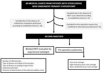
All statistical tests were bilateral, and a significance level of 5 per cent (p < 0.05) was adopted. Data were collected and tabulated in Microsoft Excel® spreadsheet software (2016 version). For statistical analyses, R statistical computing software (version 4.0.2) was employed. A graphical summary of the study design is shown in Figure 2.
Study flow chart. HRCT = high-resolution computed tomography

Results
Eighty-nine patients had their medical records analysed, and 40 met the eligibility criteria for the study and had their clinical, audiometric and tomographic data studied. None of them underwent bilateral stapedotomy within the study time frame, so the final sample included 40 ears.
Before the procedure, the mean values for PTA, high-frequency PTA and bone conduction PTA were 55.83, 55.54 and 27.45 dB HL, respectively. Thirty-six ears (90 per cent) showed findings compatible with otosclerosis on radiological analysis, and four were considered normal.
Number of regions affected by otosclerosis
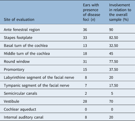
Eight ears (20 per cent) in the case group presented three or fewer regions affected by otosclerosis foci. A total of 24 ears (60 per cent) had 4 to 6 sites of involvement, and the remaining 8 (20 per cent) had 7 or more regions with findings consistent with the disease on their high-resolution CT scans. Table 1 shows the number of ears with findings compatible with otosclerosis for each site within the otic capsule.
High-resolution computed tomography involvement of each anatomical site

The mean pre-operative PTA was 51.87 (SD: 8.65) dB HL for the group with equal to or less than 3 affected sites, 55.28 (SD: 9.20) dB HL for the group with 4 to 6 affected sites, and 61.46 (SD: 20.63) dB HL for the group with 7 or more affected sites. Despite the increasing mean for the PTA according to the number of affected regions, there was no statistical difference when comparing the groups (p = 0.281; ANOVA test).
For the pre-operative high-frequency PTA, the mean value was 56.67 (SD: 16.76) dB HL for group with equal to or less than 3 affected sites, 51.25 (SD: 18.48) dB HL for the group with 4 to 6 affected sites and 67.29 (SD: 27.46) dB HL for the group with 7 or more affected sites. There was also no statistically significant difference among the groups for the results obtained (p = 0.163, ANOVA test).
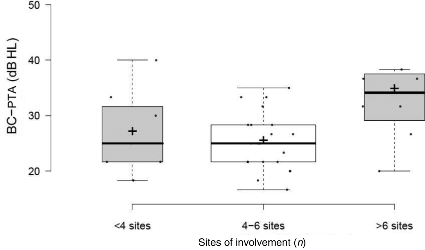
For the pre-operative bone conduction PTA, the mean value was 26.87 (SD: 7.15) dB HL for the group with equal to or less than 3 affected sites, 25.28 (SD: 4.81) dB HL for the group with 4 to 6 affected sites and 34.58 (SD: 10.22) dB HL for the group with 7 or more affected regions on high-resolution CT scan. There was a statistically significant difference in the results found (p = 0.006; ANOVA test, Figure 3).
Distribution of the bone conduction pure tone average according to the number of sites of involvement for otosclerosis on high-resolution computed tomography scans. BC-PTA = bone conduction pure tone average

After post-hoc analysis, it was observed that the aforementioned significant difference occurred between the group with three or less and the group with seven or more regions affected (p = 0.031, Tukey test) and between the group with four to six and the group with seven or more sites of involvement (p = 0.008, Tukey's Test). In these cases, the group with the highest number of affected regions presented significantly increased values for the variable. There was no significant difference between the groups with three or less and those with four to six sites with pathological high-resolution CT findings (p = 0.848, Tukey test).
Disease pattern according to disease foci location
As for the classification of the disease according to the pattern of location of the foci on high-resolution CT scans, 14 (35 per cent) ears had purely fenestral otosclerosis, and 22 (55 per cent) were diagnosed with mixed disease. Four ears had normal high-resolution CT scans and were therefore excluded from this analysis.
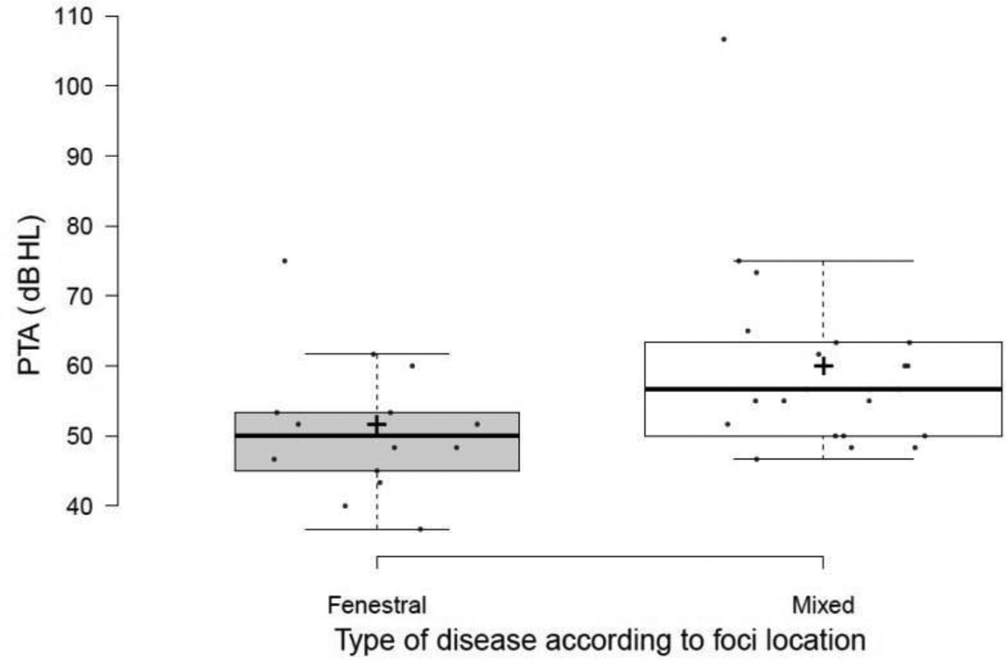
When comparing the pre-operative PTA, the group with mixed findings (mean: 59.47, SD: 13.0 dB HL) had significantly increased results (p = 0.019, Mann–Whitney test, Figure 4) than those in the group with fenestral-only disease (mean: 51.07, SD: 9.75 dB HL) on high-resolution CT assessment.
Simple pure-tone average distribution according to the disease involvement pattern on high-resolution computed tomography scan, in relation to being fenestral or mixed. PTA = pure tone average

Ears with purely fenestral involvement had mean high-frequency PTA and bone conduction PTA of 52.74 (SD: 19.48) and 26.19 (SD: 3.72) dB HL, respectively. For ears with mixed high-resolution CT findings, these values were 56.06 (SD: 21.97) dB HL and 27.95 (SD: 8.88) dB HL, respectively. There were no statistically significant differences in the comparison of these variables between the groups (p-value for high-frequency PTA = 0.682; p-value for bone conduction PTA = 0.912; Mann–Whitney test).
Disease pattern according to type of lesion
As for classification of the disease according to the type of lesions defined on the high-resolution CT scans, 18 (45 per cent) ears presented findings of otosclerosis exclusively in the active stage (otospongiosis), and 18 (45 per cent) were diagnosed with mixed disease. Four ears had normal high-resolution CT and were therefore excluded from this analysis.
When comparing the pre-operative audiometric data, there was no statistically significant difference among the ears with active-only foci and those with mixed findings on high-resolution CT. For these groups, the mean PTA reached 52.87 (SD: 6.97) dB HL and 59.54 (SD: 15.65) dB HL, respectively (p = 0.242, Mann–Whitney test). Concerning the mean high-frequency PTA, the values found were 51.02 (SD: 14.22) dB HL for those with active-only foci and 58.52 (SD: 25.68) dB HL for those with both active and inactive foci (p = 0.143, t-test). The mean bone conduction PTA for the group with only active foci was 25.74 (SD: 5.40) dB HL, whereas for the group with mixed disease this value reached 28.80 (SD: 8. 68) dB HL (p = 0.267, Mann–Whitney test).
Extension to the cochlear endosteum
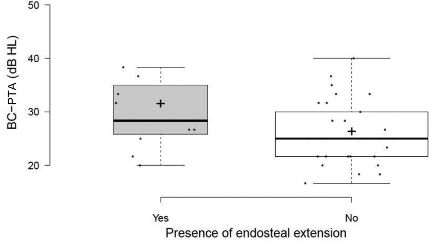
Eleven ears (27.5 per cent) showed radiological signs of endosteal extension of the disease. Among them, the mean values for pre-operative PTA, high-frequency PTA and bone conduction PTA were 60.30 (SD: 17.06) dB HL, 62.42 (SD: 25.19) dB HL and 31.21 (SD: 9.78) dB HL, respectively. For the remaining group, without endosteal involvement, the respective values were: 54.13 (SD: 9.66) dB HL, 52.93 (SD: 18.51) dB HL and 26.03 (SD: 5.90) dB HL. There was no statistically significant difference between these groups regarding the PTA (p = 0.379, Mann–Whitney test) and the high-frequency PTA (p = 0.332, Mann–Whitney test). Pre-operative bone conduction PTA values among ears with endosteal extension were worse, even though they did not reach strict statistical significance (p = 0.047, t-test, Figure 5).
Distribution of the mean bone conduction PTA according to the presence of endosteal extension. BC-PTA = bone conduction pure tone average

Presence of internal auditory canal diverticula
Eight (20 per cent) ears presented internal auditory canal diverticula. Within this group, the audiometric data had a mean PTA of 59.37 (SD: 11.92) dB HL and high-frequency PTA of 58.96 dB HL (SD: 15.56) dB HL, similar to those of the group of ears without the finding (PTA: 54.95, SD: 12.32; high-frequency PTA: 58.98, SD: 15.56 dB HL). Furthermore, no statistical significance was reached when comparing the values of these variables between the groups (PTA, p = 0.211; high-frequency PTA, p = 0.368; Mann–Whitney test). Mean pre-operative bone conduction PTA of the group of ears with diverticula was 25.41 (SD: 5.69) dB HL, lower than that found for the rest of the sample (mean: 27.97, SD: 7.78 dB HL). Nonetheless, there was also no significant difference among the values obtained for the groups (p = 0.379, Mann–Whitney test).
Discussion
Otosclerosis is a condition that primarily leads to hearing loss in working-age adults. Improving the management of this disease is therefore a public health issue, which might be reached by refining the current understanding of its pathophysiology and clinical evolution. The use of high-resolution CT has the potential to enhance not only the diagnostic performance but also the investigation of the particular manifestations of the patients. These advances in the understanding of the value of high-resolution CT in otosclerosis might in turn potentially contribute to the judicious establishment of prognoses, prediction of surgical outcomes, and the indication of current and future clinical therapies. So far, however, the association between radiological findings and the degree of hearing loss is controversial, and there is a considerable heterogeneity across the studies in the methods used for disease grading and for obtaining the radiological parameters used for comparison.Reference Wolfovitz and Luntz4 As a result, carrying out further investigations on the subject is paramount.
Otosclerosis typically affects the temporal bone in an irregular, heterogeneous and particular way. Thus, there are cases in which the problem affects limited sites in the otic capsule, without associated clinical manifestations (the so-called ‘histological otosclerosis’)Reference Silman and Silverman5. When the foci impair the functioning of the peripheral hearing organ, however, the classical auditory complaints occur.
One might suppose that the presence of an increasing number of affected regions in the otic capsule would lead to more severe audiometric manifestations. This hypothesis was tested in the present study by comparing the audiometric variables among groups of ears established by the number of sites of the otic capsule with disease foci in high-resolution CT. It should be highlighted that the dimension or characteristics of the foci were not considered, which could have an impact on the results. As for the stratification obtained, a symmetrical pattern was noted, since 60 per cent of the ears showed moderate spread (4–6 sites), with another 20 per cent showing mild spread (3 or fewer sites) and the remaining 20 per cent with severe spread (more than 6 sites). As expected, the most frequent site of injury was the fissula ante fenestram, which was present in all cases (90 per cent) with high-resolution CT compatible otosclerosis. Furthermore, in general, patterns of involvement were more frequent in the more lateral and anterior structures of the otic capsule (fissula ante fenestram, oval and round windows, promontory), with relative preservation of those that were more medial and posterior (semicircular canals, internal auditory canal, cochlear aqueduct).
Neither the pre-operative PTA nor the high-frequency PTA were significantly different among the groups obtained after stratifying the sample by the number of sites with disease foci. On the other hand, when assessing the bone-conduction thresholds, ears with seven or more sites of involvement showed significantly higher values for the bone conduction PTA compared with the other groups. Taken together, these data show that the radiological spread of otosclerosis seems to hinder, particularly among those with more disseminated conditions, the mechanisms of mechano-electrical transduction. In these cases, it could be assumed that the involvement of multiple regions of the otic capsule would impair the function of the cochlear hair cells or auditory nerve. Consistent with the findings, the increasing involvement of the otic capsule would not per se affect the mechanical transmission of the sound to the cochlea, as this kind of impairment seems to rely only on the involvement of particular sites involved in this mechanism (e.g., the footplate and round window).
Otosclerosis is more frequently divided into two types: fenestral and retrofenestral. The former is more typical and determined by the presence of lesions located on the lateral wall of the otic capsule. In the retrofenestral type, only otosclerotic foci in medial structures of the labyrinthine capsule, including the pericochlear region, semicircular canals, internal acoustic canal, vestibule, and cochlear and vestibular aqueducts are seen.Reference Swartz, Faerber, Wolfson and Marlowe6,Reference Valvassori7 A theoretical relationship between the site of involvement and the pathophysiological mechanisms of hearing loss presented in otosclerosis would be probable. Thus, the involvement of the annular ligament would lead to stapes fixation and explain the conductive component of the typical hearing loss of the disease observed in the fenestral type.Reference Swartz, Faerber, Wolfson and Marlowe6 The pathological process would gradually extend to involve the footplate and subsequentially progress medially, affecting the inner ear. As a result of the involvement of the cochlea and surrounding structures, there would be mixed hearing loss or, more rarely, isolated sensorineural hearing loss observed in the retrofenestral and mixed types of otosclerosis.Reference Causse, Uriel, Berges, Shambaug, Bretlau and Causse8
The pattern of radiological involvement of the temporal bone in otosclerosis can be easily defined according to the high-resolution CT data of the location of the foci. In the present study, no pure retrofenestral patterns were observed, which possibly reflects the study sample. A total of 55 per cent of the ears evaluated in the case group had mixed disease. This data reflects a relevant finding in the ears of patients with an indication for stapedotomy: most of them also have signs of disease in more medial portions of the otic capsule. The surgery in these very frequent cases would not impact the long-term audiological prognosis in terms of sensorineural impairment, a situation both surgeon and patient should be aware of.
Significantly higher values of PTA were obtained among those with mixed disease, which could reflect more advanced disease. However, this result was not replicated when analysing high-frequency PTA and bone conduction PTA. Thus, a puzzling fact could be seen: the presence of more medial foci in the otic capsule would not itself indicate greater involvement of the cochlear hair cells or auditory neurons. In other words, these foci might be present without causing sensorineural damage. However, retrofenestral involvement appears to be a marker of greater damage to the air-conduction mechanisms. Previous studies on this subject have controversial results, which usually vary according to their methods, especially in the definition of ‘retrofenestral involvement’. In general, studies in which this medial involvement of the otic capsule is defined by stricter criteria, such as loss of integrity or anatomical distortions of the cochlea, usually demonstrate a greater influence of the findings on audiometric parameters.Reference Kiyomizu, Tono, Yang, Haruta, Kodama and Komune9–Reference Shin, Fraysse, Deguine, Cognard, Charlet and Sévely13
• High-resolution computed tomography scans might help explain some aspects of hearing impairment in otosclerosis
• Ears with disease and the presence of endosteal extension showed a trend towards reduced average bone-conduction hearing thresholds
• Ears with disease and a higher number of affected sites within the otic capsule present significantly worse average bone-conduction hearing thresholds
• Ears with disease and associated retrofenestral involvement demonstrated higher values for the pure tone average
• Presence of internal auditory canal diverticula does not seem to impact the audiometric findings in otosclerosis
• These results might mirror pathophysiological phenomena and could be useful when managing the patients
Similarly, it was investigated whether the types of radiological foci, active, inactive or mixed, would influence the clinical manifestation of the disease. The means of pre-operative audiometric parameters were higher among ears with mixed disease, which could reflect diseases with a longer evolution time in which otospongiotic and otosclerotic phases would coexist. However, when comparing the values found for such variables between the groups, there was no statistical difference. Thus, it is not possible to say that the presence of more mature disease foci has a considerable impact on the peripheral mechanisms of hearing or that diseases with longer evolution necessarily present worse hearing thresholds.
The endosteal extension of the foci located in the fissula ante fenestram is sometimes referred to as a possible factor implicated in the manifestation of the disease. Histopathological studies of temporal bones associated this finding with the occurrence of greater damage to the organ of Corti and cochlear neuroepithelium, with consequent diminished bone-conduction thresholds.Reference Kwok and Nadol14,Reference Parahy and Linthicum15 This relationship is, however, a matter of discussion and has not been replicated by others.Reference Schuknecht and Barber16,Reference Elonka and Applebaum17 The association of findings from histological studies with audiometric data is somewhat biased as there is often a large and varied difference between the time of the last audiometric assessment and the individual's disease. Few investigations have used high-resolution CT to assess this correlation in vivo.
In this study, the mean pre-operative values of PTA, high-frequency PTA and bone conduction PTA were higher among ears with endosteal extension. Nevertheless, after statistical analysis, only the values obtained for the bone conduction PTA were significantly different for those with this radiological finding. This result replicates those from a study with a large number of patients conducted by Dudau et al.Reference Dudau, Salim, Jiang and Connor18 Furthermore, it indicated that the cochlear extension of ante fenestral foci would possibly indicate greater damage to the neuroepithelium and consequent sensorineural hearing loss. The mechanism of these damages has been widely hypothesised and might entail, for instance, the effect of inflammatory cytokines, the action of proteolytic enzymes and the involvement of the spiral ligament.Reference Causse, Uriel, Berges, Shambaug, Bretlau and Causse8,Reference Chole and McKenna19,Reference Sziklai, Batta and Karosi20
One fifth of the ears in this study had internal auditory canal diverticula on high-resolution CT scan assessment. It is believed that they might be a local manifestation of the disease. In accordance with the results obtained by Wang et al. and Puac et al., the presence of this finding did not significantly impact pre-operative audiometric findings.Reference Wang, Yoshida, Shimono, Sugimoto, Teranishi and Naganawa21,Reference Puac, Rodríguez, Lin, Onofrj, Lin and Hung22
In general, it could be said that the location and size of otosclerotic foci seem to be worse predictors of sensorineural damage than the endosteal involvement and loss of cochlear wall integrity on high-resolution CT scans.Reference Lee, Aviv, Chen, Nedzelski, Fox and Symons1,Reference Marshall, Fanning, Symons, Shipp, Chen and Nedzelski23 In addition, the existence of histological otosclerosis without clinical correspondence is in line with these observations.Reference Silman and Silverman5,Reference Iyer and Gristwood24
There are several limitations one should bear in mind when analysing our findings. First, it is a retrospective study. As such, it relies on the analysis of medical records, which are often subject to failures and inconsistencies. The strict criteria for study eligibility aimed to reduce the heterogeneity of inclusion, measurement and follow up in order to obtain a uniform sample. However, there was a large sample loss as a side effect. Thus, because of the lack of follow up, many potential participants could not be included, which might result in selection bias. Furthermore, there is a relevant sample heterogeneity, which aimed to include ears in different phases and with a broad range of disease severity. This feature, however, more effectively reflects the context of clinical practice.
When selecting the ears, only those with surgical indication were investigated. Therefore, one must understand the results obtained within this scenario and avoid generalising the conclusions to all cases of otosclerosis. Thus, for cases of mixed or conductive hearing loss with lower air–bone gaps or subclinical otosclerosis, the results presented are possibly less accurate.
Finally, it should be considered that the study did not aim to investigate the reproducibility and agreement of the high-resolution CT analysis performed. Thus, whenever possible, standardised and quantitative investigations were favoured. In addition, we submitted all scans to the same experienced radiologist. Yet, it is reasonable to ponder the possibility of measurement bias.
Conclusion
High-resolution CT scans might help explain some clinical features in otosclerosis. Ears with the disease and the presence of endosteal extension or with a greater number of affected sites within the otic capsule showed worse average bone-conduction hearing thresholds, although the results only strictly reached statistical significance for the latter. Moreover, ears with associated retrofenestral involvement had higher values for the PTA. These results might reflect pathophysiological phenomena and be helpful when evaluating an affected patient. Prospective studies with larger samples and carried out by other groups are required to corroborate the obtained data.
Competing interests
None declared