Introduction
Obesity is a chronic non-communicable disease of multifactorial origin that affects more than one-third of the world’s population.Reference Stevens, Singh and Lu1 Obesity markedly increases the risk of other co-morbidities such as depression, type 2 diabetes, hypertension, cardiovascular disease, and some types of cancer and is typically considered to be caused by an imbalance between energy intake and expenditure.Reference Romieu, Dossus and Barquera2 Adipose tissue is a metabolically active tissue that produces many adipokines with important physiological functions. Changes in adipokine profile have been related to metabolic changes making an individual more prone to the development of obesity, type 2 diabetes, hypertension, and cardiovascular disease.Reference Deng and Scherer3 Visceral adipose tissue, which plays a major role in obesity, expands mainly due to the hypertrophy of preexisting adipocytes, an event often associated with metabolic dysfunction.Reference Wajchenberg4,Reference Hajer, van Haeften and Visseren5 An increment in visceral adipose tissue is associated with increased release of pro-inflammatory adipokines.Reference Fontana, Eagon, Trujillo, Scherer and Klein6 This would explain, at least in part, why central obesity is strongly linked to metabolic diseases.Reference Jensen7
In humans and rodents, consumption of a high-fat diet (HFD), along with decreased physical activity,Reference Friend, Devarakonda and O’Neal8 reduced daily sleep,Reference Spaeth, Dinges and Goel9,Reference Wang, Carreras and Lee10 increased exposure to bright lights during the night,Reference Fonken, Workman and Walton11,Reference Albreiki, Middleton and Hampton12 and eating late at nightReference Bandín, Scheer and Luque13 have all been linked to the development of obesity.Reference Delezie and Challet14 The intake of a HFD induces excessive food consumption and weight gain due to its low satiety and high caloric density properties.Reference Golay and Bobbioni15
It has been observed in humans and animal models that a prenatal insult during fetal development is strongly associated with increased risk of developing chronic disease over a lifetime, such as obesity and other metabolic diseases.Reference Waterland and Michels16 Besides, exposure to prenatal stress (PS) has been associated with the incidence of a wide range of psychiatric disorders.Reference Harris and Seckl17,Reference Maccari, Polese, Reynaert, Amici, Morley-Fletcher and Fagioli18 It has been proposed that programming effects are caused by exposure to high levels of glucocorticoids.Reference Seckl19 Moreover, environmental insults during gestational time can disturb the hypothalamic–pituitary–adrenal axis of the fetus,Reference Sandman, Wadhwa and Dunkel-Schetter20 which is involved in metabolic pathways,Reference Nieuwenhuizen and Rutters21 altering the metabolic functionality of the entire life.
A recent meta-analysis conducted by our group showed that, in humans, PS was associated with increased body mass index in exposed offspring.Reference Burgueño, Juarez, Genaro and Tellechea22
It has been proposed that when the postnatal environment matches the prenatal one, adaptations of the phenotype of the offspring are beneficial; however, when both environments do not match, these adaptations may lead to the development of different pathologies.Reference Boersma and Tamashiro23 In this regard, in a recent meta-analysis of rodent studies, we found that birth weight was decreased in offspring exposed to PS, but then in the absence of a challenging environment, catch-up growth is prevented.Reference Burgueño, Juárez, Genaro and Tellechea24
Sex is an important variable that seems to confer a differential vulnerability to stress. Men and women differ in physiological and behavioral responses to stressors and the epidemiological patterns of stress-related diseases.Reference Frankenhaeuser, Dunne and Lundberg25 Animals also show sexual differences in sensitivity to stress.Reference Taylor, Klein, Lewis, Gruenewald, Gurung and Updegraff26 For example, recent evidence indicates that PS may program persistent alterations in placental gene expression, which depends on the fetal sex.Reference Williams, Carlsson and Bürkner27 In addition, a differential sex-dependent response and reprogramming have been observed in rats exposed to PS, which is evidenced after exposure to restraint stress in adulthood.Reference Bowman, MacLusky, Sarmiento, Frankfurt, Gordon and Luine28
In this context, the present study aimed to analyze the impact of a challenge with HFD in prenatally stressed mice. More specifically, we aimed to study the impact on body weight, adipose tissue content, and biochemical parameters. Additionally, we aimed to study changes in gene expression profile of adipokines (leptin, resistin, and adiponectin; because of its key role in regulating metabolism), sirtuin-1 (SIRT1; for its role in regulating adiponectin gene expression), and interleukin-1 beta (IL-1β; a leading inflammatory cytokine) in visceral adipose tissue.
We hypothesized that the underlying metabolic alterations produced by PS exposure could become evident when the offspring is exposed postnatally to an energy-rich diet.
Methods
Experimental animals
The use of experimental animals was in compliance with NIH Guidelines for the Care and Use of Laboratory Animals, and the study was approved by the institutional animal care and use committee at the Biomedical Research Institute (BIOMED, N°007/2016). Twelve-week-old male and female BALB/c mice bred in our Institute were housed on a 12/12 light/dark cycle (lights on at 07:00 AM) under controlled temperature (21±2°C) with ad-libitum access to food and water. Female mice were mated with males (ratio 2:1) for 2 days. We did not monitor the estrous cycle, to avoid exposing the animals to stress before starting the experiment. Regardless, our fertility ratio (pregnant/mated × 100) was 75%. Pregnant females were weighed at gestational day (GD) 14 and randomly assigned to a non-PS (NPS) or PS group (n = 8 per group).
Stress protocol
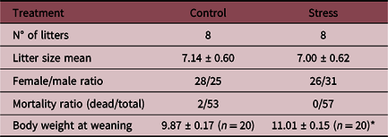
The stress procedure consisted of one 120-min restraint session in a plastic cylindrical device, starting at 10:00 AM every day from GD 14 to delivery (GD 20–21), as previously described.Reference Pascuan, Di Rosso, Pivoz-Avedikian, Wald, Zorrilla Zubilete and Genaro29 Non-stressed dams were left undisturbed in their home cages. This type of stress was selected because it influences the fetus indirectly via direct stress on the mother.Reference Ward and Weisz30 This protocol has been employed in several previous studies and has shown to significantly affect cardiovascularReference Igosheva, Klimova, Anishchenko and Glover31 and neuroendocrine stress reactivity in adult offspring.Reference Sternberg and Ridgway32,Reference Szuran, Pliska, Pokorny and Welzl33 The protocol used did not affect the number of offspring born, the male: female ratio, or the mortality ratio (see Table 1).
Litter parameters analyzed on 8 litters of each group. Litter size was analyzed using a t-test, female/male ratio, and mortality ratio with a Mann–Whitney U test. Body weight at weaning was analyzed using a two-way ANOVA with sex and prenatal treatment as factors because we found no differences between males and females, means are shown without separating by sexes.

*p < 0.0001.
On postnatal day 5, litters were sexed and culled to 6 pups, retaining an equal number of male and female pups, when possible. Discarded animals were euthanized by decapitation.
Pups were weaned at postnatal day 21 and housed in groups by litter and sex under standard conditions. To prevent litter effects from biasing the outcome of observations on adult offspring, only 2 males and 2 females from each litter were used in these experiments (1 animal in each group). Four-week-old offspring were randomly divided into two diets: standard chow diet (SD, Cooperación, San Nicolás, Buenos Aires, Argentina) and homemade HFD and continued on these diets until euthanasia at 28 weeks old (after 24 weeks of diet). Diet composition is available in Supplementary Table S1. (Experimental design and timeline are shown in Fig. 1). We chose to begin the diet at 4 weeks of age since it has been reported that promote greater weight gain in these animals.Reference Nishikawa, Yasoshima, Doi, Nakayama and Uetsuka34 Mice had ad libitum access to water and food, and body weight was recorded once a week.
Experimental design (a) and timeline (b). Pregnant BALB/c mice were divided into two groups: one group received restraint stress daily for 2 h once a day from gestational day 14 (GD 14) until delivery (GD 20−21), the other was left undisturbed. At 4-week-old offspring were distributed in two diet groups: high-fat diet (HFD) and standard diet (SD). After 24 weeks of life (20 of diet), a glucose tolerance test was performed, one week later, the same animals were subjected to an insulin tolerance curve. At 28 weeks of age, animals were euthanized (after 24 weeks of diet).

Intraperitoneal glucose tolerance test
At 24 weeks of age, experimental subjects underwent an intraperitoneal glucose tolerance test. Briefly, after 6 h of fastingReference McGuinness, Ayala, Laughlin and Wasserman35 (10:00 AM–04:00 PM), basal blood glucose concentration was measured (time point 0) by tail nick using a commercial glucometer (OneTouch UltraMini, LifeScan Inc, Johnson & Johnson). Immediately after, animals were intraperitoneally injected with 2 g/kg body weight of glucose (Sigma, St. Louis, MO, USA), and glycemia was measured at 15, 30, 60, and 120 min after the injection.
Intraperitoneal insulin tolerance test
One week after the intraperitoneal glucose tolerance test (week 25 of life), all groups underwent an insulin tolerance test without fasting.Reference McGuinness, Ayala, Laughlin and Wasserman35 Animals were intraperitoneally injected with 1 UI/kg body weight of human recombinant insulin (Insuman R, 100 UI/ml, Sanofi-Aventis Argentina SA), and glycemia was measured at time point 0 (baseline immediately before the injection), 15, 30, and 60 min following the injection using a commercial glucometer (OneTouch UltraMini, LifeScan Inc, Johnson & Johnson, Malvern, PA, USA).
Tissue collection
At 28 weeks of age, body weight was recorded, and animals were euthanized following retro-orbital bleeding after being anesthetized in a CO2 chamber. Plasma was collected, and nose-to-tail length was recorded. Visceral adipose tissue was carefully dissected, weighted, and stored at −80°C until RNA extraction.
Plasma metabolic parameters
Total cholesterol was quantified using Colestat enzimático Reagent (Wiener Lab, Rosario, Argentina) and triglycerides levels using TG Color Reagent (Wiener Lab, Rosario, Argentina). Plasma insulin concentration was determined by an enzyme-linked immunosorbent assay (Mercodia Mouse Insulin ELISA, Uppsala, Sweden) according to the manufacturer’s protocol. The detection limit was 0.2 ug/l with a coefficient of variability of 3%.
Quantitative assessment of mRNA expression by real-time polymerase chain reaction
Total RNA was isolated from visceral adipose tissue using Transzol according to manufacturer’s instructions (Transgenbiotech, Beijing, China). RNA was converted to cDNA using real-time polymerase chain reaction (RT-PCR) with oligo(dT)18 primers and M-MLV reverse transcriptase with deficient RNase H activity (Transgenbiotech, Beijing, China).
RT-PCR was performed for quantitative assessment of mRNA expression with an ABI PRISM 7500 Real-Time PCR System (Applied Biosystems, Foster City, CA, USA) using FastStart Universal SYBR Green Master (Rox) (Roche Applied Science, Mannheim, Germany). All reactions were run in duplicate. The gene expression levels were normalized using glyceraldehyde-3-phosphate dehydrogenase (GAPDH) mRNA as an internal control. GAPDH was found to be the most stable reference gene for testing adipose tissue mRNA expression among other housekeeping genes tested before starting the experiment (β-actin, cyclophilin B, and β2-microglobulin). Primer sequences are summarized in Supplementary Table S2.
Statistical analysis
Data were expressed as the mean ± standard error of the mean (SEM) for each group. All the data were analyzed using STATISTICA 7.0 software (StatSoft, Inc., Tulsa, Oklahoma, USA). The normality and homogeneity of variance for the dataset were tested using the Shapiro–Wilk test and Levene’s test, respectively, and transformed as appropiate. To analyze litter size, we used a t-test, and for other litter parameters Mann–Whitney U test was used. Data were analyzed with the General Linear Model with sex (female and male), prenatal treatment (non-PS or PS), and diet (standard or high-fat) as factors. P < 0.05 was considered statistically significant.
Results
PS together with HFD feeding increase body weight and fat content in females
At weaning, prenatally stressed animals were heavier than the control group, regardless of sex (Table 1, F(1,52) = 23.974; p < 0.0001). One week later, mice were assigned to one of two diets: SD or HFD. The HFD was used to study if there is any metabolic effect produced by stress that only becomes evident when animals faced a metabolic challenge.
As shown in Fig. 2a and b, body weight increased during the experiment with a significant interaction between sex, diet, prenatal treatment, and time (F(24,816) = 1.8791, p < 0.01). Using planned comparisons, we observed that after 5 weeks of HFD intake, PS females developed overweight (Fig. 2a), as a result of both prenatal treatment (p < 0.001) and diet (p < 0.0001). Among PS males (Fig. 2b), HFD produced a rise in body weight only after 14 weeks of HFD feeding (p < 0.05).
Body weight curve (a and b) and adipose tissue content (c and d). Body weight was recorded weekly from week 4 to 28 (from the beginning of HFD until the time of sacrifice). a: females, b: males. A three-way ANOVA with repeated measures was performed to analyze the effect of diet, sex, prenatal treatment, and time on body weight. Non-prenatally stressed + standard diet (NPS+SD, circles), non-prenatally stressed + high fat diet (NPS+ HFD, squares), prenatally stressed + standard diet (PS+SD, diamonds), and prenatally stressed + high fat diet (PS+HFD, triangles). Results from planned comparisons are shown as: #p < 0.05, ###p < 0.0001 PS+SD vs PS+HFD; & p < 0.05 NPS+SD vs NPS+HFD; $$p < 0.001 NPS+HFD vs PS+HFD. c: visceral adipose tissue weight/body length ratio (%). d: visceral adipose tissue weight/body weight ratio (%). For c and d, a three-way ANOVA was conducted to study the effect of diet, sex, and prenatal treatment on visceral adipose tissue content. All values are presented as mean ± standard error (n = 5–6 mice in each group). Results from planned comparisons are shown as: *p < 0.05, **p < 0.001, ***p < 0.0001.

Visceral adipose tissue weight/body length ratio (VAT/L) and visceral adipose tissue weight/body weight ratio (VAT/BW) analysis showed a significant interaction between sex, diet, and prenatal treatment (VAT/L: Fig. 2c; F(1,34) = 4.8883, p < 0.05 and VAT/BW: Fig. 2d; F(1,34) = 9.9218, p < 0.05). Similar results were observed when planned comparisons were performed. Adipose tissue content was significantly increased in PS-females fed with HFD, either compared to PS-females fed SD (p < 0.0001 for both ratios) or to NPS-females fed with HFD (p < 0.001 for VAT/L and VAT/BW, respectively). In males, we observed an increase in fat content associated with HFD consumption, regardless of prenatal treatment (between diets in NPS or PS for VAT/L p < 0.001 and VAT/BW p < 0.0001).
PS along with HFD intake promotes hyperglycemia and hyperinsulinemia in both sexes
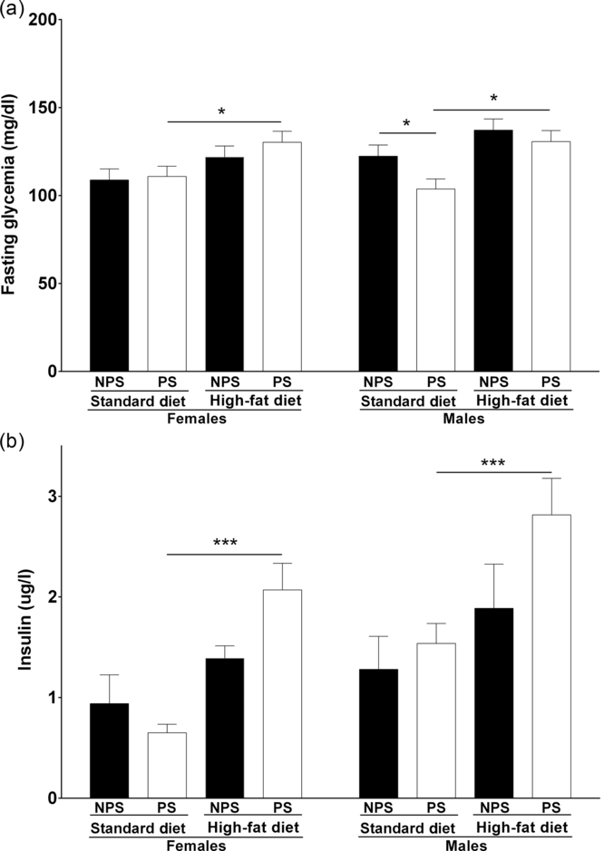
In the glucose tolerance test (Fig. 3a and b), a significant effect of sex was detected (males showed higher glucose levels than females, F(1,34) = 21.92, p < 0.0001). On the other hand, significant differences between diets were observed in the insulin tolerance test (Fig. 3c and d), with higher blood glucose levels in HFD-fed animals (F(1,33) = 8.7013, p < 0.05). Post hoc analysis showed no difference between groups. However, a significant interaction between sex, diet, and PS (F(1,34) = 5.8439, p < 0.05) was observed in basal glycemia (Fig. 4a). Planned comparisons showed that in PS-females, HFD produced an increase in basal glycemia when compared with the SD group (p < 0.05). Intriguingly, between males fed SD diet, PS produced a drop in glucose levels (p < 0.05). Finally, among PS-males, HFD proved to increase glucose levels (p < 0.05).
Panels a and b: Glucose tolerance test: Blood glucose measured during the glucose tolerance curve, for females (a) and males (b). Panels c and d: Insulin tolerance test: Blood glucose measured during the insulin sensitivity test, for females (c) and males (d). Non-prenatally stressed + standard diet (NPS+SD, circles), non-prenatally stressed + high fat diet (NPS+ HFD, squares), prenatally stressed + standard diet (PS+SD, diamonds), and prenatally stressed + high fat diet (PS+HFD, triangles). A three-way ANOVA with repeated measures was performed to analyze the effect of diet, sex, prenatal treatment, and time on glucose levels. All values are presented as mean ± standard error (n = 5–6 mice in each group).

Fasting glycemia (a) and non-fasting plasmatic insulin levels (b). A three-way ANOVA was performed to analyze the effect of diet, sex, and prenatal treatment on glucose and insulin levels. All values are presented as mean ± standard error (n = 5–6 mice per group). NPS: non-prenatal stress; PS: prenatal stress; SD: standard diet; and HFD: high-fat diet. Results of planned comparisons for glucose levels and post hoc analysis for insulin are shown as: *p < 0.05, ***p < 0.0001.

Non-fasting insulin levels were increased in animals fed with HFD (Fig. 4b; F(1,34) = 10.2053, p < 0.05). Post hoc analysis showed a similar response for both sexes with a significant increase in insulin levels in PS-animals owing to HFD intake (p < 0.0001 for both sexes).
Prenatally stressed males exhibit high cholesterol and triglyceride levels
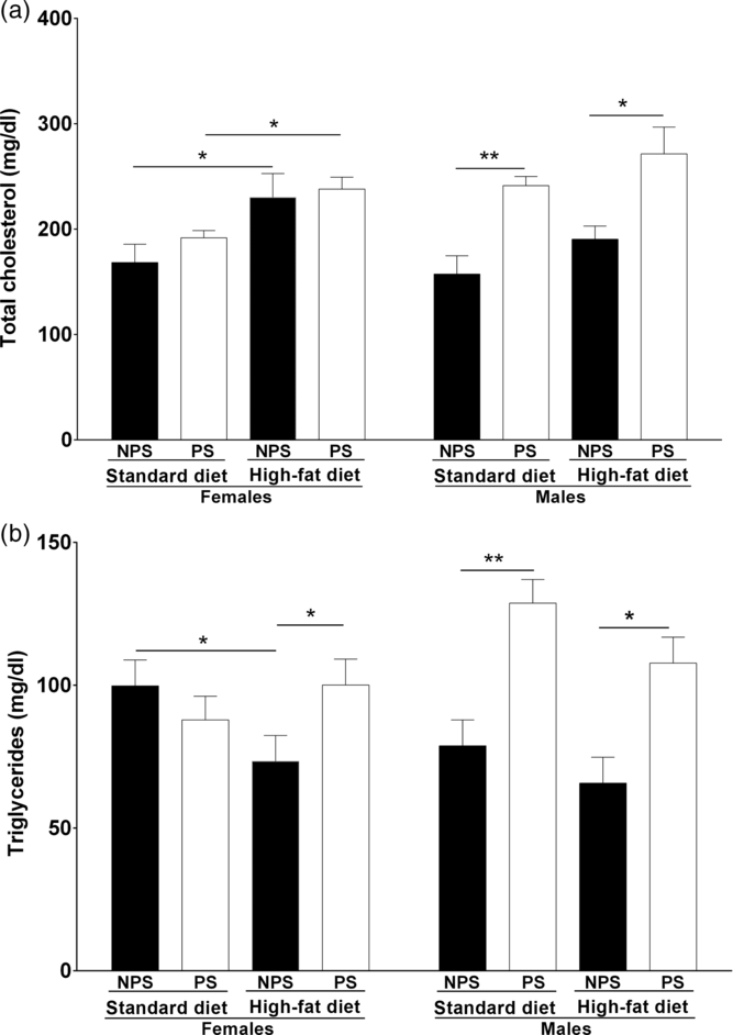
A significant interaction between sex and prenatal treatment on total cholesterol (Fig. 5a, F(1,34) = 8.89, p < 0.05) and triglyceride levels (Fig. 5b; F(1,34) = 9.4475, p < 0.05) was detected. For total cholesterol, planned comparisons showed that females fed with HFD had higher cholesterol levels regardless of prenatal treatment (p < 0.05). On the other hand, PS-males showed increased cholesterolemia, regardless of the diet (p < 0.001 and p < 0.05 for SD and HFD groups, respectively). Planned comparisons revealed that NPS-females fed with HFD exhibited a decrease in triglyceridemia (p < 0.05) which was not observed in PS-females. In males, prenatal exposure to stress produced an increase in triglyceride levels, regardless of diet intake (p < 0.001 and p < 0.05 for SD and HFD groups, respectively).
Total cholesterol (a) and triglyceridemia (b). All values are presented as mean ± standard error (n = 5 mice in each group). NPS: non-prenatal stress; PS: prenatal stress; SD: standard diet; and HFD: high-fat diet. A three-way ANOVA was conducted to study the effect of diet, sex, and prenatal treatment on total cholesterol and triglyceridemia. Results from planned comparisons are shown as: *p < 0.05 and **p < 0.001.

Exposure to PS and postnatal HFD intake leads to an imbalance in the mRNA expression of adipokines
A significant interaction between sex, diet, and prenatal treatment on leptin gene expression was detected (Fig. 6a; F(1,34) = 5.1323, p < 0.05). Planned comparisons revealed that HFD consumption irrespective of prenatal treatment resulted in increased mRNA leptin expression in both sexes (between diets: NPS-females p < 0.05 and NPS-males p < 0.0001; PS-females p < 0.001 and PS-males p < 0.0001). It was also observed that PS-mice fed with HFD exhibit an enhanced expression of leptin transcript in comparison to NPS-mice under the same diet (p < 0.05 for both sexes).
Adipokine gene expression in visceral adipose tissue a: leptin, b: resistin, and c: adiponectin. A three-way ANOVA was conducted to study the effect of diet, sex, and prenatal treatment on leptin, resistin, and adiponectin gene expression. All values are presented as mean ± standard error (n = 5–6 mice per group). NPS: non-prenatal stress; PS: prenatal stress; SD: standard diet; and HFD: high-fat diet. Results of planned comparisons for leptin and resistin and post hoc analysis for adiponectin are shown as: *p < 0.05, **p < 0.001 and ***p < 0.0001.

Resistin mRNA expression (Fig. 6b) showed a significant interaction between sex and diet (F(1,34) = 28.2693, p < 0.0001) and between sex and prenatal treatment (F(1,34) = 5.0637, p < 0.05). Planned contrasts showed that in female mice, PS produced an increase in the expression of resistin only if combined with HFD (p < 0.05 PS+HFD vs NPS+HFD). Males fed with HFD, regardless of the prenatal treatment, showed increased resistin expression levels (p < 0.0001 for both NPS and PS groups). Nonetheless, markedly high levels were observed in the NPS group (p < 0.05 NPS+HFD vs PS+HFD).
Adiponectin gene expression (Fig. 6c) was affected by sex (F(1,33) = 6.6422, p < 0.05) and diet (F(1,33) = 19.5407, p < 0.001). Post hoc analysis revealed that HFD intake in NPS-females is associated with increased adiponectin gene expression (p < 0.05). In males, this increase was independent of prenatal treatment (p < 0.05 for both NPS and PS).
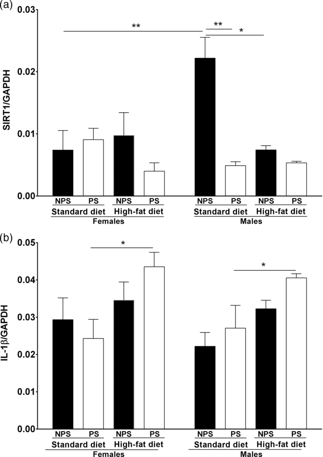
Given the contradictory effects reported for SIRT1 on adiponectin expression (reviewed in Liu and LiuReference Liu and Liu36), we found it interesting to study what happens with its expression in this model. In the present study, SIRT1 gene expression presented a significant interaction between diet, sex, and prenatal treatment (Fig. 7a, F(1,33) = 8.4143, p < 0.05). Planned comparisons indicated that SIRT1 expression was found to be decreased in both PS-males (p < 0.001) and NPS-males exposed to HFD (p < 0.05). Significant differences were also detected between sexes in control subjects (with males showing higher SIRT1 expression levels than females, p < 0.001).
Visceral adipose tissue gene expression. a: Interleukin 1-β; b: Sirtuin-1. A three-way ANOVA was conducted to study the effect of diet, sex, and prenatal treatment on IL-1β and SIRT1 gene expression. All values are presented as mean ± standard error (n = 5–6 mice per group). NPS: non-prenatal stress; PS: prenatal stress; SD: standard diet; and HFD: high-fat diet. Results of post hoc analysis for IL-1β and planned comparisons for SIRT1 are shown as: *p < 0.05.

Since SIRT1 has an anti-inflammatory effect,Reference Yoshizaki, Milne and Imamura37,Reference Yoshizaki, Schenk and Imamura38 and there is also increasing evidence suggesting that IL-1β is strongly implicated in the progression of obesity-related inflammation in insulin resistance in rodent models,Reference Tack, Stienstra, Joosten and Netea39,Reference Wen, Gris and Lei40 we decided to analyze IL-1β gene expression. We found that IL-1β expression levels were affected by diet (Fig 7b, F(1,34) = 13.5271, p < 0.001). Post hoc analysis showed that specially PS together with postnatal HFD produced an increase in IL-1β gene expression levels in both sexes (p < 0.05).
Discussion
Disruptions suffered in the intrauterine environment can lead to long-lasting consequences for the health of exposed offspring.Reference Entringer, Buss and Swanson41 Numerous epidemiological studies have shown an association between intrauterine disturbances and increased incidence of obesity, type 2 diabetes,Reference Reynolds42 and hypertension in adulthood.Reference Yu, Tain and Tiao43 Various studies have shown that PS is associated with a significant decrease in birth weight, as a result of intrauterine growth retardation (IUGR).Reference Drago, Di Leo and Giardina44,Reference Lesage, Del-Favero and Leonhardt45 It has been proposed that low birth weight does not itself increase the risk of non-communicable diseases directly, but rather favors accelerated postnatal growth. In this regard, a systematic review highlights the importance of rapid postnatal growth in underweight babies.Reference Kelishadi, Haghdoost, Jamshidi, Aliramezany and Moosazadeh46 Rapid catch-up growth can be considered a risk factor for the development of cardiovascular diseases and associated phenotypes.Reference Dulloo, Jacquet, Seydoux and Montani47,Reference Berends, Fernandez-Twinn, Martin-Gronert, Cripps and Ozanne48 Of note, in the present study, birth weight was not recorded in order to avoid excessive handling of the animals. We have previously performed a meta-analysis in rodents showing that birth weight is significantly decreased in prenatally stressed animals.Reference Burgueño, Juarez, Genaro and Tellechea22
At weaning, PS-offspring of both sexes showed increased body weight in comparison to unstressed controls, supporting recent findings indicating that prenatal environment may influence the likelihood of developing overweight and obesity.Reference Salsberry and Reagan49 Restraint stress would modify maternal behavior contributing to the long-term effects observed in offspring.Reference Maccari, Darnaudery, Morley-Fletcher, Zuena, Cinque and Van Reeth50 However, in a recent meta-analysis, we demonstrated that cross-fostering to non-stressed dams had the same effects on body weight than continuing with the same stressed mother,Reference Burgueño, Juárez, Genaro and Tellechea24 suggesting that maternal stress does not influence body weight of these animals.
In both male and female mice, body weight and adiposity showed no significant variation in animals exposed to stress during gestation. In males, an increase in body weight and adiposity was observed related to HFD-intake, albeit not to prenatal treatment. On the other hand in females, PS was associated with increased body weight and adiposity in HFD-fed mice, suggesting a predisposition to the obese phenotype in PS-female offspring triggered after a challenge with HFD. Controversial results have been found in the literature on this behalf. It has been shown in Sprague Dawley rats that the combination of PS (either induced by exogenous GCs or by a variable stress paradigm) and HFD intake determines the development of obesity and leads to the increase of adipose tissue.Reference Tamashiro, Terrillion, Hyun, Koenig and Moran51,Reference Tsai, Tiao and Sheen52 In contrast, when PS was due to movement restriction, some studies showed a reduction in body weight and adiposityReference Baran, Campbell and Kleen53,Reference Burkuš, Kačmarová and Kubandová54 while others reported that restraint stress does not produce any change in phenotype.Reference Bruder-Nascimento, Campos, Alves, Thomaz, Cicogna and Cordellini55
Regarding glucose metabolism, increased basal blood glucose and insulin were found in PS-mice fed with a HFD in both sexes, which suggests the presence of insulin resistance in this animals. Of note, alterations in glycemic control were not observed in NPS-offspring under a HFD regime, suggesting that a HFD per se may not induce this phenotype in BALB/c mice.
In general, no alterations were found in the glucose tolerance test or in the insulin sensitivity curve. Different studies have reported alterations in the glucose tolerance curve, especially after HFD intake, however, these studies were mainly conducted on rats.Reference Panetta, Berry and Bellisario56-Reference Abildgaard, Lund and Hougaard58 Besides, some authors have found, in rat models of PS, high glycemia during the insulin tolerance test, especially when the animals consumed HFD.Reference Panetta, Berry and Bellisario56,Reference Sheen, Hsieh and Tain59 The presence of hyperinsulinemia has been also described in PS animals, both mice and rat fed different obesogenic diets.Reference Boersma, Moghadam, Cordner and Tamashiro57,Reference Balasubramanian, Varde, Abdallah, Najjar, Mohan Kumar and Mohan Kumar60,Reference Paternain, de la Garza, Batlle, Milagro, Martínez and Campión61 It may be hypothesized that the observed hyperinsulinemia is predetermined by PS, which has been also shown to reprogram the activity of the HPA axis, by increasing the basal levels of GCs. This induces insulin resistance by reducing the expression and phosphorylation of insulin receptor substrate 1 (IRS-1) and thus decoupling intracellular signaling from insulin receptors and preventing the decrease of glucose levels to normal values.Reference Sakoda, Ogihara and Anai62
In the present study, HFD-fed female mice showed an increase in blood cholesterol levels. Besides, plasma triglyceride concentration was found to be decreased in NPS-females fed with a HFD, but not in PS-animals. In contrast, PS-males, regardless of the diet, presented hypercholesterolemia and hypertriglyceridemia, both associated with the presence of obesity and the development of insulin resistance.Reference Haffner, Valdez, Hazuda, Mitchell, Morales and Stern63 Accordingly, it has been reported that PS-animals showed higher triglyceride values.Reference Wyrwoll, Mark, Mori and Waddell64-Reference Othman, Ammari, Sakly and Abdelmelek66 On the other hand, a recent study conducted in BALB/c mice at 60 days of age showed that PS applied between GD 8 and 21 was not associated with alterations in triglycerides, but increases cholesterol in females.Reference Luft, Levices, Pedrazza, de Oliveira and Donadio67
Leptin and resistin are known to be associated with an increase in the number and size of fat cells and, consequently, with an increase in body weight or fat content.Reference Owecki, Miczke, Nikisch, Pupek-Musialik and Sowiński68,Reference Könner and Brüning69 Both adipokines have been proposed to be involved in the development of insulin resistance.Reference Barnes and Miner70 In contrast, adiponectin plays the opposite role, sensitizing tissues to the effects of insulin. In the present study, it was observed that leptin mRNA expression is increased in both male and female mice fed with HFD, and this increase is higher if the animals had been exposed to PS. As far as we know, there are no published studies measuring gene expression of leptin in adipose tissue in PS-animals. Tsai and colleaguesReference Tsai, Tiao and Sheen52 reported that, in a model mimicking PS by exogenous GCs administration during gestation, prenatally treated animals had decreased expression of leptin in the retroperitoneal adipose tissue, while this expression was increased when the animals were fed with HFD postnatally. In agreement with the results presented here, many authors have reported that circulating leptin levels increases as a result of HFD intake but not in PS-animals.Reference Tamashiro, Terrillion, Hyun, Koenig and Moran51,Reference Tsai, Tiao and Sheen52,Reference Panetta, Berry and Bellisario56,Reference Paternain, de la Garza, Batlle, Milagro, Martínez and Campión61 The mRNA expression of resistin was found to be increased in PS-females under a HFD regime, whereas in males increased resistin mRNA expression was found in HFD-fed mice. Finally, concerning the expression of adiponectin, we observed that in NPS-females, there was an increase in the mRNA levels caused by the intake of a HFD. In contrast, males fed with HFD showed a higher expression of this gene independently of prenatal treatment. Contrary to our results, it has been reported that in a PS model due to sleep fragmentation, male offspring showed a lower expression of adiponectin in the visceral adipose tissue, which was additionally associated with higher body weight, higher food intake, and insulin resistance.Reference Khalyfa, Carreras, Almendros, Hakim and Gozal71 Another study that measured circulating adiponectin found that it was increased due to the intake of HFD and was unaffected by PS in both sexes.Reference Panetta, Berry and Bellisario56
SIRT1 is a NAD+-dependent deacetylase protein and a key metabolic factor connecting environmental nutrient signals with energy homeostasis.Reference Schug and Li72 Among many functions, SIRT1 controls inflammatory responses, reduces adipocyte secretion, and maintains glucose and lipid homeostasis. Moreover, SIRT1 participates in glucose metabolism by increasing insulin secretion by pancreatic β cells and modulating insulin signaling.Reference Llorente, Brito, Machado and González73 It is still controversial the role SIRT1 plays on the expression of adiponectin. It has been described that in a caloric restriction setting, both SIRT1 and adiponectin are increased; however, SIRT1 has been also reported to inhibit the expression of PPARγ, a known positive regulator of adiponectin synthesis and secretion. It has been described that adipose-tissue-specific SIRT1 knockout mice show severe glucose intolerance under HFD feeding.Reference Chalkiadaki and Guarente74 Tsai and colleaguesReference Tsai, Tiao and Sheen52 observed that the amount of SIRT1 protein (measured by western blot) decreased by HFD intake in both control and prenatally dexamethasone-treated animals. In accordance, we found that male mice exposed to PS or HFD presented lower levels of mRNA SIRT1 expression, suggesting that downregulation of SIRT1 in the adipose tissue may be protective against obesity. However, this was not observed in animals exposed to both PS and HFD evidencing more complex underlying mechanisms. Further research will be necessary to elucidate these and other mechanisms that may be at work.
Since 1) an anti-inflammatory role has been proposed for SIRT1, 2) inflammation associated with obesity is considered to play a role in the development of comorbidities such as metabolic syndrome, and 3) the elevation of inflammatory cytokines is associated with insulin resistance and type 2 diabetes;Reference Schuster75-Reference Emanuela, Grazia, Marco, Maria Paola, Giorgio and Marco77 we decided to analyze IL-1β gene expression. Interestingly, an upregulation of the mRNA expression of IL-1β was observed in PS-mice fed with HFD in both sexes. We may hypothesize that PS programs and the HFD triggers this increased expression, which is not observed in mice exposed only to HFD. Mark and colleaguesReference Mark, Wyrwoll, Zulkafli, Mori and Waddell78 reported that, in a rat model that mimics PS through the exogenous administration of GCs during gestation, adipose tissue gene expression of IL-1β was increased in both sexes. Our results may not match with those reported by these authors presumably because we use a model of PS, which may not be as strong as prenatal dexamethasone administration.
This work has some limitations that should be addressed and taken into account in future studies. First, corticosterone concentration was not assessed; however, several authors have previously shown that stressed mothers had significantly higher levels of corticosterone than non-stressed mothers.Reference D’mello and Liu65,Reference Ward and Wainwright79 Second, we used females, which were studied without performing vaginal cytology, so precise stages of the estrous cycle could not be identified, this is something to be amended in the following studies. Finally, regarding gene expression analysis, although we tested several housekeeping genes and selected the most stable one, only one reference gene was used. Moreover, protein expression has not been analyzed, and conclusions were drawn only from mRNA analysis of selected genes.
In the present work, we used BALB/c mice, a strain rarely used for metabolic research owing to its low sensitivity to the development of metabolic alterations under HFD.Reference Montgomery, Fiveash and Braude80,Reference Montgomery, Hallahan and Brown81 Despite its higher sensitivity to stress,Reference Savignac, Dinan and Cryan82,Reference Razzoli, Carboni, Andreoli, Ballottari and Arban83 we found increased body weight and adiposity in PS-female mice fed with HFD, suggesting that females are prone to develop obesity under a HFD if previously exposed to stressful conditions during development. In contrast, in males, exposure to HFD was condition enough to attain increased body weight and adiposity. In addition, an increase in mRNA expression of leptin and resistin in the adipose tissue was observed. We unexpectedly detected an increase in the transcript levels of adiponectin in males fed with HFD, which requires further research. We would propose that, in females, PS is a predisposing factor that would operate through epigenetic mechanisms, although this specific hypothesis was not tested in this study. Insulin and glycemic levels were increased in male and female PS-mice fed with HFD, which together with the increase in body weight and adiposity may suggest the presence of insulin resistance. Insulin resistance may also be related to the increase in IL-1β and the decrease in SIRT1 expression observed in males.
We conclude that in BALB/c, males have greater susceptibility to PS than females. However, PS would predispose females to a greater metabolic decline than males if they were fed with HFD postnatally.
Supplementary materials
For supplementary material for this article, please visit https://doi.org/10.1017/S2040174420000987
Acknowledgments
The authors would like to thank Johnson & Johnson for the kind donation of the glucose test strips and to Maria Rosa Gonzalez Murano and Maria Agustina Vidal for their invaluable technical assistance.
Financial Support
This work was partially supported by Agencia Nacional de Promoción Científica Tecnológica (MLT, PICT 2015-1567), (AMG, PICT 2016-2727), and Consejo Nacional de Investigaciones Científicas y Técnicas (AMG, PIP 2015-2017 N° 11220150100163). MW, MLT, AMG, and ALB are members of Consejo Nacional de Investigaciones Científicas y Técnicas.
Conflict of interest
YRJ is currently employed by AstraZeneca. The other authors declare that they have no conflict of interest.
Ethical standards
The authors assert that all procedures contributing to this work comply with the ethical standards of the relevant international guides on the care and use of Laboratory animals (“Guide for the Care and Use of Laboratory Animals” (NIH) (revision 2011) and to the EC Directive 86/609/EEC (revision 2010)) and has been approved by the institutional committee (CICUAL BIOMED Res N°007/2016).